Tooth extrusion – what is this procedure? The essence of the method and indications for its use
Tooth extrusion is a separate dental procedure. Nowadays it is performed quite rarely, but in some clinical cases it is precisely this that makes it possible to preserve the root in case of severe destruction of the visible part of the tooth and subsequently, based on it, restore the crown using modern methods of restoration and prosthetics. Extrusion was first introduced to the world in 1973. The author of the technique was Dr. GS Heithersay, who proposed this procedure as an alternative to removing roots with fractures1. Read more about the essence of this method and how such treatment is carried out further in this article.
Why might extrusion develop?
Disc extrusion can begin if the protrusion has not received the necessary treatment for a long time. The main causes of this disease are usually osteochondrosis and related diseases. But this list is not limited to them. Here are other possible causes of extrusion:
- completeness;
- regular strong physical activity;
- age;
- metabolic problems;
- spinal injuries;
- lack of movement.
Strong physical exertion can lead to the development of extrusion
What is the essence of the procedure
Tooth extrusion is the procedure of pulling a tooth or remaining root out of the gum in a vertical direction. According to this definition, extrusion allows a slight elevation of the root and creates more suitable conditions for a reliable and durable crown restoration. As part of this procedure, the tooth is usually pulled out by 3-4 mm. But to carry it out, it is important that at least the dentinal rim is preserved. Its thickness must be at least 1 mm, and its height must be at least 2 mm. If there is no such rim, the specialist may try to pull it out from under the gum.
Tooth extrusion is a procedure for pulling out a tooth or remaining root from the gum.
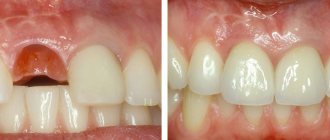
This procedure in some cases allows you to avoid root removal and subsequently restore the crown on it. However, if the root is damaged, its complete extraction followed by replacement with an implant will be the best solution. By the way, extrusion promotes the formation of bone tissue, so the procedure can also be performed as an alternative to artificial expansion of the jawbone for subsequent installation of implants.
Tips for preventing illness
In the ranking of the importance of medical actions in relation to extrusion, one of the first places is occupied by the prevention of this disease.
To effectively prevent extrusion or its complications, certain measures can be taken:
- do physical exercise regularly. Overworking is harmful, but in order for the spine to be healthy, sufficiently trained back muscles are necessary;
- stop smoking and drinking alcohol;
- when working in a job that is harmful to the spine, it is better to change profession;
- take care of your posture, avoid wearing shoes with high (more than 40 mm) heels;
- organize your workplace wisely;
- take timely measures against infections and inflammations in the body;
- If you experience the slightest discomfort, consult a doctor immediately.
To prevent the disease from occurring, it is important to carry out prevention
Trying to treat yourself on your own is dangerous: it can lead to severe complications, as a result of which the person will lose his ability to work and may even remain disabled. At the first signs of discomfort, you should immediately contact a specialist.
Indications for extrusion
As mentioned above, this method is indicated for severe destruction and fracture of the crown of the tooth, as well as for a root fracture, but only if the fracture is in the gum. For reference: according to ICD-10, the root fracture is assigned the number S02.5. Among other indications for the use of this method, dental experts highlight retention. This diagnosis is made when the coronal part does not completely erupt or remains in the gum. This situation is especially typical for wisdom teeth.
“They performed extrusion on me. There was a tooth that was constantly being destroyed. The doctors increased it several times, but it became so dark and chipped all the time. When I once again came to the appointment, they again removed all the composite, they cleaned something there for a long time, and then they said that’s all, I can’t build it up anymore. Instead of removing it, they suggested pulling out the root a little. I agreed. The procedure went well and there was no pain. In the end, they installed a crown, and it’s holding up so far.”
Ira, review on the website www.32top.ru
This procedure can be life-saving if the carious cavity is localized below the gingival level. By removing the affected surface, it is possible to prevent the influence of harmful microorganisms on the mucous membrane and its inflammation.
Traction is also prescribed for retention
Another indication, which was already mentioned above, is insufficient thickness and height of the jaw bone to securely fasten the implant. Extrusion of the tooth root allows you to free up some space between it and the alveolus, which will promote the growth of bone tissue cells and connective epithelium.
Orthodontic extrusion for complex smile rehabilitation
Orthodontic extrusion allows for significant process optimization during periodontal or orthopedic treatment of patients. In some cases, orthodontic extrusion even allows one to avoid soft tissue and bone augmentation procedures.
In this article we will describe a case in which orthodontic intervention allowed the formation of the necessary functional and aesthetic contour for further rehabilitation with the help of dental implants in the area of the central incisors of the upper jaw. The entire treatment process included the implementation of the orthodontic and periodontal phases, extraction, installation of implants and fixation of first temporary and then final restorations. Thus, we were able to achieve not only an aesthetically acceptable result, but also stabilize the position of the gums without compromising the quality of the supporting bone tissue, and improving the overall occlusal pattern.
Case review
A 28-year-old female patient sought dental care due to problems with her maxillary central incisors (Figure 1). After falling off a bicycle, she underwent endodontic treatment with further crowns on teeth 8 and 9. Ultimately, she was unhappy with the appearance of her front teeth and wanted rehabilitation with dental implants. During the diagnosis, it was discovered that these teeth were indeed hopeless for therapeutic treatment, however, additional orthodontic intervention was necessary to install implants. In addition, orthodontic treatment would reduce the level of treatment-associated complications and optimize the achievement of the most acceptable aesthetic results. In the same way, the doctor would be able to reduce the total duration of treatment.
Photo 1. View of the patient’s smile before treatment.
General somatic and dental history
The patient’s general somatic history was not burdened, except for the presence of a previous history of smoking. According to the American Society of Anesthesiologists (ASA) classification, it could be classified as class I. At least one study suggests a correlation between smoking and periodontitis, which should be considered during diagnosis. The patient was also very afraid of any dental procedures, especially anesthesia procedures. Subjectively, she noted sensitivity to temperature stimuli and the possibility of a certain migration of the lower jaw to achieve the position of maximum contact between antagonist teeth.
Diagnosis, risk assessment and prognosis
Periodontal assessment: The patient had not previously visited the dentist regularly and showed signs of periodontal disorders. Thus, in the area of some teeth, a slight loss of periodontal attachment was recorded, recession in the area of teeth No. 3, 6, 10, 13–15, 24 and 29; periodontal pocket 4 mm deep around the 18th tooth and bleeding when probing 11 teeth. The patient's previous smoking habit was considered as a potential risk factor.
Risk of periodontal complications: low (American Academy of Periodontology [AAP] II) Prognosis: moderate to good.
Biomechanical assessment: The patient had active caries in the area of teeth 13 and 29. The crowns in the area of 8 and 9 teeth were defective with signs of secondary caries in their area and traces of chipped ceramics (photo 2). Problematic amalgam fillings were noted in areas 2-4 and 13. A structurally compromised condition was typical for teeth 18 and 31, given the too large size of the ceramic inlays fixed on them. In the area of teeth 8 and 9, the radiograph also showed bone loss and the formation of periapical lesions. The roots of teeth 7 and 10 were inclined mesially.
Risk of biomechanical complications: high. Prognosis: poor for carious-affected teeth 8, 9, 18 and 31; satisfactory for teeth 2 - 4 and 13.
Photo 2. X-ray of area 8 and 9 before treatment.
Functional assessment: Signs of wear were noted in the area of the anterior teeth. In the area of teeth 8 and 9, ceramic chipping was recorded. The patient reported having problems with the temporomandibular joint. During the examination, a range of motion study, loading and immobilization tests were performed. In addition, the patient also noted a migration of mandibular positions to achieve a state of maximum contact between the teeth. All this indicated the presence of occlusal dysfunction in the patient.
Risk of functional complications: moderate. Prognosis: satisfactory.
Assessment of maxillofacial parameters: The position of the lip line, even in a relaxed smile, provoked a significant exposure of the gingival profile. With a full smile, the teeth and gums of the upper and lower jaw were completely visualized (Figure 3). The patient was dissatisfied with the appearance of the crowns on teeth 8 and 9, given the signs of chipped ceramics. She was also concerned about signs of crowding in her lower front teeth. In addition, the patient was informed of the sloping smile line from right to left, which was also noticeable when she smiled. However, this violation of aesthetics did not bother her too much.
Risk of maxillofacial complications: high. Prognosis: bad.
Photo 3. External profile view of the patient with a full smile: excessive exposure of the gum area.
Treatment Goals
The main goal of the treatment was to create adequate space and an optimized aesthetic profile for the placement of dental implants in the area of 8 and 9 teeth. Because the roots of the lateral incisors were mesially inclined, the decision was made to correct this condition orthodontically. Considering that after the removal of teeth 8 and 9, a noticeable loss of the bone tissue supporting them is possible, it was planned to carry out orthodontic extrusion of these teeth, which would allow modifying the appearance of the gingival contour in the frontal area. In addition, controlled orthodontic treatment would also normalize the patient's occlusal pattern, reducing the risk of developing occlusal dysfunction.
Sequence of treatment
Stage 1: orthodontic and periodontal treatment.
The patient underwent scaling and root cleaning procedures with further monitoring of the periodontal condition after 2 months. A follow-up visit was also scheduled after 4 months. The crowns of teeth 8 and 9 were removed and replaced with temporary crowns (Luxatemp, DMG America) using universal composite cement (RelyX Unicem, 3M ESPE). The orthodontic stage of treatment (extrusion of teeth 8 and 9) took approximately 12 months (photo 4). During extrusion, the temporary crowns were adjusted to maintain adequate occlusal relationships. At the same time, the inclination of teeth 7 and 10 was corrected to create at least 1.5 mm of bone space between the roots and the planned position of the implants.
Photo 4. Fixation of braces on provisional crowns to ensure extrusion.
Stage 2: extraction and installation of implants
After achieving the required level of extrusion (photos 5-6), a minimally traumatic extraction of teeth 8 and 9 was performed. The orthodontic treatment allowed us to achieve the formation of an ideal alveolar contour (photo 7). The implants used were Astra Tech OsteoSpeed TX 4.0 SX 11 mm (Dentsply Sirona), which were installed with minimal bone augmentation of the socket using a bone graft (Puros Cortico-Cancellous Particulate Allograft, Zimmer Biomet). Thanks to the modified Essix retainer, it was possible to ensure the installation of implants 4 mm below the free edge of the gums. X-ray data indicated excellent quality of bone tissue in the intervention area. The roots of the extracted teeth were shortened and modified to an ovoid shape - they were fixed along with temporary crowns over the implantation area to maintain an adequate level of soft tissue (Figure 8).
Photo 5. Completion of the extrusion process.
Photo 6. Dynamic contouring of the lingual surface of teeth during extrusion.
Photo 7. Minimally invasive removal of teeth 8 and 9.
Photo 8. Modification of provisional structures over the area of installed implants.
Stage 3: temporary and final restorations
After 8 weeks of healing, the temporary structures fixed to the retainer were removed. The mucosa successfully migrated into the areas between the implants and the overhanging crowns (Figure 9). Next, the screw-retained temporary restorations were secured to the implants (Figure 10) for a period of 4 weeks. Thus, it was possible to more accurately modify the aesthetic profile of the gums, after which the doctor began taking impressions. Milled lithium disilicate structures were used as abutments (photo 11), which were covered with lithium disilcate crowns (e.max, Ivoclar Vivadent) (RelyX Unicem).
Photo 9. Migration of the soft tissue area around provisional restorations.
Photo 10. Provisional crowns with screw fixation.
Photo 11. View of lithium disilicate abutments on the model.
Discussion
Despite the result of the preliminary treatment, thanks to the complex intervention, the doctor was able to achieve not only an aesthetically acceptable appearance of the frontal area of the upper jaw, but also normalization of function and occlusal relationships (photo 12), without compromising either the condition of the gums or the quality of the supporting bone tissue. Thanks to a step-by-step protocol for extrusion and augmentation of the sockets, the doctor was able to ensure that the implants were installed in sufficiently dense surrounding bone tissue. The extrusion procedure also allowed the reconstruction of an adequate soft tissue profile and the required papilla height (Figure 13). This rehabilitation protocol also made it possible to reduce the risk of developing biomechanical complications due to the removal of two problematic teeth, however, the patient still needed to at least treat caries in the area of teeth 13 and 29. Additionally, she was prescribed the use of fluoride gel (CTx4, Carifree). At the time of this writing, large questionable restorations were noted in the area of teeth 18 and 31, although all amalgam fillings had been replaced. This also improved the biomechanical prognosis. The risk of functional complications was reduced due to the normalization of occlusal relationships due to orthodontic treatment. Subjectively, the patient noted the disappearance of the symptom of migration of the lower jaw upon reaching the state of maximum contact of the teeth of the upper and lower jaw. As for the aesthetic parameters, they will remain somewhat compromised, given the high position of the upper lip. Implant-supported restorations provided an appropriate external contour while visualizing the health of the surrounding soft tissue.
Photo 12. View of the final restorations.
Photo 13. View of the patient’s smile after treatment.
Conclusion
In conclusion, it is important to note that the implementation of the three stages of the described treatment protocol was ensured through the collaboration of three different specialists in one clinic (periodontologist, orthodontist and orthopedist). Thus, the number of required visits was reduced to a minimum, while increasing the overall effectiveness of treatment. The financial benefits of such a rehabilitation algorithm, which eliminates the need for soft tissue and bone augmentation, should also be taken into account.
Authors: Scott L. Rice, DDS Taylor S. Rice, DDS Nicolas A. Ravon, DDS, MSD
Contraindications to the use of the method
Like any other dental procedure, extrusion has its contraindications. For example, it can only be carried out if the adjacent soft tissues and teeth are in satisfactory condition, since they will act as supports for fixing the traction structure.
The procedure will not be possible if the roots are too short or severely curved, and the condition of the canals and crown does not allow the installation of at least one of the elements of the corrective system: ligatures, a button or a pin with a hook.
Therefore, before prescribing extrusion, the doctor must assess the condition and length of the roots, their location. If the patient is diagnosed with an occlusion pathology in which there is no free space for traction, the procedure will not be possible. And one more thing: the method is indicated exclusively for pulpless teeth, so the nerves will first have to be removed if this has not been done previously.
How to recognize the disease
The symptoms of this disease are determined by which part of the spine begins to suffer from it. Extrusion involves incomplete prolapse of the central core of the disc, but this does not affect the nerve roots. So the disease usually does not cause noticeable symptoms such as pain. And only a pinched nerve will “inform” about the presence of problems and the need to consult a specialist.
In the case of cervical extrusion, if a nerve is pinched, the person will begin to have a headache, especially the back of the head. When turning the head, convulsions may occur. However, most often extrusion develops in the lower back. In this case, the symptoms of the disease are as follows:
- numbness of the lower extremities;
- tingling in the fingers of the lower extremities;
- buttocks or thighs go numb;
- pain is localized in a place “subordinate” to the affected nerve.
With this disease, sharp painful attacks occur quite rarely. The doctor makes a diagnosis based on completely different manifestations and symptoms. For example, testing the patient's knee reflex.
Back pain accompanied by the same sensation in the legs is a clear symptom of extrusion
Prices for orthopedic corsets and posture correctors
In most cases, a disc in the lumbar region falls out in fairly elderly people. At this age, the tissue becomes thinner, so extrusion is not uncommon in people over 60 years of age. At the same time, the thigh along the sciatic nerve may also hurt. The latter is characterized by an extremely high sensitivity to pain, so that the pain in the knees and legs can be very severe.
Young people often experience neck extrusion caused by poor posture when sitting at a desk. In this case, the following symptoms are often encountered:
Methods of carrying out the procedure
There are two main methods of extrusion. Let's look at each type in a little more detail:
- orthodontic is a more gentle method, which is often combined with correction with a brace system. This method is indicated for impacted teeth, but it requires more time and patience to achieve results. Pulling is carried out by installing an appropriate structure,
- surgical is a rather radical method, which is fraught with serious complications if errors are made on the part of the doctor. But if everything is done correctly, the result can be obtained much faster. The method does not require long-term wearing of the traction system. We will talk further about how extrusion is carried out in this case.
In the meantime, it should be noted that after surgical traction, the patient is given a temporary restoration for the period of tissue and ligament restoration. It allows you to quickly restore aesthetics.
Types of extrusion
Doctors perform two types of extrusion – orthodontic and surgical.
The orthodontic method is more gentle and can be combined with wearing braces; it is often used if there are impacted elements, but it requires more time and patience from the patient, as well as more visits to the clinic.
The surgical method is more radical, and if performed incorrectly, complications can occur. But with a technically purely performed operation, the result can be achieved faster. At the same time, you do not have to wear orthodontic devices, which during the treatment period violate the aesthetics of the smile and cause some discomfort.
During the period of tissue healing and ligament restoration after surgical extrusion, the patient is given a temporary restoration that makes the smile attractive - this is impossible with the orthodontic method.
Preparatory stage
Before the procedure, regardless of the chosen method, it is important to undergo a full diagnostic examination to ensure that the patient has no contraindications. You will need to take an X-ray or undergo a computed tomography scan, and a procedure performed by a prof. hygiene to remove plaque and deposits, as well as remove the nerve from the causative tooth if the crown is preserved and depulpation has not been performed previously.
Before the procedure, the patient should undergo a computed tomography scan
How is extrusion performed using the orthodontic method?
In this case, traction is carried out using a special orthodontic system. Its design includes the following components:
- arch or bracket - one of these parts is fixed on elements adjacent to the causative tooth using a composite or special orthodontic rings. If the patient already has braces, the bracket is not installed;
- a screw or a special pin with hooks is attached to the causative tooth, that is, to the remaining crown. If there is not enough hard tissue for this, the pin is fixed in the root canal using dental cement,
- traction – they combine the above-described structural elements and create a force effect.
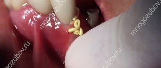
The photo shows the surgical method.
The system is fixed under local anesthesia, since to install it the doctor has to cut the fibers that connect the gum to the root. This manipulation is called fibrotomy. Due to this, the specialist gives the root mobility in order to speed up the pulling process and prevent it from returning to its original position.
Diagnostics
The doctor may suspect extrusion if the patient has the symptoms described earlier. During a neuralgic examination, he can recognize pathological reflexes. Pain often occurs when the patient performs the Lasegue test, which involves raising a straight leg. Then the result is called positive. The patient also sometimes has changes or no sensation at all in the leg, as well as in the foot.
In some cases, the doctor may prescribe blood tests to promptly recognize inflammatory processes or infection.
Ordinary radiography helps determine the extent of the spinal pathology. And yet it never helps to recognize the condition of the intervertebral discs at the moment. To find out what is happening to the discs, an MRI (magnetic resonance imaging) of the affected part of the spine should be performed. And in order to reduce the likelihood of error to a minimum, MRI examinations are performed on high-field equipment with a magnetic flux intensity of 1 Tesla, or better yet, 1.5. Using these devices does not cause any harm to the patient’s body.
If you want to learn in more detail how the MRI procedure of the spine is performed, and also consider when a magnetic resonance examination is indicated, you can read an article about this on our portal.
MRI is one of the most informative diagnostic methods
You can determine which nerve is compressed using EMG (electromyography).
How long does treatment last?
After the procedure, the patient will have to visit the orthodontist once a week so that he can monitor the process and adjust the tension of the rods. Usually, in one week, in this way it is possible to extend the root by about 1 mm. Therefore, to stretch it by 3-4 mm, you need to visit the doctor about 3-4 times, and the process itself can take up to a month.
But the treatment does not end there. It will take some time for the root and ligaments to recover. This process usually takes a couple of months. In the meantime, the patient is undergoing a temporary restoration with the removal of the causative element from the bite and its splinting - this is necessary to relieve the chewing load from it.
Expert recommendations during treatment
During the period of traction, orthodontic experts recommend increasing your oral care. You should brush your teeth not only in the morning and evening, but also every time after eating. It is also recommended to chew on the opposite side and try to eliminate any mechanical factors that could interfere with the healing process. If the structural elements rub the mucous membrane, you should resort to orthodontic wax.
Orthodontic wax will help protect the mucous membrane from damage
Corpus horizontal movement of the tooth. Elimination of three, diastema, protruding teeth.
Part 4.
aligner overview page here This article describes the solution to certain clinical situations in the oral cavity, in particular, the elimination of teeth and diastemas (gaps between teeth). The page is an excerpt from the work of Ryakhovsky A.N. and Arevadze T.Yu., for complete information, please see the source.
Most problems can be solved in different ways, the main thing is to choose the optimal one.
A rail attachment should also be used to close the diastema and trema (Photo 13).
Photo 13
Features of the surgical procedure
This method is most often used in cases where the crown part of the tooth is so damaged that it can no longer be restored in the classical way, or if it is necessary to re-prosthetize / replace the old restoration material. This procedure can also be performed if the patient wants to get results faster and without the need for temporary “aesthetic loss”.
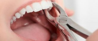
This operation is not classified as complex, but it requires appropriate experience and skill from a specialist. For pulling, the doctor uses a special scalpel, elevator and forceps. You could say he creates a dislocation and then does not completely remove the root, after which he fixes it with sutures.
The photo shows the surgical method of the procedure
Hernia formation
A spinal hernia develops in four main phases. Each of them has its own characteristics. Their treatment is also different, and the later the phase of the disease, the more difficult it is to cure it.
Phase number one. Prolapsed intervertebral disc
Degenerative phase. Now the intervertebral disc has a deficiency of fluid - this leads to a drop in pressure in the disc, loss of elasticity, softening, and greater pliability. Although there is little fluid, the volume of disc tissue, of course, does not disappear anywhere, as does the axial load. Moreover, the load may later increase altogether, for example, if the body tends to be overweight or if a person does not eat properly. Because of this, a gradual protrusion of the disc wall begins, and it protrudes beyond the limits of the spine allocated by nature. This is just the first stage of the development of the disease.
Prolapse of the intervertebral disc is the first stage of hernia formation
There are still no significant symptoms. Only slightly higher back fatigue and accelerated fatigue from physical activity are noticeable. There is no pain yet, so patients usually do not pay any attention to this phase. They feel that there is no need to seek the help of a specialist.
Phase number two. Intervertebral disc protrusion
The nucleus pulposus has almost no previous shock-absorbing properties, the fibrous ring is also weakened, and all for the same reasons - due to insufficient fluid and nutrients. However, both the core density and the load on the spine remained the same. So now the nucleus begins to shift from its nature-prescribed position, and it puts pressure on the annulus fibrosus. Then everything follows the principle of a jackhammer: under any, even the smallest load, the nucleus “beats” the fibrous ring. Of course, this is far from a full-fledged blow, this is just an increase in pressure, but gradually it brings its results. One day, the annulus fibrosus begins to recede. This leads to an increase in protrusion of the intervertebral disc.
Protrusion is the second stage of development of intervertebral hernia
Before pain appears, the only symptom that can be noticed is fatigue. Often people, wanting to help a patient, can recommend that he play sports to restore his back. However, in most gyms, trainers understand “back rehabilitation” as something completely different. As a result, the patient greatly accelerates the development of the disease with incorrectly selected exercises.
Eventually the core finds a weakness in the already severely damaged annulus fibrosus and then “wedges” into it. Because of this, the second stage of the disease begins - protrusion of the intervertebral disc. We especially note: the fibrous ring is still intact, and the nucleus pulposus is still within its boundaries. So far there is only a strong protrusion of the disc.
How can protrusion be recognized?
In cases where the bulging disc does not come into contact with the nerve roots, this phase passes without any symptoms. If there is contact, the nature of the pain, as well as where it will be felt, will be determined by the location where the bulge occurs. The back, neck, and limbs may hurt. Sometimes the spaces between the ribs or the back of the head may ache. The muscles in the areas “subordinate” to the pinched root may become slightly weaker, and this is accompanied by problems with the sensitivity of these areas.
The main symptom of protrusion is severe pain
Protrusion has other symptoms:
Recommendations after surgery
As mentioned above, after the operation the tooth is covered with a temporary restoration. About a week after surgery, the sutures are removed. Over the next 2-3 weeks, the patient must adhere to a special diet - it is recommended to eat only soft, warm food, not too cold or hot. You will also need to strengthen your oral hygiene and treat soft tissues using an antiseptic solution, for example, Chlorhexidine, as prescribed by your doctor.
To treat the mucous membrane, you can use Chlorhexidine
1.5-3 months after the operation, the root acquires sufficient stability, which allows endodontic treatment and prosthetics to begin. Some experts insist that it is better to install a permanent prosthesis only 1-2 years after traction in order to prevent the development of complications. It will take quite a long time for the periodontal ligaments to fully recover.
How impacted elements are pulled out
Extrusion in this case has its own characteristics. It can be carried out in two different ways, but each of them necessarily includes both surgical and orthodontic stages. These are the two methods:
- delayed - first the doctor dissects the gum and exposes the crown. At the next appointment, 2-3 days later, he attaches a special orthodontic button and rods to the impacted element, after which he installs brackets on the adjacent supporting elements (if there are braces, there is no need for brackets),
- one-step – in one visit, the doctor cuts the gum, fixes the orthodontic system and applies sutures.
With the one-step method, the structure is installed immediately.
During rehabilitation, the specialist prescribes antibiotics and antiseptics to the patient. Again, you will have to adhere to a special diet and exclude any traumatic factors. Pulling in this way can last up to 1 year or even longer.
How to Treat Disc Extrusion
Sufficiently small defects, up to 0.5 cm, can be cured using traction of the spinal column and performing therapeutic exercises. For extrusion greater than 0.8 cm, the doctor may prescribe a comprehensive treatment, which includes massage, physiotherapy, and acupuncture. It is possible to use exercise therapy in a gentle manner. There is no need for surgery now.
If the protrusion reaches more than 1.2 cm, special treatment methods are required. In order to correctly choose what and how to treat such a patient, it is imperative to conduct an inpatient examination. This is the only way not to make a mistake with the selection of treatment. First, an attempt is made to cure the disease with conservative therapy, however, if there is no improvement in the patient’s condition, surgical intervention may be performed. Surgery is usually done when a patient has cauda equina syndrome. That is, if the patient is in great pain from compression of the nerve endings, the functions of his legs and pelvis are impaired. Such large extrusions pose a great threat: they can lead to paralysis. It is not worth treating such a patient using outpatient methods.
Conservative methods can only get rid of small extrusions
If the extrusion is localized in the lumbar region, a sick person can be treated in different ways. When selecting treatment, you should look at how severe the disease is. When the size of the protrusion is less than or equal to 0.2 cm and the complete absence of cauda equina syndrome, conservative methods are used. If the protrusion is larger than 0.6 cm, surgery is required.
Conventional treatments for the disease
The first step is to stop the inflammatory processes in the tissues. If necessary, then relieve the pain. After this, the back muscles should be strengthened to form a strong muscle corset. In order to achieve this, physiotherapy and therapeutic exercises are prescribed.
If nerve compression occurs, non-steroidal drugs that relieve inflammation are very popular. With their help, you can relieve inflammation and relieve pain without spending a lot of time.
Advantages and disadvantages of the method
The main advantage of the method is the ability to preserve the root and subsequently carry out prosthetics based on it. Here is a list of other obvious advantages of this procedure:
- the ability to avoid expensive treatment, for example, prosthetic bridges or implantation,
- the ability to extend the life of a living tooth for up to 5-10 years and even more,
- the ability to avoid traumatic surgery to build bone tissue before installing implants.
The main thing is that an experienced, highly qualified doctor undertakes the treatment, since much here depends on the quality and accuracy of the manipulations performed. If a specialist makes mistakes, they can result in resorption and a decrease in root stability, and the development of ankylosis.
Among the disadvantages, patients highlight the duration of treatment and the inconvenience associated with it. Partly, it is the fact that pulling takes quite a long time, during which you have to adhere to strict dietary restrictions and be careful not to accidentally damage the system. But the result of such a correction can be considered the preservation of a living root, the opportunity to avoid more expensive and, by the way, no less lengthy implantation.
1Persii L.S. Orthodontics. Diagnosis and treatment of dental anomalies: a guide for doctors, 2004.
Types of orthodontic tooth movement
Depending on the direction of the acting force, the movement of the teeth can be inclined-rotational (see Fig. 149,150), body (see Fig. 152), which also includes vertical, i.e. dento-alveolar lengthening or shortening (see Fig. 153-156) and rotational (rotation around the longitudinal axis, see Fig. 157).
One of the most common types of orthodontic movement is oblique-rotational. During this movement, the tooth does not move body-wise, when parallelism with its original longitudinal axis is maintained, and the crown with part of the root tilts in the direction of the acting force, while the apical part of the root moves in the opposite direction. Thus, 4 zones of tissue transformations are formed: two pressure zones (see Fig. 149.6 - zones 1 and 4) and two traction zones (see Fig. 149.6 - zones 2 and 3). This type of movement includes vestibulo-oral tilt (torque or inclination) and mesiodistal (angulation).
With this type of movement, a certain place in the tooth root does not move and rotation occurs around it (see Fig. 150, a - 0). The location of the rotation axis depends on a number of circumstances: the place of application of force to the crown, its clinical height, the magnitude of the acting force, and the anatomical structure of the socket. But most often the axis of rotation is located between the middle and apical third of the root (see Fig. 149, b, c and 150, a). At this point, the periodontal fissure retains its original size, periodontal tissue is not compressed and, therefore, no movement occurs. If you trace from this point “0” (see Fig. 150, a) towards the neck and apex of the tooth to the points of contact of the root with the wall of the socket, then you can clearly see that the degree of compression of the periodontium gradually increases (see Fig. 150, a - 2 and 6).
When studying the topographic relationships of the tooth in the alveolus in a transverse section in the cervical area (see Fig. 150, b), it is clear that the root is in contact with the wall only in a small area (see Fig. 150, b - 1). In adjacent areas (Fig. 150, b - 2) very slight compression of the periodontium can be seen, and at point 0, which is the center of resistance or rotation, it is completely absent.
For the body movement of the tooth, it is necessary to create such a force that its resultant passes through the center of rotation or, at least, in the immediate vicinity of it (Fig. 142,152). The solution to this issue can be twofold: first, move the point of application of force closer to the center of rotation, which is difficult to do directly in relation to the root, but you can lengthen the rigid fastening from the vestibular side of the apparatus used towards the apex of the root, creating a couple of forces; the second is to create, by combining two apparatuses, a pair of oppositely acting forces of equal magnitude. For example, if, during retrusion of a front tooth, a force is applied to any removable apparatus in the cervical area to move it labially, then a second force can be created closer to the cutting edge on the vestibular side, directing the force to the oral side.
Tissue changes during vertical movements of teeth are subject to general laws, namely, when there is a load along the axis of the tooth in the direction of the root apex, bone tissue is resorption at the bottom of the socket, and with the opposite effect - tooth extension - new bone is formed there. It should be noted that during intrusion, it is undesirable to carry out simultaneous extrusion of the neighboring one, since the latter will predominate.
Dental alveolar lengthening (“extension”, extrusion) under the influence of orthodontic appliances is most often planned for the lateral teeth in the treatment of a deep bite or for the anterior teeth in the treatment of an open bite, as well as in the elimination of supra- or infraocclusion of individual teeth. Under the influence of traction force, new bone is built in the area of the crest of the alveolar process (i.e., at the edges of the socket), the bottom of the alveoli (see Fig. 153, 154) and its entire internal surface.
Extrusion of single-, double- and multi-rooted teeth occurs according to the same laws; only in the last two groups of teeth is the formation of new bone at the dome of the interroot septum clearly visible (see Fig. 153, b - IV). In all cases, bone is also formed at the edges of the alveoli. Tissue transformations are identical both during tooth extraction with mechanically operating devices and with functional guides; the difference can only be quantitative. After extrusion, as a rule, minor correction of the gum contours and sometimes the bone socket is required.
Dentoalveolar shortening (immersion, impaction, intrusion) can be straight, purely vertical, or inclined, depending on the point of application of force. The practical application of this type of tooth movement occurs in relation to the anterior teeth when treating a deep bite and in relation to the lateral teeth when correcting an open bite. With increased pressure (the action of the orthodontic apparatus), the periodontium is compressed mainly in the area of the root apex (pressure zone) and less along the rest of the inner surface of the socket, and according to this, bone tissue resorption occurs (Fig. 156).
A special situation is created in the interroot septa, on the surface of which a wide zone of pressure appears and not only their dome, but also the entire surface is resorbed (see Fig. 156, IV). On the frontal histological section in the area of two incisors, one of which was subjected to intrusion (Fig. 155), the septum near the latter was compressed. No changes were noted on the cancellous bone beams lying immediately below the circular ligament, but a small number of osteoclasts were noticeable on the deeper ones. According to the movement of the septum, the periodontal space of the displaced tooth on the traction side was one third wider than that of the neighboring one. The cervical ligament of the tooth being moved is lowered, while that of the symmetrical tooth is elevated.