According to antiplagiat.ru, the uniqueness of the text as of October 16, 2018 is 90.3%.
Key words, tags: tooth extraction, wisdom tooth, bone tissue, implant installation, augmentation, implantation.
Despite the rapid development of modern dentistry, tooth extraction is still a very common operation. When a doctor removes a tooth, be it a dystopic wisdom tooth or a severely rotted molar, in its place there remains a hole, a cavity, which is called the alveolar socket. Previously, the affected tooth was simply removed, and the resulting cavity, the socket, healed on its own, with a significant decrease in bone tissue. But today, when talking about qualified dental care, we should talk about preserving the alveoli in its original state, even in the absence of a tooth.
Anatomy of the alveolar process
The alveolar, or dental, process (from Latin - processus alveolaris) is the part of the upper and lower jaws that extends from their bodies and contains teeth. The development and normal functioning of this structure is ensured by the roots of the teeth located in it. The alveolar process appears only after teeth erupt and almost completely disappears with their loss. After a tooth is removed, the corresponding area undergoes resorption (resorption). Dental alveoli, or sockets, are individual cells of the alveolar process in which teeth are located. They are separated from each other by bony interdental (interalveolar) partitions. Inside the alveoli of multi-rooted teeth there are also internal (intra-alveolar) interradicular septa, which extend from the bottom of the alveoli and divide the alveoli into chambers (according to the number of roots). The alveolar socket of the tooth has clear, defined boundaries, and it has all the conditions for bone regeneration; you just need to help it maintain its contour.
What is around the tooth?
The tooth does not just sit in the soft flesh of the gum, held only by its tissue. In a healthy state, the tooth interacts with both the nervous and circulatory systems of the body. In addition, the roots of the tooth fit into their corresponding “slots” in the jawbone. Therefore, the tissues surrounding the tooth are not just “pulp”, but its functional support and protection: the gums and periosteal tissues hold the tooth in its intended position, conduct blood to it through the vessels and connect the nerve endings of the dental unit with the entire nervous system.
The root of the tooth is held in the jaw by a bonding material - natural dental cement. The tissue surrounding the root is called periodontium. And the notch in the jawbone where the tooth is inserted is the alveolar foramen, or tooth socket.
Archaeologists have long known this fact: when human remains are discovered, it is the teeth that usually turn out to be the part of the skeleton that is preserved better than other bones. Even when, after thousands of years, skulls are destroyed, the teeth from them are often returned to scientists of our time unchanged - in the form in which they were during the life of a person or other creature.
All this suggests that with proper daily care and timely treatment using modern technologies, the teeth of each of us have every chance of being preserved into old age. And, unlike our ancestors who lived centuries and millennia ago, people of the current generation have every opportunity to come to the VivaDent clinic and use implantation and prosthetic technologies if any of the teeth could not be saved.
Hole preservation procedure
Preservation of the socket of an extracted tooth is a fairly simple and effective operation for preserving bone volume and maximizing the preservation of the natural contours of the alveolar socket. This surgical procedure is performed under local anesthesia and does not pose any risk.
Usually the operation is carried out in several stages:
- treatment of the hole after tooth extraction with special antiseptic compounds;
- installation of a membrane to protect the vestibular wall;
- filling the socket cavity with granular bone-forming substance;
- fixation of the operating surface by tensioning the free edges of the connective tissue;
- applying a bandage or a thin, neat suture.
Complete healing of the postoperative area occurs by the fourth week, and after 3–4 months, in some cases 6 months, if the condition is satisfactory, this area can be used to install an implant.
Treatment quality criteria
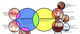
The main criterion for the quality of an alveolar socket preservation operation is its independent complete healing and maximum preservation of bone tissue, preservation of the natural contour and volume of the alveolar ridge, improvement of the condition of soft tissues and simplification of further stages of treatment. If the operation is successful, there will be enough bone tissue in the socket to install a dental implant. Separately, it is worth highlighting the advantages of condomization for the doctor and the patient - this is improved long-term treatment results, more predictable aesthetics and, of course, saving time for the doctor and the patient.
Treatment of alveolar sockets
Treatment is carried out strictly by a dentist. Remember that when treating alveolitis yourself with rinsing and antibiotics, you will not get rid of it, all these measures are useless! The main treatment procedure can only be performed by a dentist. Treatment at the dentist goes something like this:
- Removal of the blood clot under anesthesia, removal of food debris and necrosis from the socket (if all this is not removed, any treatment will be null);
- The hole is disinfected and washed with an antiseptic; antiseptics are placed in it, which are changed from time to time by the dentist at separate appointments;
- The dentist will prescribe antiseptic baths and an antibiotic, and painkillers for pain;
Moscow metro station Zvezdnaya, Danube Avenue, 23
Indications for condom
Preservation of the socket of an extracted tooth is indicated for everyone who in the future wants to install a dental implant in its place. The process of loss and resorption of the jaw bone at the site of the extracted tooth begins immediately after the operation. In the first year, bone tissue resorption will be 25% in volume, and over the next 3 years the loss in width will be 40 - 60%. In the case of socket preservation after tooth extraction, a larger volume of bone tissue in the jaw and the most natural contours of the alveolar process are preserved. And then, subsequently, the likelihood of such a complex operation as alveolar bone augmentation when installing an implant will most likely not be required.
What does the crown of a tooth consist of?
The part of the tooth that rises above the gum is not a monolithic piece made of homogeneous bone material. It is something like a nut in a shell. On top of the tooth is covered with a harder layer designed to protect against various influences - this is tooth enamel. And underneath it lies a softer and more vulnerable layer - it is called “dentin”. And even deeper, under the dentin, going into the root part, lies the “core” of the tooth - a cavity called the pulp.
Let's consider each component separately.
Enamel
Tooth enamel is de facto one of the hardest tissues inside the human body. Even large bones, designed to bear a load of tens of kilograms, are actually looser in structure than tooth enamel - after all, they do not need to constantly encounter various bacteria, constantly grind anything foreign (that is, food), and even collide with antagonist teeth.
Healthy enamel has a light (although not completely snow-white) color and a glossy surface texture; it is very smooth and dense.
Dentine
Dentin is also a type of bone tissue: that is, in an adult, dentin, like enamel, does not grow or change (unless we talk about cases of carious lesions). It is looser and more fragile compared to enamel - but it is still a “non-living” material that does not have nerves or capillaries. Dentin is something like a backup protective layer between the enamel and pulp.
Pulp
Under the dentin layer in the tooth there is a cavity, securely closed on all sides by hard “walls”. But this is not an “air bubble”, but a kind of chamber where the finest blood vessels and nerve fibers are concentrated. The pulp is very necessary during the period when the tooth is growing - all nutrients are transferred to it through this living core.
However, when a person becomes an adult and his teeth are finally formed, the role of the pulp ceases to be so important - it ceases to nourish anything. However, it may still be useful: after all, if the patient feels toothache, it means that it is the nerve fibers in the pulp that make it known about some kind of inflammation. However, when treating pulpitis, dentists often remove the pulp with all its contents - the adult patient’s tooth does not suffer from this, either functionally or aesthetically, and in the future such a pulpless tooth will definitely not cause pain.
Contraindications
Preservation surgery is subject to the same restrictions as any osteoplastic surgery. Separately, I would like to note that it is not recommended to preserve a hole after tooth extraction in a state of acute pain, since the risk of complications increases. But in each specific clinical case, the actions of a professional dentist are strictly individual. Sometimes situations arise when you have to take risks, but this is due solely to medical indications. In any case, it is necessary to understand that performing a tooth extraction operation and subsequent preservation on a planned basis is better than as part of emergency care.
Symptoms of alveolitis and dry socket
The diagnosis of “alveolitis” is made quite simply. With alveolitis, the following symptoms are observed:
- Outwardly, it may be empty, with a yellowish coating on the walls of the hole and traces of food debris, and festering blood clots are also visible. The gum next to the hole is usually inflamed, red, swollen, and hurts when touched.
- The pain associated with this disease varies and can be both acute and mild. Some people also experience pain in the head when the socket becomes inflamed.
- When a blood clot festers, it always begins to smell unpleasant, and the hole that is inflamed also has an unpleasant odor. It can be described as the smell of rotting, decay. A clot that festers leads to intoxication of the body, which is expressed in the person’s poor condition, as well as weakness and fever.
- In most cases, alveolitis occurs without swelling of the soft facial tissues due to the fact that the infection and pus come out through the sore hole. But there are cases when this does not happen, the facial tissues and gums swell, all this is accompanied by high fever and acute pain.
Restrictions
It is logical to perform a preservation operation after the removal of permanent molars, within the so-called sevens, second molars (except for wisdom teeth, eights), since when baby teeth fall out, bone tissue does not resorption. There is no upper age limit for performing an operation aimed at preserving the natural contours of the alveolar process.
Price
According to the classifier of surgical interventions in the oral cavity, condomation is not among the standard surgical interventions and this, of course, affects the cost of its implementation. In addition, the price of osteoplastic materials and related products is quite high. Some materials are used only in conjunction with a special membrane to avoid ingrowth of soft tissue into the alveolar socket. The price is also affected by the method of tooth extraction. Removal of a tooth or tooth root is sometimes still a traumatic procedure, leading to direct loss of alveolar bone and soft tissue. With atraumatic removal, the alveolar bone is preserved in a larger volume, which reduces the final cost of the operation.
Preservation of the socket after tooth extraction is an important procedure when planning the installation of an implant. Timely preservation of the socket will allow you to preserve such valuable bone tissue and avoid a much more complex and expensive operation to build it up - augmentation, as well as significantly reduce the overall treatment time. The cost of preserving the natural contours of the alveolar socket pays off, because you thereby provide the teeth surrounding it with a reliable position, and as a result, during implantation, you get an imitation of the removed tooth root. This procedure applies even to those patients who do not intend to restore or replace a lost tooth with an implant, because in this case the gingival contour will have the most aesthetic appearance, without failures, and will allow for the most aesthetic bridge prosthetics. Therefore, reservation is gaining increasing popularity in dental dentistry.
According to antiplagiat.ru, the uniqueness of the text as of October 16, 2018 is 90.3%.
Key words, tags: tooth extraction, wisdom tooth, bone tissue, implant installation, augmentation, implantation.
*Images: Astra Tech Dentsply Implants; Principles of hard tessue regeneration and implant therapy.
Tooth tissue
- Enamel: The most durable tissue in the human body, covering the surface of the dental crown. In terms of hardness, enamel is equivalent to crystal, since 97% of this tissue is inorganic.
- Dentin: the main hard tissue of the tooth. It consists of 70% hydroxyapatite, as well as water and organic material, fibers consisting of collagen, and dentinal tubules.
- Cement: tissue that covers the surfaces of the root and neck of the tooth. It is similar in hardness to bone. Cementum connects the alveolar bone to the tooth via the periodontal ligament.
- Dental pulp: loose tissue consisting of connective tissue fibers located in the tooth cavity. Rich in nerves and vessels (lymphatic and circulatory). Supplies dentin with nutrients.
Considering how a tooth is structured, one cannot exclude nearby tissues that support and strengthen the tooth.
- Periodontal ligament: fibrous tissue connecting the tooth root and alveolar bone.
- Alveolar bone: The jawbone that supports the tooth - it contains the sockets where each tooth is seated.
- Gums: soft tissue covering the alveolar bone.
- Gingival sulcus: a small space between the tooth and gum, the depth of which is between 1 and 2 mm. If this space deepens due to inflammation, a gum pocket is formed.
Based on their chewing function, teeth are classified into 4 types:
- The front teeth are incisors, used for cutting and grasping food.
- Fangs are cone-shaped teeth located next to the incisors, involved in holding and tearing food.
- The back teeth are small molars (premolars) and large molars (molars) that grind food.
Knowing the structural features of teeth, it is easier to find a common language with a dentist. You will feel confident and better understand the essence of the manipulations being performed.
Structure of the human tooth: