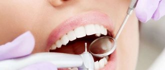
Fissure sealing is an extremely effective method of caries prevention and the main etiotropic method for the prevention of fissure caries. It involves the prevention of caries of the chewing surface of molars by filling fissures and other anatomical recesses of healthy teeth with adhesive materials - sealants in order to create a barrier to external cariogenic factors (microorganisms and carbohydrates)
Sealing tasks:
— eliminating the local risk of caries;
— creating conditions for the death of microorganisms remaining in the deep areas of the fissures; — elimination of potential reservoirs for the accumulation of cariogenic microorganisms; — acceleration of enamel mineralization in the fissure area when using glass ionomer cements.
Currently, glass ionomer cements, compomers, and composites are used for sealing.
Based on their chemical composition, sealants can be divided into groups:
1. Composite sealants: - classic, - sealants-ormokers, - flowable composite materials. 2. Compomers. 3. Glass ionomer cements Composite sealants can be: self-hardening and photo-hardening, for example: Estiseal LC, Fissurit, Helioseal.
Composite sealants and compomers are used to seal the fissures of teeth that have completely erupted.
Glass ionomer cements can be used for prophylactic coating of fissures in teeth at the eruption stage (Fuji VII, Fuji Triage).
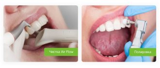
Techniques for using sealants: - cleaning teeth with a regular brush, - cleaning with a circular brush with antiseptics, - applying material to fissures, - correction of teeth closure, - insulating the glass ionomer surface with varnish, - monitoring the safety of the sealant every 3-6 months.
Toothpastes for children
Requirements for children's toothpastes: Fluoride content. Daily use of fluoride toothpaste in combination with proper brushing techniques is the basis for caries prevention! The use of low fluoride toothpastes containing less than 500 ppm F in the formula as a preventive measure is considered unsatisfactory. One of the problems associated with the use of fluoride toothpaste in children is the ingestion of the toothpaste by children with the subsequent risk of dental fluorosis.
Parents are advised to monitor the amount of toothpaste and the process of brushing teeth in children under seven years of age. Low abrasiveness. For temporary teeth (baby teeth) and newly erupted permanent teeth, the use of gel pastes is optimal. The RDA value for baby toothpastes should not exceed 50.
The absence of flavoring additives that can make a child want to eat pasta. It is preferable to use neutral, mint or fruit flavors that do not cause rejection in the child.
Examples of toothpastes for children aged 2 to 3 years: - Blend-a-med (0.005% NaF-250 ppmF) - My first Colgate (NaF) - Elmex enfant (aminofluoride - 250) - Lacalut Baby (aminofluoride – 250 ppm F, vitamins A, E) – Lacalut Kids 4-8 (aminofluoride – 500 ppm F, vitamins A, E) – Oral-B Stages fruity ( NaF – 500 ppm F) – Pokemon ( Na2PO3F- 500 ppm F)
Toothpastes for children from 2 to 6 years old: - Blend-a-med Junior Gel - Colgate junior (0.15% NaF- 680 ppm F-) - Colgate junior super star (0.76% Na2PO3F - 1000 ppm million F-) - Lacalut Kids 4-8 (aminofluoride - 500 ppm F-) - Lacalut Teens 8+ (aminofluoride and NaF -1400 ppm F-) - Dental dream for children (0.5% Na2PO3F -660 ppm F-) – Four Fruit – Mildfresh junior (0.76% Na2PO3F – 1000 ppm F) – Sanino Junior – New Pearl Junior 7-12 years (0.76% Na2PO3F – 1000 ppm F, oil tea tree) – Children's pearl complex – Prodent for teenagers
Palatine blind fossae
6. Histopographic studies of edentulous jaws have shown that: a) the prosthetic bed is constantly changing due to bone resorption that occurs after the loss of teeth, and continues even after the disappearance of the alveolar process (type III), although the intensity of atrophy in the latter case slows down;
b) the apex of the alveolar process is covered with a mucous membrane with a thick layer of fibrous tissue, poor in blood vessels, and tightly fused with the periosteum. On the slopes of the alveolar process, the layer of mucous membrane is thinner, and closer to its base the amount of loose connective tissue rich in small blood vessels increases. After complete atrophy of the alveolar process, the excess fibrous tissue at its apex disappears and the mucous membrane covering the bone becomes thinner;
c) atrophy of the bone substance of the alveolar process is of the lacunar type, and of the hard palate is of the smooth type of resorption:
d) on the hard palate, starting from the zone of the former first premolars, distally to the soft palate, the thickness of the soft tissues gradually increases due to mucous glands, loose connective tissue and an increase in the vascular network. A small dense vascular network is located laterally between the median suture and the base of the palatine slope of the alveolar process, which allows selective compression to be applied to these areas when taking a functional impression.
7. The palatine blind fossae are located 1-2 mm anterior to the distal edge of the horizontal plate of the palatine bone and cannot serve as an accurate clinical landmark for the line of transition of the soft palate to the hard palate.
8. The relationships between the planes of the hard and soft palate are characterized by three types.
In the first type, the surface of the hard and soft palate is almost in the same horizontal plane (35%). In the second type, the surface of the soft palate in relation to the hard palate forms a pronounced angle (44.5%). In the third type, the soft palate hangs almost vertically in relation to the hard palate (19.5).
9. Compression of the mucous membrane of the prosthetic bed of the edentulous lower jaw in the vertical direction is impossible, but it is possible and advisable in the vestibulo-oral direction at the base of the alveolar process. This will help create a closing valve.
10. The fibers of the mental muscle change their direction due to atrophy of the alveolar process. In the first type, they bend around the transitional fold, in the second, they become horizontal, and with complete atrophy they even rise upward. The nature of the muscle attachment also changes - it becomes aneurotic in nature, which makes the mucous membrane of this area unyielding.
Cavity formation in caries of natural pits
Cavity formation in caries of natural pits
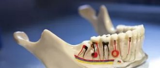
The natural pits on the chewing teeth include the depressions present on the buccal surface of the molars, as well as blind pits formed by the dental tubercles on the palatal surface of the front teeth (see Fig. 6, b, c).
Among the anterior teeth, blind fossae are more common on the small incisors, less common on the central incisors of the upper jaw, and almost never occur on the canines and lower incisors.
If the carious process that arose in the natural fossa on the buccal surface of the molar has not affected the entire fissure up to the chewing surface, then it is possible to create a box-shaped cavity without bringing it to the chewing surface. It is necessary that the cavity, as in all cases, be located not only in the enamel layer, but must penetrate into the dentin. During work, you should remember the proximity of the pulp chamber.
The carious process on the buccal surface of the molars creates a symmetrical round or diamond-shaped destruction of the tooth tissue. When forming a cavity for the tab, it is necessary to make it asymmetrical in order to facilitate the correct insertion of the tab. To do this, it is best to give it the shape of a trapezoid with rounded corners (Fig. 18, a). In some cases, it is enough to additionally include a nearby fissure in the cavity, which will serve as a guide when introducing the insert into the cavity (Fig. 18, b).
Formation ends with the creation of a fold along the entire edge of the cavity, which is best done with a small carborundum head.
The direction of wax removal should be perpendicular to the buccal surface of the tooth. Consequently, all work must be done with a fissure bur in a contra-angle handpiece and the direction of this bur must be perpendicular to the buccal surface of the tooth.
In cases where caries does not spread towards the pulp chamber, you can use a bur to follow the path of development of the carious process, excising
softened dentin. In this case, the drill can be given a direction slightly deviating from perpendicular, then all the walls of the cavity should be created at the same angle. The wax model should be drawn out in the same direction, and then the finished inlay should be inserted.
When the carious process spreads along the buccal fissure of a molar, a cavity is formed depending on the strength of the enamel roof hanging over it. If the roof remaining on the chewing surface above the cavity is thin, then in order to avoid breakages under the force of chewing pressure, it may be recommended to bring the cavity to the chewing surface.
Often the fissures of the chewing surface of the molars and the fossa on the buccal surface are simultaneously affected by the carious process. In this case, it is necessary to connect all the affected areas of the tooth into one common cavity. The direction of wax removal should be the same as for fissure caries, i.e., parallel to the long axis of the tooth (Fig. 18, c).
In this case, the contra-angle handpiece with the fissure bur should be held strictly parallel to the long axis of the tooth. Having created a cavity on the chewing surface, as described above, they move to the buccal surface of the tooth. The working part of the bur in this case will be the side surface.
While sparing the tooth tissue, you need to deviate from the strictly vertical direction of the bur, giving it a slightly inclined position, parallel to the groove on the buccal surface of the tooth (Fig. 18, d).
A fold is created along all edges of the cavity on both the chewing and buccal surfaces of the tooth.
When forming a cavity on the palatal surface of the upper anterior teeth, care must be taken, since in this area the pulp is located close to the surface of the tooth. Nevertheless, the rule of cavity formation not only in the enamel layer, but also in dentin, remains in force.
By excising tooth tissues affected by caries, they simultaneously create an asymmetrical cavity shape, for which they use natural depressions that are often present on the palatal surface of the incisors. These depressions can serve as retention areas where secondary caries may occur.
In its finished form, such a carious cavity on the palatal surface of the incisors most often represents a rounded cavity with one or two spurs (Fig. 18e).
The direction of wax removal should always be perpendicular to the palatal surface of the incisors. It is best to form a cavity using a fissure bur No. 1-3 in an angled tip directed perpendicular to the palatal surface of the tooth.
When, simultaneously with caries of the blind fossa on the palatal surface, there are carious cavities on the proximal surface of the incisor, it is advisable to combine both cavities into one common one or connect them together with a bridge.
The fold is made only on those edges of the cavity or bridge that extend onto the palatal surface of the tooth.
Sealants for fissures and blind pits of teeth
In 1923, Hyatt introduced the concept of prophylactic odontotomy. Since then, various filling materials have been placed directly into the fissures and natural pits of teeth to prevent the development of caries.
Early specialty sealants included cyanoacrylates and polyurethanes.
However, cyanoacrylate resins were quickly destroyed in the oral cavity, and polyurethane resins had low adhesion to tooth tissue. The most successful composition was Bowen resin (Bis-GMA). The monomer was easily cured chemically, under the influence of ultraviolet irradiation or visible light. Bis-GMA was an elastic liquid, which had to be diluted with monomers for easier penetration into fissures and pits. The first Bis-GMA sealant was the UV-curable Nuva Seal (Caulk/Dentsply, Milford, DE).
The first generation of sealants was polymerized under ultraviolet irradiation with a wavelength of 356 nm.
They were classified as "temporarily adopted" by the ADA in 1972 and "adopted" in 1976. The first generation of sealants were not effective due to a combination of factors. For example, Alphaseal (Amalgamated Dental Co., London) significantly absorbed ultraviolet light, which prevented its complete polymerization at depth. “Nuva Lite” (Caulk/Dentsply) had a significant dependence on the power of the lamp luminous flux (at least 10 mW per 1 cm2). The power of ultraviolet lamps was not always the same, and the area of the light beam was small. Failures were also due to insufficiently developed methods of their application and a lack of understanding of what a decisive unfavorable factor is the contact of saliva on the enamel after acid treatment. Insufficient rinsing and drying of enamel were the most common errors in clinical practice.
The second generation of sealants was based on chemical curing (self-curing).
The most widely and successfully used were Concise White Sealant System (3M Dental Product, USA) and Delton (Johnson and Johnson Dental Products, Co, USA). Most are unfilled, clear, tinted or opaque (opaque) sealants. Opacity was achieved by including white pigment or paint in their composition.
Third generation sealants polymerize when exposed to visible light
with a wavelength of 430-490 nm. Due to ease of use and better physical and mechanical properties, this group of materials is currently most widely used.
Currently, in order to seal blind pits and fissures of teeth, sealants belonging to three classes of materials are used:
composite materials, compomers and glass ionomer cements.
Glass ionomer cements
They have a cariesstatic effect due to the release of fluoride, are chemically fixed on the tooth surface, and do not require etching of the enamel before the procedure, but they are not strong enough and wear out quickly. For sealing purposes, glass ionomer cements of the second type (intended for filling teeth) can be used for loaded restorations. Some studies suggest that the use of glass ionomer materials as fissure sealants may be appropriate in newly erupted teeth where fissure mineralization is extremely low. The difficulty in such cases is associated with the need for longer etching of the enamel followed by the use of composite sealants.
Compomers
— light-hardening composite materials, which, due to their composition, have some of the properties of glass ionomer cements, namely, slightly greater hydrophilicity than composites and the ability to release fluoride in small quantities upon contact with oral fluid. The compomer sealant is Dyrect Seal (Dentsply). It is used with NRC (Non-Rise Conditioner) leave-in conditioner and the fifth generation Prime&Bond NT adhesive system, which ensures deeper sealing of fissures with polymer. NRC conditioner simultaneously partially dissolves mineral components and primes tooth tissue; Prime&Bond NT is applied on top of it, to which the sealant itself is fixed. The developers of this system consider this technique as an alternative to invasive fissure sealing.
As mentioned, the most numerous is the group of composite sealants, which are a composite material of higher fluidity due to the lower content of inorganic filler.
Composite sealants can be divided according to a number of characteristics.
- By type of hardening: self-hardening (chemical hardening)
- light-curing
- based on BisGMA (bisphenol glycidyl methacrylate)
- unfilled (containing less than 26-28% filler)
- transparent
- containing mineralizing components
UDMA-based sealants have slightly higher fluidity, lower viscosity, but at the same time higher shrinkage. Filled materials are more resistant to wear and abrasion, but unfilled materials penetrate better into narrow fissures and pits. Transparent sealants make it possible to judge the changes occurring underneath them (for example, the development of caries, the appearance of pigmentation), so their use is advisable in people with a pronounced cariogenic situation. Colored (most often chalky white) sealants allow the patient or his parents to control the retention of the material on the tooth surface. Opaque tooth-colored sealants are the most aesthetically pleasing, but they are difficult to maintain because they blend in with the color of the surrounding tooth surface.
It is worth noting the recently developed method of non-invasive mineral sealing of fissures by deep fluoridation with the addition of copper.
The method involves two-phase impregnation of enamel with the preparation “Enamel-sealing liquid” (Humanchemy, Germany). The enamel is first treated with solution No. 1, containing magnesium salts, copper-fluoride and silicon complexes. In this case, a fluorosilicate complex is formed in the pores of the enamel. After wetting with solution No. 2 (an alkaline suspension of highly dispersed calcium hydroxide), the fluorosilicate complex disintegrates. As a result, crystals of calcium fluoride, magnesium fluoride and hydroxyfluoride copper-P are formed. Their sizes are extremely small (about 50 A0), which allows them to freely penetrate the pores of the enamel. Fluoride crystals are immersed in a silicic acid gel formed during the interaction of solutions, which protects them from destruction. Long-term release of fluoride from crystals provides a caries-preventive effect. It is believed that highly dispersed calcium fluoride in enamel pores has 5 times greater solubility than calcium fluoride formed when applying simple fluorides. The effectiveness of this prevention method will be determined over time.
Medical Internet conferences
Functions of fissure sealing: creates a barrier for cariogenic bacteria; has a remineralizing effect on enamel if the sealant contains active fluoride ions. Four types of fissure structure : Funnel-shaped fissures are more open, well mineralized, they do not retain food debris due to free washing with oral fluid, and are caries-resistant. Cone-shaped - mainly mineralized due to oral fluid, but conditions appear for the retention of food debris and microorganisms. Mineralization of drop-shaped and polyp-shaped fissures occurs mainly from the pulp of the tooth. This process is less intense than mineralization due to oral fluid, and the fissures remain hypomineralized for a long time. Considering the high caries resistance of hard tissues, fissure sealing is not recommended in teeth with a high initial level of mineralization (ILM). General hygiene measures are sufficient. For teeth with average IUM, immediately after eruption, it is recommended to conduct a month-long course of local application of calcium - phosphate-containing and fluorine-containing preparations, followed by sealing with a composite sealant. For teeth with low fissure IUM, it is not recommended to use composite sealants using 38% phosphoric acid as an etching agent. In this case, glass ionomer sealants are used, or invasive sealing with a composite sealant, or, if indicated, a preventive filling method. The presence of pigmented fissures and natural depressions in teeth at the maturation stage, in contrast to teeth with mature enamel, indicates an actively ongoing process and requires invasive sealing methods. Initial caries is an indication for invasive sealing with composite sealants. Contraindications to sealing: the presence of intact wide, well-connected fissures; teeth with healthy pits and fissures, but with carious lesions on the proximal surfaces; pits and fissures that remain healthy for 4 years or more do not require sealing; poor oral hygiene. Indications for sealing and preventive procedures for fissures of erupting teeth with immature enamel based on fissure enamel electrometry indicators (μA): Low IAM (up to 8 μA) - hygienic measures, observation; average IUM (from 9 to 20 μA) - hygienic measures, a course of fluoride and calcium phosphate-containing drugs, fissure sealing; high IUM (up to 20 μA) - hygienic measures, a course of fluoride and calcium phosphorus-containing drugs, fissure expansion, preventive filling. For fissures after enamel maturation: 0 μA, healthy enamel - hygienic measures, observation 1-2 μA, initial caries - hygienic measures, course of fluoride and calcium phosphate-containing drugs, fissure sealing; up to 8 μA, progressive initial or superficial caries - hygienic measures, a course of fluoride and calcium phosphate-containing drugs, fissure expansion, preventive filling.
This comprehensive approach has been taken into account by the world's leading manufacturers of preventive products. The VOCO company (Germany, Cuxhaven) produces a fluoride-containing rinse aid "Profluoride M", a gel for applications "Profluoride Jelly", a two-component self-curing multi-purpose fluoride-containing varnish "Bifluoride 12", a series of sealants "Fissurit" and a unique highly filled sealant based on ormoker "Admira Seal" "
High efficiency (preventive effect of fissure sealing) is estimated by different authors from 55% (Going, Coti, Hough, 1976) to 99.1% (Buonocore 1974) and the low cost of the fissure sealing method in combination with general comprehensive prevention of dental diseases will significantly reduce the growth rate dental caries in the area of fissures and pits.
Currently, in order to seal blind pits and fissures of teeth, sealants belonging to three classes of materials are used: composite chemo- and photocurable materials, glass ionomer cements and compomers.
The material used to seal fissures (sealant), as a rule, is a special composite resin that is cured chemically or with the help of light. Due to their high fluidity, unfilled sealants easily penetrate even very narrow and deep fissures to their very bottom, leveling the chewing surface of the tooth and facilitating its hygiene. They have better marginal adaptation, longer retention, and wear out faster. Filled sealants have a smaller penetration depth, a smaller micromechanical adhesion area, shorter retention times, but are more resistant to abrasion. They are used for invasive sealing technology, but their application process is relatively complex, time-consuming and moisture-sensitive. Sealants do not adversely affect the normal mineralization process of enamel. Mineral elements from the oral fluid can diffuse freely along the edge and partially through the coating substance itself. This makes it possible to ensure a physiological level of metabolic processes in the hard tissues of the tooth under the coating, while simultaneously preventing the penetration of large protein molecules. The material is moisture-proof and very durable, which allows you to protect your teeth from fissure caries for a long time (up to 5-8 years). In addition, the sealant contributes to the saturation of the tooth enamel and periodontal environment with fluoride in the ion exchange reaction due to the soluble salt (fluorides) added to the composition for 1-28 days.
Types of composite sealants : 1) Self-polymerizing or chemo-curing “Concise White Sealant” (3M, USA), “Delton” (Johnson and Johnson), “Delton”, “Fis Seal” (Russia); 2) Photopolymerizable “Estisial LC” (Kulrer), “Sealant” (Bisco), “Fissurit”, “Fissurit F” (Voco), “Delton-S”, “Fis Sil-S” (Russia), Helioseal, Prisma Sheild .1. Opaque (not transparent) - easy to control, but does not imitate tooth color and it is impossible to monitor the condition of the enamel underneath; 2. Transparent – aesthetically pleasing, allowing observation of the condition of the enamel underneath, but difficult to distinguish when monitoring its safety; a) Colored (chameleons) have a bright color only at the time of polymerization, and after that they correspond to the natural tone of the tooth or are transparent “ClinPro Sealant” (3M ESPE, USA), “Helioseal Cler Chroma” (Ivoclar Vivadent); b) Not painted. A. Containing fluorine (Fissurit); B. Fluoride-free (Fissurit F).
The third generation of CPM - materials that harden under the influence of visible light with a wavelength from 430 to 490 nm (“Fissurit”, “Helioseal”, “Estisial LC”), one-component, long working time, completeness of polymerization is determined by light exposure, risk of destruction during testing curing is minimal. These materials are based on low-viscosity methacrylic acid derivatives. Borosilicate glass with a particle size of 99% less than 1 micron is used as a filler in the preparations, which provides good penetrating properties. At the same time, the release of fluoride from Fissurit F and its entry into the enamel continues for more than 190 days. During this period, Fissurit F releases 4-5 mg of fluoride to strengthen dentin enamel. Another preparation from VOCO (Germany, Cuxhaven) with fluorine, the light-curing sealant “Admira Seal” contains spatially inorganic - organic copolymers (ormokers), providing excellent mechanical properties and ideal biocompatibility (no toxic resin).
The fissure sealing procedure begins with thoroughly cleaning the tooth from plaque with a brush and paste, and then air drying. Next, the fissures are treated with 32% phosphoric acid for etching (a process in which the core or shell of enamel prisms is destroyed by acid) for 30-40 seconds, washed with distilled water and dried again. They are then filled with the liquid phase of the composite filling material. Under the influence of a special lamp, the material hardens in 40-45 seconds, after which the excess is removed with a hard carborundum head and the material is ground on the chewing surface.
The preventive effectiveness of materials is determined by the degree of their preservation in the fissures and the retention of this class of sealants ranges from 20 to 90% and depends on the accuracy of the sealing technology.
Glass ionomer cements - Dyract seal (Dentsply), Prima flou (DG), Vitacryl (Medpolymer), ASPA (Dentsply), Fuji (WHS), Glass Ionomer (Shofu Inc.), Alfa-dent, Aqua Ionoseal (Voco) have a cariesstatic effect , thanks to the F, Al, Zn, Ca contained, due to the release of fluoride, these materials have a pronounced cariesstatic effect. GIC is chemically fixed on the tooth surface, does not require etching of the enamel before the procedure, has high biocompatibility, is less demanding than CPM for drying the working area, but has a number of technological inconveniences (the need for mixing, difficulty in placement, short working time, long curing), low aesthetic properties, low fluidity, large edge leakage, are not strong enough compared to composites, and wear out quickly. For sealing purposes, glass ionomer cements of the second type (intended for filling teeth) can be used for loaded restorations. Some studies suggest that the use of glass ionomer materials as fissure sealants may be appropriate in newly erupted teeth where fissure mineralization is extremely low. The difficulty in such cases is associated with the need for longer etching of the enamel followed by the use of composite sealants. If it is necessary to carry out preventive filling (when, when examining a fissure, the tip of the probe gets stuck in it), we offer condensable, highly aesthetic glass ionomer cement - “VOCO Ionofil Molar” - which has three excellent properties. They are easy to use and less sensitive to execution techniques, which allows them to be used without etching or the use of adhesive. Plastic-free classic glass ionomers have a coefficient of thermal expansion similar to dentin, in addition, they have the so-called “battery” effect of constantly releasing significant amounts of active fluorides. Preservation of GIC after 1, 6, 12, 24 months. is 90, 80, 60 and 20% respectively, after 3 years - 10% (composite sealant - 90%), but, nevertheless, GICs provide a high level of caries reduction of occlusal surfaces - 80-90% in 2 years, teeth , even after macroscopic loss of material, have half the risk of caries than teeth not coated with GIC.
Compomers are light-hardening composite materials that, due to their composition, have some of the properties of glass ionomer cements, namely, slightly greater hydrophilicity, fluidity, and the ability to release fluoride in small amounts than in composites when in contact with oral fluid. The compomer sealant is Dyrect Seal (Dentsply). It is used with NRC (Non-Rise Conditioner) leave-in conditioner and the fifth generation Prime&Bond NT adhesive system, which ensures deeper sealing of fissures with polymer. NRC conditioner simultaneously partially dissolves mineral components and primes tooth tissue; Prime&Bond NT is applied on top of it, to which the sealant itself is fixed. The developers of this system consider this technique as an alternative to invasive fissure sealing. The wear of compomer sealants is higher and retention is lower than that of CPM. Over 2 years, the safety of the sealant-composite is 32%, the sealant-compomer is 0%; complete loss - 10 and 38%, respectively, but after the loss of the compomer, caries develops less frequently than after the loss of the compomer. The effectiveness of caries prevention has been confirmed by many studies. Coating teeth with fluoride-containing varnish led to a reduction in the increase in caries on treated surfaces by up to 70% and a decrease in the risk factor by up to 35%. The highest effectiveness of caries prevention is ensured by the fissure sealing method: the reduction in the increase in fissure caries over the year was 92.5%. Achieving high results of prevention through sealing is due to the performance of two main functions of sealants: 1. Creation of a physical barrier to cariogenic factors on the tooth surface. 2. Remineralization of enamel in the fissure area, if the sealant contains active fluoride ions.
Research results
An assessment of the effectiveness of sealants showed that the reduction in the increase in dental caries depends on the retention of sealants on the occlusal surfaces of the teeth, the ability to release fluoride ions into the tooth tissue and oral fluid, and the effectiveness of caries prevention in permanent teeth increases significantly when sealing fissures and pits is combined with local fluoride prevention and hygiene oral cavity.
Scientific studies have proven that a correctly performed procedure is 100% effective in protecting tooth surfaces from caries, since it serves as a physical barrier to possible destruction. The effectiveness of the procedure is suspended or stopped when the adhesive substances between the film and the tooth are destroyed or part of them is lost. However, teeth that are sealed are significantly less likely to develop caries in the future than those that were never treated. The seal is effective for 5 years but can retain its properties for up to 10 years. Doctors' reports show that 7 years after sealing, about 49% of teeth remain completely sealed. But sealing should not be considered a permanent procedure. Regular visits to the dentist are necessary for preventive examinations, which will allow you to monitor the condition of sealed teeth.
Anatomy: Internal base of the skull (basis cranii interna)
The inner base of the skull (basis cranii interna) has a concave, uneven surface corresponding to the shape of the base of the brain. There are three cranial fossae: anterior, middle and posterior. The posterior edges of the lesser wings (ala minor) and the tubercle of the sella turcica of the sphenoid bone (tuberculum sellae turcicae ossis sphenoidalis) separate the anterior cranial fossa (fossa cranii anterior) from the middle one (fossa cranii media).
The border between the middle and posterior cranial fossa (fossa cranii posterior) is the upper edges of the pyramids of the temporal bones (margines superiores partis petrosae) and the back of the sella turcica of the sphenoid bone.
When examining the internal base of the skull, numerous openings for the passage of arteries, veins, and nerves are visible.
Cranial fossae. The inner base of the skull is deepened; there are three cranial fossae: anterior, middle and posterior. These depressions deepen from the forehead to the back of the head, forming terraced structures. • The anterior cranial fossa is formed by the orbital parts of the frontal bones, the cribriform plate of the same bone and the large wings of the sphenoid bone (and is limited from the middle fossa by the small wings of the sphenoid bone and the tubercle of the sella). • The middle cranial fossa is formed by the body and greater wings of the sphenoid bone, the anterior surfaces of the pyramids and the squamosal parts of the temporal bone. • The posterior cranial fossa is formed by the occipital bone, the posterior surface of the pyramids and the internal surfaces of the mastoid processes of the temporal bones, the posterior part of the body of the sphenoid bone (dorsum sella).
1. The anterior cranial fossa (fossa cranii anterior) is formed by the orbital parts of the frontal bone (pars orbitalis ossis frontalis), on which the cerebral eminences and finger-like impressions are well defined, and by the ethmoidal plate of the ethmoid bone (lamina cribrosa ossis ethmoidalis), through the openings of which numerous bundles pass olfactory nerve fibers. In the center of the cribriform plate rises the cock's crest (crista galli), in front of which there is a blind foramen (Moran's foramen, foramen caecum), surrounded by the pterygoid processes of the cock's crest of the ethmoid bone and the legs of the frontal crest (Morand Sauveur Francois, 1697-1773 - French surgeon and anatomist), and frontal crest.
Near the crest of the ethmoid bone there is Palfyn's sinus - a space connecting to the frontal and ethmoidal cells (Palfyn Jean, 1650-1730 - French physician and anatomist).
2. The middle cranial fossa (fossa cranii media) is much deeper than the anterior fossa. The walls of the middle fossa are formed by the body and large wings of the sphenoid bone (corpus et alae majores ossis sphenoidalis), the anterior surface of the pyramids and the scaly part of the temporal bones (facies anterior partis petrosae et pars squamosa ossis temporalis). In the middle cranial fossa, the central part and lateral sections can be distinguished. The central part is occupied by the sella turcica with its pituitary fossa. At the bottom of the pituitary fossa of the body of the sphenoid bone there may be a non-permanent formation (found in 0.3% of adults) - Landucert's canal (syn.: craniopharyngeal canal, canalis craniofaryngealis). It penetrates the body of the sphenoid bone and opens on its lower surface (near the junction of the wings of the vomer) with a “pharyngeal” opening.
The canal contains a continuation of the dura mater of the brain in the form of a fibrous coupling containing connective tissue and blood vessels (veins) (Landuzert Fedor Pavlovich (1833-1889) - professor of the St. Petersburg Medical-Surgical Academy).
Anterior to the pituitary fossa, the chiasm groove (sulcus hiasmatis) is visible, leading to the right and left optic canals (canalis opticus), through which the optic nerves pass. On the lateral surface of the body of the sphenoid bone there is a well-defined carotid groove (sulcus caroticus), and near the apex of the pyramid an irregularly shaped foramen (foramen lacerum) is visible. Here, between the lesser and greater wings and the body of the sphenoid bone, there is the superior orbital fissure (fissura orbitalis superior), through which the oculomotor, trochlear and ophthalmic nerves pass into the orbit. Posterior to the superior orbital fissure there is a round foramen for the passage of the maxillary nerve, then an oval foramen for the mandibular nerve.
At the posterior edge of the greater wing of the sphenoid bone, the foramen spinosum is visible, through which the middle meningeal artery passes into the skull. On the anterior surface of the pyramid of the temporal bone there is a trigeminal depression (impressio trigemini) - Meckel's fossa (Meckel Johan Friederich (senior), 1724-1774 - German anatomist), lateral to it is the cleft of the canal of the greater petrosal nerve ( hiatus canalis nervi petrosi majoris) - Taren's foramen - an opening on the anterior surface of the pyramid of the temporal bone, through which the greater petrosal nerve exits, and the groove of the petrosal nerve (Tarin Pierre (1725-1761) - French physician and anatomist). Even more lateral and anteriorly there is a cleft (hole) of the canal of the lesser petrosal nerve and a groove of the lesser petrosal nerve.
Lateral and posterior to these formations, the roof of the tympanic cavity (tegmen tympani) and the arcuate eminence (eminentia arcuata) are visible. Between the carotid canal and the trigeminal ganglion - the Gasserian ganglion (syn. ganglion of the trigeminal nerve, ganglion trigeminale) on the pyramid of the temporal bone there is Gruber's notch (syn.: jugular notch, inciscura jugularis), covered with a thin bone plate (Gasser Johann Laurentius, 1723 -1769) - Austrian doctor and anatomist; Gruber Wenceslau Leopoldovich (Gruber WL, 1814-1890) - Austrian anatomist who worked in Russia). In the pyramid of the temporal bone, under the dura mater of the brain, there is a Dorello canal formed by it and the groove of the inferior petrosal sinus - a canal through which passes the inferior petrosal sinus, vessels and abducens nerve leading to the cavernous sinus (Dorello Paolo, born in 1872 .) - Italian anatomist). In the area of the apex of the pyramid of the temporal bone there is Princeteau's tubercle - an elevation to which the superior petrosal sinus adjoins (Princeteau Laurent (1858-1932) - French doctor and anatomist).
The topographic-anatomical landmark during surgical interventions on the labyrinth, less often on the cerebellum, is Trautmann's triangle - the region of the skull limited behind by the sigmoid sinus of the dura mater of the brain, in front - by the posterior semicircular canal of the inner ear, above - by the upper edge of the petrous part of the temporal bone (Trautmann Moritz ( Trautmann Moritz F., 1832-1902) - German surgeon).
3. The posterior cranial fossa (fossa cranii posterior) is the deepest. It is formed by the occipital bone, the posterior surface of the pyramids and the inner surface of the mastoid processes of the right and left temporal bones, as well as the posterior part of the body of the sphenoid bone and the posteroinferior angles of the parietal bones. In the center of the fossa there is a large (occipital) foramen, in front of it is the Blumenbach clivus (syn. slope of the skull, clivus), formed by the bodies of the sphenoid and occipital bones fused in an adult, on which the bridge (brain) and medulla oblongata lie (Blumenbach Johann ( Blumenbach Johann Friedrich, 1752-1840) - German physician and anatomist, zoologist and anthropologist).
Between the bodies of the occipital and sphenoid bones there may be an additional bone - Albrecht's bone (Albrecht Karl Martin Paul, 1851-1894 - German anatomist). In the posterior edge of the large foramen of the occipital bone, during development, the Kerckring bone stands out - the ossification point of the occipital bone (Kerckring Theodor, 1640-1693 - Dutch physician and anatomist).
Posterior to the foramen magnum (occipital) along the midline are the internal occipital crest (crista occipitalis interna) and the cruciate eminence (eminentia cruciformis). On the back surface of the pyramid, on each side, the internal auditory opening (porus acusticus in tern us) is visible, leading to the internal auditory canal (meatus acusticus internus). In its depths, the facial canal begins, in which the facial nerve passes. The vestibulocochlear nerve emerges from the internal auditory opening. At the bottom of the posterior cranial fossa behind the pyramids there is a paired jugular foramen (foramen jugulare), through which the glossopharyngeal, vagus and accessory nerves pass, and medial from it is the hypoglossal canal for the nerve of the same name. Through the jugular foramen, the internal jugular vein also exits from the cranial cavity, into which the sigmoid sinus continues, lying in the groove of the same name.
On the surface of the cranial vault, 3 cm posterior and above the upper edge of the external auditory canal, there is a Keen point, which is a topographic and anatomical landmark for puncture of the lower horn of the lateral ventricle of the brain (Keen William Williams, 1837-1932 - American surgeon).
On the inner base of the skull, in the region of the posterior cranial fossa, there is a Mouret zone - an area of the skull bounded above by the inferior petrosal sinus of the dura mater, behind - by the transverse sinus, in front and inside - by the internal auditory canal on the pyramid of the temporal bone; this area is a common site for cerebellar abscesses.
The boundary between the vault and the inner base of the skull in the region of the posterior cranial fossa is the groove of the transverse sinus (sulcus sinus transversi), which passes on each side into the groove of the sigmoid sinus (sulcus sinus sigmoidei).
Vestibule of the oral cavity on the upper jaw
The vestibule of the oral cavity (vestibulum oris) has the appearance of a narrow horseshoe-shaped gap; it is limited externally by the mucous membrane of the lips and cheeks, and on the internal side by the labial and buccal surfaces of the alveolar process of the jaw. On the mucous membrane in this area there are three frenulums: labial and two buccal.
The superior labial frenulum (frenulum labii superioris) occupies a central position and is a vertical fold of the mucous membrane connecting the lip to the corresponding part of the gum. The labial frenulum can move along with the muscles of the oral cavity.
Buccal frenulums (frenulum buccales) are located in the area of the transitional fold at the level of the canines or premolars. They can be single or multiple. The buccal frenulum is directly connected to the canine muscles (m. caninus). When interacting with the orbicularis oris muscle (m. orbicularis oris), these muscles cause the lips to move forward, and when interacting with the buccal muscles (m. bussinator), they pull the lips back. These movements are used in the functional design of the impression in the area of the labial frenulum, buccal frenulum, and also the muscles located here (Fig. 14).
The labial section of the vestibule of the oral cavity corresponds to the anterior portion of the transitional fold, located between the buccal frenulum. The mucous membrane of the labial region is closely connected with the orbicularis oris muscle and the muscles intertwined with it, elevating the upper lip (m. levator labli superioris), lowering and retracting the wings of the nose (mm. depressor alae nasi). These muscles begin from the nasal process of the maxillary bone and from the lower orbital edge of the body of the upper jaw, descend vertically down and attach to the skin of the upper lip along the nasolabial fold and partially in the skin of the wing of the nose.
The canine muscle (m. caninus s. levator anguli oris), levator anguli oris, is located directly under the quadratus muscle of the upper lip; begins at the bottom of the fossa under the infraorbital foramen and is woven into the skin of the corner of the mouth (Fig. 15).
The buccal sections of the vestibule of the oral cavity correspond to the areas of the transitional fold located distal to the buccal frenulum. Beneath the mucous membrane in this area (bilaterally) is the buccal muscle, which forms the lateral wall of the cheek. The buccal muscle extends with a wide base from the outer surface of the upper and lower jaw against the sockets of the molars, and behind it from the pterygomandibular ligament (lig. pterygo-mandibularae) and is directed forward to the corner of the mouth. Here it merges (above and below) with the orbicularis oris muscle. At the back, the buccal muscle is covered by the masticatory muscle (m. masseter), from which it is separated by a fat pad.
The masticatory muscle, starting from the zygomatic process (proc. zygomaticus) of the upper jaw and attached to the outer surface of the angle of the lower jaw, during contraction presses, displacing inward, the buccal muscle and the mucous membrane of the transitional fold of this area.
The buccal part of the vestibule of the oral cavity, especially with severe atrophy of the jaw, is reflected on the impression in the form of a wide and softly defined notch, corresponding to the densest and therefore easily vulnerable area of the transitional fold. Anatomically, this notch corresponds to the location of the base of the zygomatic process of the upper jaw.
The distal sections of the buccal sections of the vestibule of the oral cavity are under the direct influence of the posterior fibers of the buccal muscles, which are attached here to the maxillary tuberosities and the hooks of the pterygoid processes of the main bone.
The vestibular space in the area of the maxillary tuber (tuber maxillae) is usually narrower than the remaining areas of the vestibule of the oral cavity, especially with the mouth open.
The well-developed tendons of the temporal muscle (t. temporalis), attached to the area of the coronoid process of the mandible, and the ascending branch of the mandible itself have a great influence on the shape and volume of the distal buccal vestibule of the oral cavity. The impression according to this zone should be formed by movements of the lower jaw, mainly from side to side, as well as forward.
In addition, the shape of the edges of the prosthesis is determined here by the configuration of the lower jaw itself.
Palatal part of the upper jaw . Distal from the alveolar tubercles are the maxillary-pterygoid notches (incisurae pterigomaxillary), limited posteriorly by the hooks of the pterygoid processes (Fig. 16). The bottom of the maxillary-pterygoid notch is formed by the lower fibers of the internal pterygoid muscle (m. pterygoideus interrius), attached to the alveolar tuberosities and to the posterolateral surface of the horizontal part of the palatine bone. The submucosal layer in this area contains mucous glands and fatty tissue, which forms a layer between the mucous membrane and the underlying muscles.
The posterolateral edges of the prosthesis should be located in the middle of the pterygomaxillary notches. The medial borders of these notches are formed by the tendons of the muscles that stretch the soft palate (m. tensor veli palatini). Both mentioned muscles, like the muscles that lift the soft palate (m. levator veli palatini), are removed from the edge of the prosthesis, which distinguishes this zone from other areas of the periphery of the prosthetic field and creates the possibility of lengthening its boundaries in the case of large atrophy of the bone and mucous membrane.
The middle part of the posterior edge of the hard palate ends with a more or less pronounced protrusion (spina nosalis posterior). This area is the site of attachment of the aponeurosis of the soft palate and the levator soft palate muscles. The area of bone above the protrusion is covered with a thin layer of easily vulnerable mucous membrane. Even slight pressure usually leads to bedsores.
On the sides of the posterior nasal protrusion there are palatine (blind) fossae (fov. coecum). They represent a fusion of the excretory ducts of the mucous glands, the openings of which are located in close proximity to the so-called vibrating zone “a”. The blind fossae are a convenient landmark for determining the boundaries of the posterior edge of the prosthesis.
The vibrating zone “a” is a section of the mucous membrane identified when pronouncing the sound “a”. The direction of the vibrating zone usually varies according to the shape of the palate: the higher the palatine vault, the more anteriorly this line is located and the sharper its bend. With a flat palate, the vibrating zone “a” usually extends further posteriorly with a gradual, smooth bend; in this case, its wide posterior edge is formed (Fig. 17).
The vibrating zone is located on the soft palate; one should distinguish between this zone and the border between the hard and soft palate located in front of it. The border of the prosthesis should pass within the vibrating zone and cover the blind fossae.
The location of the muscles distant from the posterior edge of the hard palate determines some features of determining the boundaries of the prosthesis in this area. The posterior border of the complete denture of the upper jaw, in contrast to the rest of the periphery of the prosthetic field of the upper or lower jaws, which must be designed functionally, is determined by the above landmarks, the main of which are the blind fossae and the vibrating zone.
The degree of possible lengthening of the distal edge of the prosthesis also depends on the angle of inclination of the soft palate in relation to the pharynx. There are three forms of the slope of the soft palate: steep, gentle and average (Fig. 18).
With a steep, steep palatine slope, the posterior edge of the hard palate corresponds to the place of almost direct transition of the fixed mucous membrane into the moving tissues of the soft palate.
In such cases, the possibility of lengthening the distal edge of the prosthesis is very limited and the palatal part of the valve appears as a narrow strip. With a gentle slope of the soft palate, the width of the palatal part of the valve is large. With an average slope, the width of the palatine valve has an average value.
The mucous membrane of the posterior parts of the palate was studied in detail by Sh. I. Gorodetsky and he called it “a zone of mucoglandular tissues not subject to muscular action.” This section of the mucous membrane of the palate is free of muscles and is an elastic, pliable tissue with vessels and nerves deeply located in it. The pliability of this zone is determined by well-developed layers of fatty tissue and glandular tissue, thickening towards the pharynx. A different point of view is shared by E.I. Gavrilov, who connects the vertical compliance of the mucous membrane of the jaws with the state of the vascular network. According to his concept, a well-developed vascular network in certain areas of the hard palate, called buffer zones by this author, imparts spring properties to the soft tissues, which are important for prosthetics.
Histological and topographic-anatomical studies have established that the mucous membrane covering the alveolar processes and hard palate in the area of the sagittal suture is characterized by small vascular fields and therefore does not have buffering properties. In areas of the mucous membrane located between the base of the alveolar process and the middle zone, dense vascular fields are found, especially abundantly represented in zone “a”. Accordingly, the buffering properties of the mucous membrane of the hard palate increase towards zone “a”.
The data obtained by V. I. Kulazhenko on the point compliance of the mucous membrane of the hard palate, obtained using an electron-vacuum apparatus, coincide with the topography of the buffer zones according to E. I. Gavrilov. The buffering properties of the mucous membrane of the prosthetic bed of the upper jaw change throughout a person’s life, depending on his age and state of health. The width of muscle-free areas of the palatal aponeurosis (between bone and muscle) ranges on average from 2 to 6 mm.
The study of the mucoglandular zone allowed Sh. I. Gorodetsky to justify the use of the so-called laboratory edging, the formation of which is carried out on a model. The width and depth of the edging is determined by the degree of compliance of the mucous membrane in various parts of the mucoglandular zone.
It is considered, however, more rational to design the edging for the palatine valve directly in the oral cavity using special impression compounds (Dentafol, etc.).
So, the distal boundaries of the prosthesis for the edentulous upper jaw and the size of the palatine valve vary in shape and location depending on the bony contours of the posterior edge of the hard palate, the shape and direction of the slope of the soft palate, pharynx and the structure of the tissues of the mucoglandular zone.
The essence of the system
Black's classification of carious lesions is a system for dividing the disease into certain classes depending on the location of the destroyed area of hard tissue and the coverage of certain elements of the jaw row.
Despite the fact that this scale was developed more than a hundred years ago and does not include secondary and root caries, its use is widespread in modern dental practice.
Dr. Black identified 5 main classes of caries, to which another degree of development of the disease was later added.
The purpose of creating this classification was to select the optimal method of therapy - selection of a suitable material for filling and a method of preparing the affected surface.
The topography of types of cavities according to Black is well demonstrated in the following diagram: