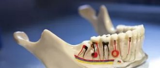
Central upper incisor
Average age of eruption: 7-8 years
Average age of root formation: 10 years
Average length: 22.5 mm
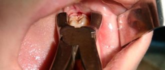
The crown of the central upper incisor, close to rectangular on the vestibular side and wedge-shaped on the proximal side, allows for convenient endodontic access and is ideally positioned for direct examination using a mirror. The tooth is especially suitable for the novice doctor, since in it the canal is directly visible for a third of its length. With fiber optic illumination, the view of the channel can be improved.
Primary opening using a fissure bur is made immediately above the enamel palatal tubercle of the equatorial third of the crown on the lingual surface of the tooth. The instrument is directed along the long axis of the root. Based on the final shape of the access cavity, a triangular hole is made. Trephination of the tooth cavity often occurs during the first implantation. After the feeling of “falling” into the pulp chamber, the fissure bur is replaced with a spherical bur No. 4-6 with an extended shank.
A ball bur is used to widen the hole towards the incisal edge. You need to make sure that the pulp cavity is completely open. A fissure bur may again be required to widen the access cavity and give it its final shape. At this time, all carious dentin, which has significantly changed its color and pulp calcifications, is removed. It is necessary to remove leaking fillings and treat proximal carious cavities with adequate temporary filling.
The root is quite characteristically cone-shaped and sharply tapering towards the apex. The cross-section of the root canal approaches triangular in the cervical part, gradually becoming rounded closer to the apical foramen. Multiple canals in the root are rare, but accessory and lateral canals are common. The apical foramen is rarely located exactly at the apex, but is usually within 2 mm laterally.
Lateral upper incisor
Average age of teething: 8-9 years
Average age of root formation: 11 years
Average length: 22.0 mm
The crown of the upper lateral incisor, approximating an oval shape, is almost ideal for endodontic access, as is the case with the central incisor. Fiber optic illumination is also helpful when accessing this tooth.
The initial opening using a fissure bur is made immediately above the enamel tubercle in the equatorial third on the palatal surface of the tooth. The access cavity is oval in shape. When performing the initial opening, the fissure bur often occupies the entire narrow cavity of the coronal pulp. After removing the roof of the pulp chamber, a ball-shaped bur No. 4 or 6 is used to clean it from carious dentin, pigmented areas and calcifications.
A fissure bur may again be required to finalize the oval shape of the access cavity.
Adequate expansion is then created using spherical burs. Care must be taken to ensure that probes, endodontic cutting instruments and condensation instruments do not come into contact with the walls of the access cavity.
To ensure the cleanliness of the canal walls and their hermetically sealed filling, all carious tissues and leaky fillings must be removed and replaced with temporary filling materials.
The cross-section of the canal varies from oval in the cervical part to round in the apical foramen. The root is slightly cone-shaped and can bend at the apical part, usually in a distal direction. The apical foramen is often located closer to the anatomical apex than the central incisor, but can be located laterally within 1-2 mm of it.
In rare cases, access is complicated by the presence of a “tooth-in-tooth” developmental anomaly, invagination of part of the lingual surface of the tooth into the crown. This creates a space in the tooth that is surrounded by enamel and communicates with the oral cavity. Tooth-in-tooth is most common in the upper lateral incisors, but can occur in other teeth. Due to anatomical developmental defects, these teeth are prone to caries and the pulp may die before the apex is fully formed. This formation (“tooth within a tooth”) is localized in the crown; it must be processed mechanically and removed or bypassed.
The neck of the tooth under the crown is exposed
Crowns must be replaced periodically; they cannot last for decades. A properly made crown extends a short distance under the gum (from 0.5 to 1 millimeter). This seals the tooth, so that nothing from the oral cavity gets under the crown onto the tooth.
Over time, the gum atrophies, that is, it begins to slide off the crown. When the intensity of this process increases, when the gum is exposed by approximately 1-2 millimeters, the tooth is exposed. It begins to get affected by various infections contained in the oral cavity. This is the cause of the development of various diseases, caries, which provoke exposure of the neck of the tooth.
If the tooth under the crown is alive, the person complains of an irritable reaction when eating hot or cold. As the inflammatory process develops, severe aching pain begins to bother you. In such a situation, it is possible to save the tooth root. To do this, remove the crown. There are also treatment methods in which there is no need to remove it. More detailed advice can be obtained from the clinic doctor.
Upper canine
Average age of teething: 10-12 years
Average age of root formation: 13-15 years
Average length: 26.5 mm
As the longest tooth, the canine has an imposing shape designed to withstand strong occlusal forces. Its long crown with a thick layer of enamel is subject to abrasion by the cutting edge. As she ages, she often has deep cervical erosions.
The access cavity corresponds to the shape of the lingual surface of the crown and is oval. To obtain direct access, the cavity must be expanded incisally, but not so much as to weaken the actively functioning cusp. The initial access is made in the middle part of the crown from the palate. If the pulp cavity is deeper, a No. 4 or 6 ball-shaped extended bur may be required. A sweeping motion with this bur will open up the oval pulp cavity.
As it moves through the cervical portion and down apically, it remains oval. Thorough cleaning of this oval-shaped canal is difficult, so attention must be paid to targeted file processing.
The root canal is quite straight and long. Most canines require instruments 25 mm or longer in length. The last 2-3 mm of the top often bends in some direction.
The morphology of the canines rarely changes radically, and lateral and accessory canals are less common than in the upper incisors.
The vestibular cortical plate over the apex of the tooth root is often destroyed to form a fenestration. The apical foramen is usually located close to the anatomical apex, but can be located laterally, especially if there is an apical bend of the root.
The neck of the tooth is exposed, how to treat it
After the doctor has diagnosed the cause of the exposed cervix, he recommends the patient one of the following treatment methods:
- Filling the exposed area. This procedure is indicated at the initial stage of the pathological process. When a wedge-shaped defect has formed, there is nothing to attach the filling material to, so filling is irrelevant.
- Remineralization of enamel. A product containing calcium is used. The patient will not feel pain during the procedure. The process lasts about 10 minutes. A drug is applied to the affected tooth to help strengthen the enamel, protect the roots from exposure, and prevent tooth loss. In addition, the advantage of remineralization is the return of the enamel to its former shine. How many sessions a person needs is determined by the doctor.
- The enamel is coated with fluoride (fluoridation). Special varnishes or gels that contain fluoride are used. Fluoridation can be simple or deep. With the help of such methods, the dental neck is restored and tooth sensitivity is reduced. With deep fluoridation, the drug strengthens the enamel as it penetrates inside it.
- Installation of veneers. This is a thin ceramic plate. It is applied to a previously ground tooth. This technique is indicated for advanced cases. With the help of veneers, it is possible to completely eliminate the disease and minimize the risk of its recurrence.
- Crowns. Before installing crowns, the tooth is ground down. It is a radical way to protect the tooth root and prevent further progression of the pathology.
- Gum plastic surgery.
Drug and surgical treatment
If the cause of exposure of the neck of the tooth is a lesion or pathology of the mucous membrane, the patient is consulted by an implant surgeon. During the operation, tissue taken from the palate of the mouth is implanted under the tooth root. This helps to increase the volume of gum tissue and cover exposed areas of the tooth. At the end of the surgical intervention, sutures are placed on the gums. After this, the patient is scheduled to regularly visit the attending physician, who will monitor the healing process.
Use an antiseptic solution to rinse your mouth. You can eliminate swelling and redness of the gums with the help of medicinal gels. If pus is present, an antibacterial drug is prescribed. The choice of medication is made only by the attending physician; self-medication is prohibited.
Upper first premolar
Average age of teething: 10-11 years
Average age of root formation: 12-13 years
Average length: 20.6 mm
The first upper premolar is a transitional tooth between the incisor and molar and most often has two roots.
When molars are lost, the main chewing load falls on the premolars. In removable prosthetics, these teeth are used as supporting teeth, which increases the impact of torque on them. Additional torque forces, together with deep carious lesions, can cause severe calcification of the pulp cavity. Early molar loss often causes rotation of the premolars, which can make identification of the pulp chamber difficult.
The mouths of the canals are located below and somewhat to the center of the tops of the mounds. The initial opening is made in the central fissure, giving it an oval shape in the bucco-palatal direction. After identifying the mouth, the doctor must accurately determine the presence of an anastomosis leading to the mouth of another canal. The direction of the roots can be determined using an endodontic probe. Root bifurcation visible on a routine periapical photograph may indicate tooth rotation. With divergent roots, less expansion of the occlusal approach is required, and with parallel roots, on the contrary, it may be necessary to remove the crown tissue towards the tops of the cusps. All infected dentin and leaking fillings should be removed and replaced with suitable temporary fillings.
Options for root anatomy include fused roots with separate canals, interconnecting canals or a “web,” a common apical foramen, and the possible presence of three roots, which is rare but should always be kept in mind. In the latter case, the mouths of the buccal canals will not be clearly visible using a dental mirror. An endodontic probe or a thin file will help determine the structure of the canal. Cams and Skidmore report that maxillary premolars with three roots and three apical foramina are found in 6% of cases. The length of the root is much shorter than that of the canine, and a distal bend is not common. The apical foramen is usually located close to the anatomical apex. The length of the roots when using intact tubercles as reference points is usually the same. The apical part of the roots often tapers sharply, ending in very narrow and curved tips.
Given the possibility of vertical mesial-distal fractures of the crown or root of the first premolar, before endodontic treatment, all fillings should be removed and the crown should be carefully examined under fiber light.
To prevent vertical fractures of the crown or root after endodontic treatment, it is necessary to completely close the occlusal access cavity.
The structure of the periodontium –
As we said above, the periodontium is located between the cementum of the tooth root on one side and the compact lamina of the alveoli on the other side. It consists of loose fibrous connective tissue (FCT), the main component of which is mature type I collagen fibers. Moreover, in people under 25 years of age, in addition to mature collagen, fibers of immature collagen (procollagen) can also be found in the periodontium. Between the bundles of collagen fibers there is an intercellular substance with blood and lymphatic vessels, as well as nerve fibers.
There are no mature elastic fibers in the periodontium, and there are only a small number of immature elastic fibers (oxytalan), which are located along the walls of blood vessels. At the same time, the collagen fibers themselves are rigid and incapable of stretching - so how is the physiological mobility of the tooth formed? The fact is that collagen fibers in the periodontium have a shock-absorbing effect due to their spiral bends. These bends straighten during chewing load on the tooth, and when it stops, they curl again. Thanks to such bends, the tooth has physiological mobility.
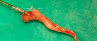
Periodontal fibers (histological specimen) –
The cellular composition of the periodontium is represented primarily by fibroblasts, cementoblasts and osteoblasts, which are involved in the construction of collagen, cement and bone tissue, respectively. In addition, Malasse epithelial cells were found in the periodontium, which can be a source of the formation of cysts and tumors. We will talk about the full composition of cellular elements below.
Types of periodontal fibers:
Collagen fibers passing nearby intertwine with each other, forming strong bundles with a diameter of 0.2 mm (such bundles are called periodontal ligaments or ligaments). There are several alternative classifications of periodontal fibers, and below we present two of them. According to the classification of I.P. Gayvoron periodontal fibers can be divided into 3 types (Fig. 2) –
- dental-gingival fibers,
- dentoalveolar fibers,
- interdental fibers.
1) Complex of dental-gingival fibers - bundles of these fibers begin from the cementum of the tooth root in the area of the bottom of the gingival pocket, and then they spread fan-shaped, weaving into the soft tissue of the gums around the neck of the tooth (marginal edge of the gums). This type of fiber ensures a tight fit of the gum to the neck of the tooth. Below you can see that the dentogingival fibers have a variety of directions and form a three-dimensional network in the gum tissue. The structure of these fibers is quite thin and not too powerful.
2) Dento-alveolar fibers (horizontal and oblique) - these fibers begin just below the fibers of the previous group. They are located in the periodontal gap between the cementum of the tooth root on one side and the compact lamina of the alveoli on the other side. Dento-alveolar fibers are usually divided into horizontal and oblique. Horizontal fibers are quite small in number, and they go in a horizontal direction from the surface of the tooth root - to the apices of the interalveolar septa (Fig. 5).
Almost the entire surface of the roots is covered with oblique dento-alveolar fibers, and it is predominantly this type of fiber that holds the tooth in the alveolus and also performs a support-shock-absorbing function. At one end these fibers are attached to the cementum of the root, and at the other to the wall of the alveoli. Due to the oblique direction of the fibers, the tooth is suspended inside the alveolus and, thus, chewing pressure is not directly transmitted from the tooth to the bone tissue of the alveolus. In general, the arrangement of bundles of dento-alveolar fibers in the lateral sections of the periodontal fissure looks like a hammock mesh (Fig. 5-6).
In the middle third of the periodontal fissure (in young people under 25 years of age) there is a dense intermediate plexus of immature collagen fibers - the so-called “Sikcher plexus”. The fibers of this plexus have a very high regenerative potential. They are of great importance for the regeneration of periodontal structures, for example, this can be important when planning orthodontic treatment. But it must be taken into account that the Sickher plexus disappears in people over 25 years of age. A number of researchers have put forward a logical explanation for the need for the presence of immature collagen in the periodontium.
The fact is that part of the periodontal fibers begins to form from the side of the cement of the tooth root, and the other part - from the side of the bone plate of the alveoli. According to a number of authors, periodontal fibers are not a single formation, and when both parts of the fibers reach the middle of the periodontal fissure, they are connected through immature collagen fibers. By the way, this theory is confirmed by the clinical observations of the author of the article, who has repeatedly carried out replantation of teeth completely removed from the socket. At the same time, fragments of periodontal fibers were preserved on the side of the socket and the root of the tooth, thanks to which the periodontium regenerated perfectly in young patients (24stoma.ru).
3) Transseptal (interdental) fibers - these fibers go from the neck of one tooth to the neck of another, and for this purpose the fibers pass over the tops of the interalveolar septa. Those. they form a “ligament” that goes from the cementum of the root of one tooth (on the side of the contact surface) to the cementum of the root on the contact surface of the adjacent tooth. These periodontal fibers perform the function of maintaining the continuity of the dentition, as well as redistributing the chewing load along the dentition.
Alternative classification of periodontal fibers –
There are several alternative classifications of periodontal fibers. These classifications may include the following division of fibers into groups:
- Circular (free) fibers - they start from the neck of the tooth, fan out, ending in the soft tissues of the gingival margin.
- Fibers of the alveolar ridge - they connect the neck of the tooth with the crest of the alveolar bone (in Fig. 4 they are called dentocomb fibers).
- Horizontal fibers are a small group of fibers that are located immediately under the fibers of the alveolar ridge (at the very entrance to the periodontal space). These fibers run horizontally, forming, together with the transseptal fibers, the circular ligament of the tooth.
- Oblique fibers are the most numerous group of fibers that connect the tooth root with the compact lamina of the alveoli (in the previous classification they were called dento-alveolar).
- Apical fibers - they diverge perpendicular to the dento-alveolar fibers from the apices of the roots to the bottom of the alveoli.
- Transseptal fibers - they run horizontally from the neck of one tooth to the neck of another, connecting adjacent teeth to each other.
Second upper premolar
Average age of teething: 10-12 years
Average age of root formation: 12-14 years
Average length: 21.5 mm
Similar to the first premolar in crown shape, the second premolar differs mainly in its root shape. Its crown is narrower in the bucco-palatal direction and somewhat wider in the mesial-distal direction. The mouth of the canal is located in the center, but it is more slit-like than oval. In the presence of a slit-like orifice, the physician should assume the presence of two canals until proven otherwise.
The external shape of the tooth is slightly oval, but wider in the mesial-distal direction than that of the first premolar.
All infected dentin and leaking fillings should be removed and replaced with temporary fillings.
The root may have two separate channels connecting into one, or two channels interconnecting in the form of a “web”. Accessory or lateral canals are possible but are less common than in incisors. Vertucci et al found that 75% of upper second premolars had one foramen at the apex, 24% had two foramina, and 1% had three foramina. Of all the teeth studied, 59.5% had additional canals. These investigators reported that when two canals are joined into one, the palatine canal is often directed toward the apex in a straight line. They further stated that “if, on a direct periapical photograph, the root canal sharply narrows or even disappears, this means that at this point it is divided into two canals, which either remain separate (type V) or, before reaching the apex, merge again (type II)".
The length of the root of the second upper molar is comparable to that of the first premolar. Apical bending is common, especially with a large volume of the maxillary sinus.
To prevent vertical coronal or crown-root fractures after endodontic treatment, complete closure of the occlusal access cavity is necessary.
Types and symptoms of apical periodontitis
Depending on how the inflammatory process proceeds, two types of disease are distinguished: acute apical periodontitis and chronic apical periodontitis. The second is always a consequence of the first: if acute inflammation is not cured in time, then it has every chance of becoming chronic.
The symptoms of acute apical periodontitis of pulpal origin are difficult to miss - they can cause the patient many unpleasant moments. The acute period is usually accompanied by the following conditions:
- nagging pain in the area of the affected tooth, which intensifies over time, becomes pulsating, and can radiate to other parts of the face;
- swelling of soft tissues, enlarged lymph nodes;
- rise in temperature;
- headache.
The duration of acute apical periodontitis ranges from 2 days to 2 weeks. In the absence of appropriate treatment, two options are possible: either the disease will progress, involving more and more tissue, or it will enter a chronic stage. The clinic of chronic apical periodontitis looks like this:
- pain in the affected area is mild or absent altogether, and can also occur when pressing or biting;
- slight tissue swelling may be observed;
- Possible bad breath;
- the diseased tooth is sensitive to temperature influences.
The chronic course of the disease may be accompanied by acute periods. With exacerbation of chronic apical periodontitis, the same symptoms are observed as in the acute form.
Upper first molar
Average age of teething: 6-7 years
Average age of root formation: 9-10 years
Average length: 20.8 mm
The largest in size, with a complex anatomy of the root and root canal system, the so-called “6-year molar” is the most frequently treated, while presenting the greatest difficulties in treatment among the posterior teeth. During its treatment, the largest number of endodontic errors and complications arise, and it is undoubtedly one of the functionally important teeth.
The three separate roots of the maxillary first molar form a trifurcation: the palatal root is the longest, and the distal buccal and mesiobuccal roots are approximately the same length.
The palatal root in the apical third often curves in a buccal direction. Of the three channels, it has the largest diameter and is the easiest to access. Its mouth is shifted to the palatal wall of the crown. The root deviates sharply from the median axis of the tooth. In cross section, the root is flattened and has a ribbon-like shape, which requires special attention when cleaning and instrumenting it. Fortunately, it rarely has more than one apical foramen.
The distal buccal root is conical and usually straight. It always has one channel.
The mesiobuccal root of the first molar has generated more research, clinical inquiry, and frustration than any other root in the oral cavity. Green showed that 14% of the mesiobuccal roots of the maxillary first molars studied had two apical foramina, and 36% of the roots had two orifices. Pineda reported that 42% of these roots had two canals and two apical foramina. Slowey confirmed Pineda's data within a few percent difference. The fact that nearly half of these roots have two canals, whether they end in a single opening or not, is reason enough to always assume two canals until careful examination proves otherwise.
The additional orifice lies centrally, between the orifices of the mesiobuccal and palatine canals. The search is facilitated by using fiber optics and by identifying the anastomosis between the orifices of the mesiobuccal and palatal canals. The second canal in the mesiobuccal root will always be narrower than the other canals, so it is more difficult to clean and shape. Access to the main mesiobuccal root canal is easier when a straight entry is properly created.
All carious tissue, leaking fillings and denticles must be removed before endodontic treatment begins.
After treatment, complete closure of the approach is necessary to prevent vertical coronal or crown-root fractures. If indicated, internal reinforcement with intraradicular pins is recommended.
Apical periodontitis of the tooth - what is it?
The diagnosis of periodontitis is made when the patient's periodontal tissues near the apex of the tooth root become inflamed, which is why it is called apical. It affects deep gingival and bone structures, so it is not easy to treat - unlike, for example, caries. Nevertheless, this disease requires immediate intervention, because if the patient is not in a hurry to treat apical periodontitis, then he risks encountering unpleasant consequences of the disease: the appearance of granulomas and cysts, destruction of the alveolar bone, and in the most severe cases, even sepsis.
Second upper molar
Average age of teething: 11-13 years
Average age of root formation: 14-16 years
Average length: 20.0 mm
The shape of the crown of the second upper molar is very similar to the first upper molar, although it is not as rectangular and massive. Adequate access on both teeth can usually be achieved without disturbing the transverse enamel ridge. The second molar is often easy to prepare due to the straightforward approach to the orifices.
A distinctive feature of the morphology of the upper second molar is the closely spaced and sometimes fused three roots. The shadows of parallel root canals often overlap on the radiograph. Its roots are usually shorter than those of the first molar and not as curved.
The three orifices may form a blunt triangle, and sometimes they are located almost in a straight line. The floor of the pulp chamber is noticeably convex, creating a slightly funnel-shaped shape of the canal orifices. Sometimes the canals extend from the bottom of the pulp chamber at an acute angle, resulting in the need to remove the edge of the dentin in order to create a straight line of access.
Complications during access occur if the molar is tilted distally. Initial exposure is performed with a fissure bur with a cutting tip, and then a short ball-shaped bur is used, which is best suited for opening the pulp chamber and forming an access cavity. Then, small hand instruments are used to establish the patency of the canal and its working length. After this, the bulk of cleaning and shaping can be done by machining the files in the endodontic handpiece.
To improve radiographic visibility, especially when layering the shadow of the process of the zygomatic bone, photographs can be taken in perpendicular and distal projections at an angle.
All infected dentin, leaking fillings and denticles must be removed before endodontic treatment begins. To prevent vertical coronal or crown-root fractures, it is necessary to completely close the access cavity. If indicated, immediately after endodontic treatment it is necessary to apply internal reinforcement with intraradicular pins.
Diagnosis of apical periodontitis
It should be remembered that the symptoms of apical periodontitis are similar to the symptoms of some other oral diseases: pulpitis, sinusitis, hilar cyst and others. To make an accurate diagnosis, the following studies are necessary:
- electroodontometry
- helps to assess the degree of pulp necrosis; - radiography
- allows you to see tissue changes in the apical region; for chronic apical periodontitis, x-ray is the best diagnostic method, but it may not detect acute inflammation at an early stage; - A blood test
is an additional diagnostic method. With apical periodontitis, an increase in the level of leukocytes and ESR is observed.