Intraoral (or dental, intraoral) radiography is one of the most effective and informative ways to diagnose diseases of the teeth and periodontal tissues. In addition, hidden and serious pathologies, such as cysts and granulomas, will never be detected during a routine visual examination.
Unlike other x-ray methods, intraoral x-rays allow you to get a complete picture of a local area of the jaw.
What is a targeted dental photograph?
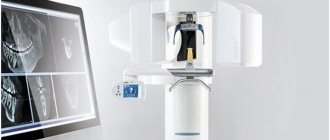
Another name for the procedure is targeted intraoral contact radiography - this is a simple and fast method of X-ray diagnostics in dental practice. Research is carried out in clinics using analog X-ray machines or digital radiovisiographs. The doctor gets the opportunity to examine both the condition of the tooth being examined and those located nearby.
- Carry out diagnostics. For example, detect the development of an inflammatory process.
- Evaluate the results of the therapy. This is necessary not only in the treatment of pulpitis, caries or other dental diseases, but also in preparation for prosthetics.
When using analog X-ray equipment, an image is created on film and then transferred to special paper. A digital device allows you to obtain an electronic photograph. If necessary, any area of the image can be enlarged on the monitor for a more thorough examination.
- Interproximal. Allows you to diagnose pathologies of the crown part of the tooth, detect the presence of carious cavities, as well as defects that can form under fillings and crowns.
- Periapical, allowing to assess the condition of bone tissue. This type of imaging helps monitor the quality of therapy provided.
The radiation dose for a dental x-ray is 2-3 μSv, which is very small. For comparison, with fluorography we receive a radiation dose of 500-800 μSv.
Indications for use
Dental radiography is used for two main purposes: to diagnose and evaluate the quality of treatment performed.
Allows you to detect the following diseases:
- hidden carious lesion;
- pulpitis (inflammation of the neurovascular bundle of the tooth);
- cyst at the roots of the teeth;
- periodontitis (purulent inflammation of the tissue between the root of the tooth and the hole in which it is located);
- gum disease, in which bone tissue atrophies (periodontitis, periodontal disease);
- neoplasm.
Intraoral radiography also helps evaluate the results of procedures such as:
- root canal filling;
- treatment of periodontal diseases.
Pros and cons of the diagnostic method
The use of a dental visiograph has a number of advantages compared to analog X-ray machines. First of all, it is an opportunity to get a clear picture of the tooth and the tissues around it. It is convenient to store the resulting images on a computer or other electronic media, printing them if necessary. A digital image allows a more detailed assessment of the clinical picture, since the image can be enlarged several times. In addition, digital dental radiography is a safe procedure. If necessary, multiple procedures may be performed. For example, during implantation, control images are taken before and after each implant is installed. And also after complete completion of prosthetics. The equipment used is characterized by a reduced level of radiation exposure. The dose of radiation that the body of the person under study receives is so small that it does not cause any harm to health. Most often, it does not exceed the natural background recorded in some megacities. Despite the large number of advantages, this method has disadvantages. For example, the image can be performed only in 1 plane covering a small area of tissue. Therefore, the maximum effectiveness of the method is achieved at the stage of early diagnosis or to control the quality of therapy already carried out.
Popular methods of radiation diagnostics
Today, the most common and popular method of radiation examination in outpatient practice is intraoral radiography of teeth, or intraoral photograph of the tooth. Sometimes intraoral photographs of teeth are called targeted, which is incorrect. A targeted photograph is a photograph taken outside the standard positioning, and standardized studies are named according to the positioning method.
At a therapeutic appointment during endodontic treatment, at least three intraoral photographs of each tooth under examination must be taken:
- A diagnostic image is necessary to assess the condition of periodontal tissues at the time of examination, make a diagnosis, determine the number and shape of roots, the direction of canals, and choose treatment tactics.
- measuring photograph - a photograph of a tooth at the treatment stage with endodontic instruments inserted into the canals with a fixed stopper length of the working part or verifiers after instrumental treatment of the canals. If the orthogonal projection is performed correctly, provided that the visiograph program is accurately calibrated and there is no projection distortion for incisors and premolars, some measurements can be taken from the diagnostic radiogram. For multi-rooted teeth, it is preferable to measure the length of the canals using endodontic instruments (Fig. 1), an apex locator or from a three-dimensional photograph.
- a control photograph is taken immediately after the end of endodontic treatment in order to determine how well the root canals are filled, as well as after a certain specified time, in order to verify the absence or detect the presence of complications (Fig. 2). When examining multi-rooted teeth and in cases where there is an additional canal, in an image taken with the orthoradial direction of the beam (direct projection), the root canals often overlap each other, which significantly complicates diagnosis and can lead to errors in the treatment process. To obtain a separate image of the root canals, radiography with an oblique (eccentric) direction of the central beam is used (Fig. 1). For each specific case, the mesial or distal inclination (angulation) of the tube in the horizontal plane is selected (for more details, see: Rogatskin D.V., Ginali N.V. The Art of Dental Radiography, 2007).
Ideally, maximum information about the topography of the roots and the condition of periodontal tissues can be obtained by performing polypositional radiography. In this case, for diagnostic purposes, three photographs are taken - one in a straight line, with an orthoradial direction of the beam, and two in an oblique projection - with a distal-eccentric (Fig. 1) and mesial-eccentric direction of the beam (respectively, straight, posterior oblique and anterior oblique projections).
The most important aspects of successful intraoral radiography are standardization and consistent manipulation. Standardization of manipulations means the ability of a specialist conducting a radiation examination to choose the optimal method for each case and take a series of identical images, regardless of the position, condition of the patient and the time separating one examination from another. That is, if a diagnostic or measurement image is considered to be of high quality, each subsequent clarifying and control image must be made with the same spatial and technical settings, and each subsequent image must be identical to the previous one (Fig. 1, 2).
Rice. 1. Diagnostic and measuring images of tooth 36, taken in direct (a) and distal-eccentric projection (b). 36 - chronic apical periodontitis (K04.5) with characteristic changes on the mesial root.
Rice. 2. A control image immediately after treatment of teeth 21, 22 (chronic periapical abscess in a state of suppuration) (a) and a delayed control image 5 months after filling the canal (b), the state of repair at the treatment stage.
Execution steps
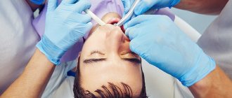
Interproximal radiography is one of the types of intraoral examination that allows you to obtain a picture of 1 section of the oral cavity with an image of the upper and lower dentition. To obtain the image, a special holder is secured between the closed teeth. It allows you to identify interdental caries and various changes in bone tissue due to gum disease. And also check the correct installation of crowns, dentures or fillings. An X-ray of the tooth is taken by a doctor in an office specially equipped for this. Before taking an X-ray of a tooth, the doctor gets acquainted with the problem and studies its location.
To obtain a clear image of the problem area, the patient's head is fixed in the required position. The process of visiography takes a matter of seconds, but during this time the patient is required to remain completely still. The video in this article shows how to take targeted photographs of teeth so that the image is as informative as possible. Using a digital sensor, the radiologist directs a beam of rays to the required area. The photograph is taken either from the inside of the mouth or from the face. During the manipulation, the patient does not feel any pain or discomfort.
The original digital image differs from the traditional film image because it is processed using a special program in a few seconds and transferred to the monitor screen.
General information
An X-ray examination that allows you to get a real clinical picture of one or more teeth located nearby. A simple and reliable method for accurate diagnosis, used in most dental clinics.
Problems that can be solved by analyzing a targeted X-ray image:
- making an accurate diagnosis;
- drawing up a treatment plan with monitoring the dynamics of the condition;
- assessment of the degree of development of pathology;
- assessment of the restoration of hard tissues and the condition of the dental canals;
- assessment of the actual condition of blood vessels, soft tissues, rudiments;
- identification of pathologies in a latent course;
- identifying the area affected by caries, as well as identifying the source of inflammation.
Targeted X-rays are done using a digital radiovisiograph, which minimizes the radiation dose. The equipment produces a direct beam beam that directly affects the area being examined. The technique is absolutely safe for health.
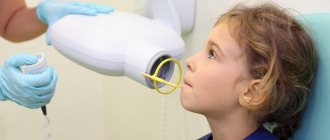
Features of the procedure in children
Contact intraoral radiography can be prescribed for children in cases where tooth damage cannot be examined in any other way. The technique allows early detection of disturbances in the process of teething, bone tissue diseases, and prescribing effective treatment. In addition, this method allows you to control the implementation of orthodontic manipulations if the child has problems with the formation of the jaw.
The study is carried out in the same way as in adult patients. Children under 2 years of age are recommended to undergo x-rays only in case of urgent need. For example, in case of injury during childbirth, to monitor the development of the jaw, or after a fall from a height, to assess the integrity of the teeth.
General overview
From a technical point of view, the procedure is an X-ray analysis of the state of one or more adjacent units, characterized by the speed of obtaining complex data and ease of execution. The resulting image allows the dentist to:
- To formulate a correct diagnosis, the formulation of which on the basis of visual examination data alone may be impossible.
- Determine and prescribe an effective course of treatment, as well as control its pace.
- Monitor the intensity of the spread of pathology formed in the oral cavity.
- To study the condition of the structure of bone tissue and root canals, dentin and blood vessels.
- Identify hidden anomalies and pathological processes, determining their localization.
To obtain targeted dental images, digital equipment is used - a radiovisiograph, one of the functional advantages of which is the minimum radiation dose to the patient during the shooting process. The directed beam flow maintains focus, affecting only a selected area of the jaw row, which makes the diagnostic procedure safe for the patient’s health.
Restrictions and contraindications
How often a dental x-ray can be taken can only be determined by a doctor, guided by the severity of the problem and the patient’s condition.
It is forbidden to take dental x-rays during pregnancy, especially in the early stages. A woman needs to take care of the condition of her teeth in advance in order to eliminate the need for treatment while carrying a child. X-rays are contraindicated for children under 2 years of age.
In case of urgent need, an x-ray can be performed, but not earlier than the second trimester. The only exception is when emergency assistance is needed. Manipulation must be carried out using all possible means of protection. The period of breastfeeding is not a limitation for the study.
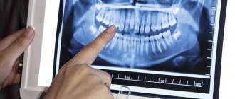
Interpretation of a dental radiograph
Only a dentist or radiologist can decipher and describe a dental image. The image shows the tooth: its root, internal canals, shape, anatomical features.
- Examines the rigidity, density and uniformity of the bone structure. Evaluates the location of each element of the dentition.
- Determines the presence of signs of clearing or darkening, indicating the development of an inflammatory process, cysts, granulomas, neoplasms.
- Depending on what the dental x-ray shows, a diagnosis is made.
Carious formations in the picture look like light areas of various shapes with unclear boundaries. The development of pulpitis is characterized by bone damage. The image shows a violation of its homogeneity in the interroot space. With the development of periodontitis, a granuloma appears in the area of the tooth root in the form of a darkened round shape with clear contours. With periodontitis, the image shows a decrease in the density of the bone structure, a decrease in the height of the partitions between the elements of the dentition, and the formation of “pockets.”
Indications for use
Factors that determine recommendations for radiovisiography include:
- Pain in the dental area.
- Routine diagnostics prescribed to detect pathologies.
- The need to identify the degree of bone loss before implantation.
- Identified diseases of gum tissue, including periodontal disease.
- Determination of the severity of mechanical damage received.
- The need to control the growth of “eights”, or wisdom teeth.
- Checking the quality of root canal cleaning.
The procedure is mandatory before installing prosthetic structures or orthodontic devices, as well as when planning surgical intervention.
Where can I take a photo and how much does it cost?
Targeted radiography can be performed in any specialized medical institution. The average cost of the procedure is about 400-450 rubles. Some clinics include in the cost of conducting several studies at once, which involve monitoring the treatment being carried out.
All categories of patients, both adults and children, can take a targeted photograph of a tooth. The procedure is prescribed with caution to pregnant women and infants under 2 years of age.
A referral for a radiovisiographic image is given by a dentist. The resulting image makes it possible to determine the presence of a problem, confirm or refute the diagnosis, and prescribe treatment. To monitor the patient’s condition during treatment, the doctor may recommend taking a second photo of the tooth.