Deep caries with complications is one of the most common dental diseases. When patients complaining of excruciating pain in their teeth come to see our dentistry specialists in Moscow, upon examination it becomes clear that the inflammatory process has begun to actively develop in the dental canals.
In such situations, dental treatment must be started immediately, otherwise the inflammation will continue to develop and can affect the jaw and periodontal tissues, which can lead to serious complications such as granuloma, gumboil, and cyst.
The occurrence of such pathologies can only be prevented by competent treatment of dental canals at an early stage of the destructive process.
TREATMENT OF TOOTH CANALS – A LITTLE ANATOMY
If you look carefully at the photo below, you can see a narrow cavity that runs along the entire length of the tooth root. This cavity is not empty - inside it there are soft tissues of different types: nervous, connective, circulatory and lymphatic. The cavity with tissues is the dental canal, which can sometimes have small branches or other structural anomalies.
During root canal treatment, the doctor removes the pulp tissue, but there is no need to be afraid of the negative consequences of this procedure. Pulp tissue plays a key role in childhood, when the process of active development of incisors and molars is underway; in an adult, pulp tissue is solely responsible for tooth sensitivity. If canal treatment is carried out professionally and the inflammation in the root cavity can be eliminated, then you will not feel the difference between a living and a “dead” tooth.
What a miracle is BeeFill Pack technology?
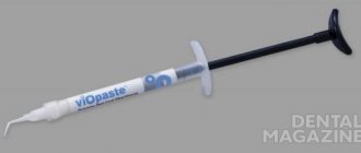
To completely seal the root canals of teeth, we use the BeeFill Pack system from the German company VDW GmbH, which completely eliminates the possibility of re-infection.
Indications for root canal filling with BeeFill technology:
- caries, pulpitis, periodontitis;
- depulpation;
- purulent abscess;
- refilling of canals as a result of poor quality treatment;
- severe dental injury;
- preparation for prosthetics.
Not all dental clinics use the technology of three-dimensional filling of the root canal system with thermoplastic gutta-percha, which meets established European standards.
HOW TO DETERMINE THAT TOOTH CANAL NEEDS TREATMENT
The need for dental canal treatment is determined only by a qualified specialist - a dentist and after a thorough examination of the oral cavity. Make an appointment with our dentistry in Moscow at the slightest manifestation of toothache; also, do not delay a visit to the dentist if you experience swelling on the gum or other discomfort in the tooth and gum area.
It is worth knowing that sometimes an infection that has entered the dental canal may not manifest itself in any way. Pulp tissue can die asymptomatically, and in such cases, only a visit to the doctor can determine the need for urgent treatment of root cavities. Traditionally, radiographic examination is used to diagnose canal infection.
Tooth canal treatment
from 2000 rub. More about prices
We have been working since 1994
we are one of the first to open private dentistry in Moscow
Best materials
only new and modern equipment for dental treatment
Free
consultation with a dentist
Payment options
- cash
- plastic cards
- cashless payments
Doctors' experience
- with great experience
- graduated
- conference participants
The main indications for dental canal therapy are:
- 1. Trauma or active inflammatory process in the nerve tissues of the tooth.
- 2. The appearance of a special type of neoplasm on the gums - fistulous tracts, which outwardly resemble pimples.
- 3. Fractures and dental root injuries.
If you are suffering from throbbing pain, you notice a swelling on the gum, darkening of the tooth crown - contact our dentistry in Moscow - Vanstom for consultation and treatment. The clinic’s specialists will be able to quickly alleviate your condition and provide high-quality dental canal treatment.
When is root canal treatment necessary?
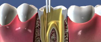
Treatment and filling of canals are stages of endodontic treatment. The doctor opens the tooth to treat the following diseases:
- deep caries - the last stage of the disease, characterized by extensive damage to dentin, accompanied by severe toothache that appears when chewing, pressing on the tooth, eating cold and hot foods and drinks;
- pulpitis - inflammation of the neurovascular bundles located in the tooth cavity, most often occurs against the background of advanced caries, characterized by paroxysmal pain, which intensifies when eating hot or cold food;
- periodontitis - inflammation of the apex or edge of the tooth root, develops due to damage to the connective tissue that holds the dental unit; symptoms of the disease are acute pain, swelling of the gums, swelling of the lip or cheek, as well as pathological mobility of the tooth.
The main reason for the development of these diseases is non-compliance with the rules of oral hygiene. If you brush your teeth twice a day and visit the dentist regularly, these problems can be avoided. Experts recommend undergoing a preventive examination every six months. In this case, the doctor will be able to detect caries at an early stage and promptly eliminate it, thereby preventing the development of pulpitis and other complications.
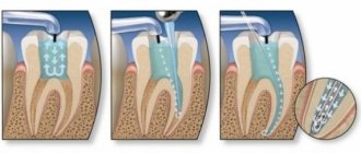
STAGES OF DENTAL CANAL TREATMENT
The process of dental canal treatment consists of the following stages:
1. Anesthesia of the area in which medical procedures will be performed. It is carried out using local anesthetic drugs.
2. Isolation of the tooth with a special rubber gasket. The cushioning material will protect the sensitive mucous membrane and prevent saliva from getting into the open area.
3. Elimination of tissue damaged by caries and removal of the apical part of the pulp chamber.
4. Measuring the length of the canal and establishing its shape. For these purposes, a special device can be used - an apes locator, and the doctor may also prescribe an x-ray of the diseased tooth.
5. Removal of pulp tissue, after which the canal is expanded for subsequent filling. Expansion of the cavity can be carried out by a doctor either manually or mechanically using dental files - a specialized, thin metal instrument.
6. Cleaning and disinfection of canals with antiseptic solutions and agents that destroy pathogenic microorganisms;
7. Drying and filling the prepared cavity.
Depending on the phase of development of the disease, the stage of its neglect and the type of procedure for the treatment of dental canals can take from 2-3 hours or be carried out over several days.
Temporary filling
In some cases, medicine is introduced into the canals for a certain period of time. Manipulation is carried out for:
- elimination of pathological microflora;
- stopping the inflammatory process;
- isolation of the canal, when it is impossible to carry out treatment in one visit.
Indications for temporary installation of a filling are injuries, perforation of walls, periodontitis in acute or chronic form.
The main active components of medicinal non-hardening pastes are antibiotics.
MATERIALS FOR FILLING DENTAL CANALS
The choice of materials for canal filling is the most important stage of treatment. The composite must firmly seal the root cavity and retain its structure for as long as possible.
Traditionally, thermogutta-percha is used to fill root cavities, which, when hardened, tightly blocks the dental canal and at the same time accurately repeats its shape. The material fills all pores and cracks in the canal, does not cause periodontal irritation and does not cause darkening of the natural enamel layer of the teeth.
Other materials and techniques can be used to fill the canals; the specific method is selected by the dentist after examining the patient, carrying out diagnostic measures and radiographic examination.
Classical treatment methods
Dentistry has long been dealing with the problem of root canal inflammation. Treatment was carried out using two methods:
- opening the tooth chamber, placing arsenic paste, removing it after the tissue has died, and filling;
- opening the dental chamber, placing a resorcinol-formalin mixture, which blocked the inflammatory process, filling.
There are classical and modern treatment methods. When comparing, it can be clearly noted that classical therapy has a number of disadvantages:
- both compositions have a toxic effect on the body;
- risk of incomplete pulp death;
- risk of residual infection and relapse;
- painful procedure.
These methods are now practically not used, since modern means and drugs make it possible to completely clean the inflamed cavity and destroy the infection painlessly, with minimal trauma and without the risk of relapse.
BeeFill Pack system capabilities
Filling root canals with gutta-percha pins is considered a successful and safe method of strengthening and preserving a tooth. With the usual filling method, it is not possible to achieve complete adherence of the material to the walls of the crown. And only the use of the unique BeeFill Pack technology ensures:
- Constant and uniform heating of gutta-percha.
- Precise delivery with optimal pressure, which allows the material to penetrate into all branches of the root canal.
- Possibility of filling difficult root canals.
- Accuracy of channel length determination.
Comprehensive control of the filling procedure eliminates the penetration of gutta-percha into the periodontal tissues. The use of the BeeFill Pack device not only makes the dentist’s work easier, but also allows the doctors of our Research Center clinics to carry out effective treatment and high-quality root canal filling.
Work in canals with pronounced curvature
The curvature of the root canals largely determines the complexity of endodontic treatment. In this article we will discuss the basic principles of working in curved canals, which a doctor can effectively implement in his practice.
Grade
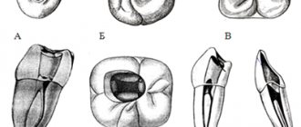
It is extremely important to evaluate the location, angle and radius of curvature before starting treatment (Figures 1 and 2). Many methods have been proposed to measure root canal curvature (Figure 1), but it has not yet been established which method is best (Hartmann et al, 2018).
Photo 1: Image taken from “Methods for measurement of root canal curvature: a systematic and critical review” by Hartmann et al, 2022 (International Endodontic Journal). This image illustrates simple methods for calculating root canal curvature. These methods help determine the level of potential treatment complexity. The physician can then decide whether he is competent to perform endodontic interventions or whether the complexity of the clinical case is beyond his skill level and warrants referral to a specialist (Figure 2).
Figure 2: Tooth UR6 with sharp apical curvature at the MB root (MB1 and MB2). Apical curvature is much more difficult to manage clinically than coronal curvature. Anatomical curves associated with longer roots or calcified canals create additional problems during the treatment process.
Complications
Curvatures can lead to the development of many iatrogenic complications (photo 3):
- formation of steps;
- excessive expansion of the canal and transport of curvature;
- perforations;
- apex transposition;
- file fracture.
Sometimes iatrogenic complications are quite difficult to correct; in some cases, they cannot be stopped at all.
Photo 3: Possible iatrogenic complications that may arise during the treatment of root canals with severe curvature.
Advice
Be patient: it is extremely important to adequately plan the treatment process and ensure sufficient time for its implementation (photo 4).
Photo 4: LL6 root canal treatment. A thorough analysis of the radiograph allowed us to evaluate the anatomical features and existing bends of the root canal. At first glance, LL6 does not present much complexity. However, upon closer inspection, two long mesial roots with apical curvatures were identified.
Straight access: It is important to provide direct access to curved canals. This reduces the stress on the endodontic files as they do not have to bend as they enter the root canal. The shape of the access cavity is corrected as conservatively as possible using ultrasound.
Coronal canal treatment: The quality of the coronal canal treatment reduces the degree of canal curvature and can help convert a C-curve to a J-curve (Figure 5). Rotary nickel titanium orifice files can be used for this purpose. Instruments such as Gates Gliddens on the posterior teeth can provoke a destructive effect on the hard tissues of the endodontic space and form steps.
Photo 5: Widening the coronal portion reduces the degree of canal curvature. The medial channels on the LR6 were originally C-shaped. Expansion of the coronal part of the canal transformed them into a J-shape, optimizing the treatment process.
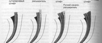
Carpet: To avoid breaking the rotary tool, it is essential to recreate the carpet using hand files. It is better to start working in canals with pronounced curvature with files with an inactive apex. The file can also be pre-bent to the shape of the canal, since a straight tool, as a rule, cannot follow the curvature of the canal and provokes the formation of a step.
The author of the article usually starts root canal treatment with a size 8 file and manually expands it to size 10. A rotary flexible file is then used to increase the size of the endodontic space. For curvature processing, it is not recommended to use hand files larger than size 15, given their rigidity. In very curved canals, it may be necessary to gradually widen the endodontic space to the level of the canal curvature (Figure 6). In such conditions, it is much easier to work in curvature in the future.
Photo 6: UR7 with very curved channels. The canals were processed in three stages (in the coronal, middle and apical parts). You should not “forcibly” push the hand file along the curvature of the canal; the movements of the instrument must be consistent. You can process the canal with a rotary file to the depth of the manual file, and then continue working with a hand tool again until the full length of the canal is reached.
Cleaning and debridement of the endospace: Use flexible rotary instruments that allow for consideration of the anatomy of the canal when debridement of the endospace (Figure 7).
Photo 7: The use of a flexible rotary system is extremely important when working in canals with severe curvature.
Irrigation: Frequent irrigation using sodium hypochlorite helps prevent debris from clogging the root canal. However, the author does not recommend endoirrigation with EDTA during the early stages of root canal treatment because EDTA is a chelating agent that can soften dentin, causing the formation of steps.
Patency and Recapitulation: It is important to continually check for patency using small hand files (size 8-10 files). Accumulation of debris can block the patency of the endospace and cause the development of iatrogenic errors (Figure 8).
Figure 8: Endodontic treatment UR6: MB1 and MB2 are calcified and distorted. Small hand files were used to maintain patency throughout the root canal procedure. This contributed to the prevention of blockage of the endospace during treatment.
CBCT: Preoperative CBCT is extremely useful in planning the treatment of curved canals. CBCT can help identify the fusion of two canals: in such cases, the canal with less curvature is treated first, and then the canal with more curvature (Figure 9).
Figure 9: A CBCT scan was performed prior to starting root canal treatment on tooth LL7. This made it possible to verify the additional distal root. A pronounced curvature was visualized in the MB and ML, but they merged in the apical third. Taking into account all these details made it possible to plan an endodontic treatment algorithm.
Anatomical curves in the mesial-distal projection can be visualized on a targeted radiograph. However, these radiographs do not allow visualization of the curvature in the buccopalatal (or buccolingual) plane. CBCT allows clinicians to see the “third dimension”, thereby facilitating proper treatment planning and implementation (Figure 10).
Photo 10: Endodontic UR7. CBCT identified severe curvature of the MB root in the bucco-palatal plane. This feature could not be verified on a targeted radiograph.
conclusions
Endodontic treatment of canals with severe curvature is a rather complex clinical task. Before treatment, it is extremely important to carefully assess the complexity of the clinical situation in order to avoid the development of iatrogenic errors and complications. This article presents some clinical tips for creating access and treating root canals with severe curvature. CBCT is a valuable tool in planning endodontic treatment of canals with complex anatomical structures.
Posted by Creena Patel