Cyst on the root of a tooth - treatment
A cyst is a local neoplasm of dense consistency, on the root of a tooth, ranging in size from 2 mm to 2 cm. If left untreated, it progresses and increases in size. The formation of a cyst is the body’s response to the inflammatory process occurring at the root. In practice, there are two reasons leading to this disease:
1. Progressive infection.
2. Mechanical damage to the tooth.
Most often, this situation occurs when timely medical assistance in the treatment of caries is not provided. Unqualified filling of dental canals, malformations, trauma and infections of the nasopharynx are factors contributing to the development of infection in the oral cavity. In rare cases, a cyst occurs with progressive sinusitis, when microbes are transferred by the bloodstream to the apex of the tooth root.
A dental cyst is dangerous because it develops without symptoms for a long time. In some cases, minor pain occurs when biting on a sore tooth, or pressure on the gum at the site of swelling. X-rays help identify the disease. In acute cases, increased sensitivity to sudden temperature changes, mechanical and chemical influences appears.
Dr. Sadov's Center for Comfortable Dentistry performs tooth root treatment using the most modern equipment. Implantologist, surgeon and orthopedist, endodontist, who have completed internships in leading clinics in Europe and America, provide high-quality and painless treatment of this pathology.
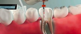
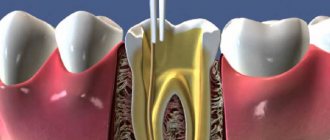
The primary task in treatment is to remove the pus and save the tooth. Highly professional doctors of our clinic, using high-quality anesthesia, will remove damaged tissue, and if necessary, it is possible to remove damaged dental pulp. If the cause of the infection is improper filling, then the doctor removes the filling, washes all existing canals with antiseptics and expands them. Antibiotics, anti-inflammatory and antiallergic drugs must be prescribed.
Three days later, when you visit the doctor again, the area is re-treated with antiseptics and closed with a temporary filling. At the next visit to the dentist, if there is no purulent discharge, a permanent filling is installed. If the inflammatory process continues, then surgical intervention is necessary. The final x-ray examination, carried out using high-precision equipment, confirms the quality of treatment carried out at Dr. Sadov’s Center for Comfortable Dentistry.
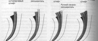
Measles canal filling
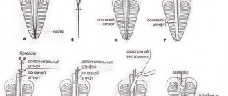
The procedure for filling root canals is carried out in various ways, for example: by the method of cold lateral (side) condensation of gutta-percha pins (reliable and affordable). Pins that resemble gutta-percha sticks in appearance are inserted into the root canal. After insertion, these sticks are compacted against one of the walls of the root canal. Between the gutta-percha there is a special paste for filling the canals.
The most modern method is filling the root canals using the vertical condensation method of hot gutta-percha. With this method, the doctor seals the top of the root canal using a heated gutta-percha pin. After this, the remaining volume is filled with hot liquid gutta-percha. This method is more expensive.
After the filling, if everything is done well, the doctor removes excess gutta-percha and sealer, and then installs a temporary filling. X-ray control is required. Permanent tooth filling in one visit is carried out only in exceptional cases, since it is necessary for the material to cool completely.
After root canal filling, discomfort may persist for some time. The tooth may be sensitive when biting. Within a few days the pain goes away.
Periodontitis of the tooth root
Periodontitis, in some cases, occurs as a complication of pulpitis. The tissue surrounding the root of the tooth, which holds it in the alveolus, is called periodontium. Accordingly, periodontitis is its inflammation; depending on the location, periodontitis is divided into:
1. Apical.
2. Marginal.
With the apical type, damage to the periodontal area located directly near the very apex of the tooth root is observed.
Marginal type - the inflammatory process originates at the edge of the gum. If medical care is not provided, the chronic disease progresses; over time, the bone tissue located in close proximity to the apex of the tooth root dissolves. The resulting cavity is filled with granulations, which lead to blockage of blood vessels. Lack of treatment provokes the formation of a fistula, or a growing granuloma that forms a cyst.
This is a disease of infectious origin when bacteria or fungi penetrate through a damaged surface to the root of the tooth. It should be noted that treatment of tooth root inflammation is a long process. The cause of the disease in some cases is:
1. Deep caries.
2. Pulpitis.
Chronic infection in the oral cavity leads to damage to the internal organs of a person, where the leading place is occupied by endocarditis, an infectious and inflammatory disease of the heart muscle.
Symptoms observed with periodontitis of the tooth root:
1. The patient feels that the diseased tooth is longer than the others.
2. Aching toothache. Increased pain with pressure.
3. Purulent periodontitis is characterized by throbbing pain, sometimes radiating to the ear, under the tongue or temple.
The chronic form is practically asymptomatic, sometimes unpleasant sensations appear when pressing on the diseased tooth. But a damaged tooth always has a grayish tint; in advanced cases, a fistula forms through which pus flows into the oral cavity.
Treatment of ongoing inflammation of the tooth root involves treatment of the root canals, with mandatory physiotherapy, which has an anti-inflammatory and analgesic effect. During this period, it is necessary to direct efforts to prevent the spread of infection into nearby cavities.
Dentistry AcademyDent, having the most innovative technologies, provides high-quality medical services until the oral cavity is completely healthy in the most advanced cases. A personalized course of treatment, based on the use of innovative technologies and high-quality materials, allows patients to get rid of progressive oral diseases and gain freedom in communication, thanks to a “Hollywood” smile.
Tooth root caries
Tooth root caries is less common, periodontitis or cysts. This disease occurs as a complication of cervical or root caries. Damages the root structure, which is located under the gum tissue itself. This pathology is almost impossible to detect during a visual dental examination.
To make a timely, accurate diagnosis, Dr. Sadov’s Center for Comfortable Dentistry has installed a universal X-ray system with the highest accuracy of the images taken - ORTHOPHOS XG 5, which has the ability to take panoramic X-rays. With a special gentle regime for children.
With the active proliferation of pathogenic microflora in the oral cavity, bone tissue rapidly loses: potassium, calcium and phosphorus. The result is a thinning of the cementum layer - the tissue covering the root and neck of the tooth and holding the tooth in its alveolar space. One of the main causes of dental caries is insufficiently performed hygienic procedures for oral care.
At AcademyDent you can undergo professional teeth cleaning. Hygienic teeth cleaning, carried out in our clinic by a highly professional dentist - orthodontist, in rare cases causes minor discomfort. But the procedure performed using ultra-modern equipment will qualitatively relieve you of: tartar, remove plaque from the dental surface, perfectly polish the enamel or perform fluoridation. These activities will help avoid diseases such as caries. Its danger lies in the fact that, as it progresses over a number of years, it does not cause pain and only a highly qualified doctor can recognize it in the early stages and prevent tooth loss.
The resulting gap between the gum and the neck of the tooth root creates a kind of pocket where food debris accumulates. Such a gum pocket is the cause of the formation of deep root caries.
The second reason for the appearance of this disease is inflammation of the gums and infectious processes occurring in the oral cavity:
1. Periodontitis.
2. Periodontitis.
3. Periostitis
4. Jaw osteomyelitis.
Inflamed periodontal and periodontal tissues often lead to detachment of the cervical part of the tooth from the gums and form a gum pocket. Where pathogenic microflora actively multiplies. In special cases, an ultra-modern method of x-ray examination is used for diagnosis - an orthopantomogram, which is successfully used by highly qualified doctors of our clinic, which allows us to obtain a detailed image of the teeth and bones of the facial part of the skeleton.
Many dental clinics consider damage to the root of a tooth by caries as a direct indication for its removal. But Dr. Sadov’s clinic has extensive experience in treating this pathology. Based on the most advanced technologies using drugs that have proven themselves in the best clinics in the world, the patient’s tooth is preserved and possible complications are effectively prevented.
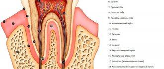
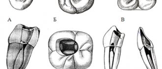
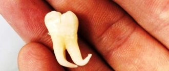
Molars
These are large molars. There are twelve molars in total, including wisdom teeth, six on each jaw. The crown of large molars is cuboidal. On the chewing surface of the crown there are from 3 to five tubercles. The presence of so many tubercles on the chewing surfaces of large molars provides numerous points of contact when closing the jaws and facilitates the grinding of food. They are located in the chewing zone and are used for primary food processing.
Perforation of the tooth root
This disease occurs as a result of cracks or holes, through which the tooth cavity or its root comes into contact with nearby tissues. This occurs as a consequence of unprofessional treatment or incomplete treatment. Tooth tissue affected by caries softens and collapses, followed by perforation (formation of a through hole).
The second reason is mechanical impact. The cause may be a blow and unskilled actions of the dentist when installing the pin associated with excessive use of force. There are often cases when perforation was discovered untimely. For a long period of time, the patient experienced aching pain and an ongoing inflammatory process in the gums surrounding the damaged tooth. Only an x-ray can confirm or refute this diagnosis.
By contacting highly qualified specialists, you can save the tooth and completely restore its function. Dr. Sadov’s Center for Comfortable Dentistry employs highly qualified doctors who have repeatedly successfully corrected the mistakes of their colleagues and restored health to patients. But in the future, the health of the oral cavity depends only on the patient himself, who is obliged to carefully monitor the cleanliness of the oral cavity and follow all the doctor’s instructions.
Dental formula
Human teeth are arranged symmetrically in the form of two dentitions - the upper dentition and the lower dentition. There are eight teeth on each side of the dentition from the median plane.
A person has 4 types of teeth:
Root resorption - treatment
Resorption is a resorption process that occurs with the roots of teeth, which is not always a consequence of caries or tooth decay. In practice, two types of resorption have been registered:
1. Internal resorption - the pathological process begins from the inner root surface of the tooth, where it borders the pulp (nerve cavity).
2. External resorption - it begins on the outer surface, in the area of the connection of the tooth root with the jaw, using the ligaments with which the tooth is held in the socket.
Both injuries are devastating and in the vast majority of cases lead to tooth loss. Treatment of this disease at AcademyDent allows you to save the tooth, although this is very difficult and in most cases not possible.
Internal resorption is the result of long-term chronic inflammation in the pulp (nerve) of the tooth, usually asymptomatic. Often the cause is improper installation of a deep filling or mechanical injury.
External resorption progresses asymptomatically. It is characterized by the rapid penetration of connective tissue and blood vessels into the root of the tooth and is localized below the gum line. The first sign is a change in tooth color, which is confirmed by an x-ray. For preventive purposes, it is recommended to take x-rays for people who want to avoid a terrible disease once every three years.
Treatment of internal resorption consists of root canal treatment - endodontic treatment. The pulp from the tooth and all inflammatory agents are removed. In the treatment of external resorption, tissue that has penetrated into the root of the tooth is removed. To prevent possible relapse, restoration material is used on the root surface itself. In particularly advanced cases, tooth extraction is recommended. An endodontist at AcademyDent Dentistry will conduct professional treatment in comfortable conditions and painlessly in order to restore the functional ability of a damaged tooth.