It's all the virus's fault!
The term "stomatitis" is derived from the merger of two Greek words: stoma (mouth) and itis (inflammation). There are a great variety of different types of disease - serous, aphthous, allergic, etc. The most dangerous is herpetic, or cold sore, stomatitis caused by a virus. Its main manifestations are painful ulcers covering the oral mucosa. The trigger mechanism of the disease is the activation of herpes simplex virus types 1 and 2. First of all, the disease threatens those who have a weakened immune system and, as a result, the body simply does not have the strength to give a worthy rebuff to viruses.
Causes of development of inflammatory diseases
The human oral cavity is inhabited by many beneficial, opportunistic and pathogenic microorganisms, the totality of which is called microflora [2]. Aerobic and anaerobic bacteria, various types of fungi, viruses, and protozoa were found in it [3]. If a person is healthy, the composition of the microflora is balanced, due to which the health of the entire oral cavity is maintained. However, under the influence of various unfavorable factors, general and local immunity can decrease, and the balance of microflora can be disrupted due to the excessive development of certain pathogenic microorganisms
Often these reasons act in a complex manner - against the background of a chronic or acute disease, the patient’s immune defense decreases, which leads to intensive reproduction of a certain strain of microorganisms, its predominance in the intraoral flora and the start of the inflammatory process. For example, candidal stomatitis is caused by fungi of the genus Candida. The causative agent of herpetic stomatitis is the herpes simplex virus.
Risk factors for oral inflammation [1]:
- systemic diseases (diabetes, chronic diseases of the gastrointestinal tract, endocrine and other disorders);
- weakened immunity (for example, due to HIV, unfavorable environmental conditions, hypo- and avitaminosis, poor nutrition);
- past infectious diseases, especially in children (ARVI, influenza, chicken pox) [4];
- long-term use of potent medications, in particular antibiotics;
- unsuccessful prosthetics;
- prolonged stress;
- lack or insufficient oral hygiene ;
- bad habits (smoking), etc.
Thus, it is believed that one of the key reasons for the development of aphthous stomatitis is chronic diseases of the gastrointestinal tract. Oral candidiasis can develop due to long-term use of antibiotics, and leukoplakia can develop in response to constant irritation of the mucous membranes of the lips and oral cavity, for example, with a poorly fitted denture [1].
Forms of the disease
Lightweight
It is considered the most beneficial for the body. In this form, people with high immunity suffer from herpes stomatitis. It flows without temperature. It is distinguished by single rashes that do not cause discomfort and disappear on their own without consequences.
Average
General disorders are added: weakness, drowsiness, fatigue, loss of appetite. Rashes appear in several places at the same time. The temperature rises to 37-37.6°C.
Heavy
This form of stomatitis indicates extremely low immunity. The rashes are multiple and painful. Severe headache, chills, and vomiting appear. The temperature exceeds 38oC.
If the disease is mild, the patient may not notice any external signs!
HIV infection
Oral ulcers are a common symptom of HIV infection and occur in approximately 30% of patients. In this case, their treatment is very specific. An infectious disease doctor selects a treatment regimen and medications for each patient individually. Sometimes ulcers are considered normal and do not require treatment, but this happens extremely rarely and only in advanced stages of the disease.
The patient can also go to any public dental clinic for surgical removal of ulcers. But before that, you need to make sure that there are no negative consequences after such surgery. If removal is justified, it is carried out in accordance with all precautions.
How to distinguish herpes from stomatitis?
Many patients try to find an answer on the Internet to the question: “Do I have stomatitis or herpes? How to recognize? Herpes stomatitis can be easily distinguished from ordinary stomatitis by 3 key signs.
- With herpes infection, the rash is localized in the gum area. Whereas with stomatitis - on the soft tissues of the oral cavity (tongue, cheeks).
- A herpes rash first appears as blisters, which then ulcerate, while stomatitis begins with the appearance of ulcers.
- Herpetic stomatitis is characterized by a stable appearance of the rash in the same places, and with ordinary stomatitis its location often changes.
Recurrent necrotizing peryadenitis
Recurrent necrotizing peryadenitis is also called Setton's aphthae. Its symptoms are:
- Seals appear in the submucosa of the oral cavity;
- Instead of compactions, ulcers with raised edges develop over time;
- The ulcers become inflamed, causing blood and lymphatic discharge.
Places where such afts accumulate are the upper and lower lips, cheeks and sides of the tongue. The pathology is characterized by extremely severe pain. Patients find it so difficult to eat that many give up eating completely. Difficulties also arise during conversation. The healing process of aphthae lasts a long time - sometimes up to several months, and the pathology itself persists for up to several years.
Herpetic or aphthous?
It will be somewhat more difficult to distinguish between herpetic and aphthous stomatitis. The latter got its name from the Greek term “aftha”, which means “ulcer”.
If with herpetic stomatitis there are many ulcers, but they are small, then with aphthous stomatitis there are few of them, and the size can reach 7–8 mm.
The second important distinguishing feature is the absence of swelling of the gums with aphthous stomatitis.
If you are looking for differences between herpetic and aphthous stomatitis, then the third thing you should pay attention to is the localization of the rash. Aphthous is characterized by the appearance of ulcers in the oral cavity, while herpes infection can spread to the border of the lips.
Symptoms of aphthous stomatitis
Usually, 1–2 days before the appearance of aphthae, areas of the mucous membrane with increased sensitivity are detected, and a burning sensation may occur. The aphthae themselves are round, have clear boundaries, and are covered with a gray or yellowish coating. Their size, as a rule, does not exceed 1 cm, and the mucous membranes around them turn red.
Such areas of erosion heal within up to 2 weeks without scarring. But in 1 case out of 10, the diameter of the ulcers is more than 1 cm, they affect deeper areas of tissue, and the borders of the pathological area may look raised. Healing in this case takes up to 6 weeks, after which a scar forms.
Aphthous stomatitis is characterized by damage to the mucous membranes of the cheeks, the inside of the lips, the soft palate, tonsils, and the lateral surfaces of the tongue. This is due to the lack of keratinization of the epithelium in these areas. Much less often, aphthae appear on the hard palate, back of the tongue, and gums.
How to distinguish herpes sore throat from stomatitis?
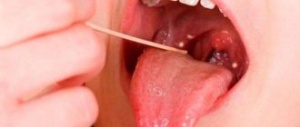
Despite the fact that these medical terms have the same grammatical root, herpetic sore throat and herpetic stomatitis are two different diseases. Unlike stomatitis, herpes sore throat occurs not due to the penetration of the herpes virus, but as a result of an adenovirus infection (in particular, the Coxsackie A virus). Children are more likely to suffer from this disease than adults. The rashes are localized, for the most part, on the soft palate and tonsils. Typical symptoms of stomatitis include pain in the abdomen and abnormal bowel movements.
Attention!
The disease begins acutely, with a jump in temperature to 40 degrees, and is severe, so differential diagnosis should only be carried out by a doctor.
Ulcers caused by trauma
The main reason for the appearance of ulcers when the mucous membrane is injured is infection in the wound. Most often, white rashes occur due to the habit of biting nails or the tip of a pencil or pen.
Fans of seeds and hard toothbrushes may also be at risk. Many people believe that hard bristles remove plaque better. Actually this is not true. If you carefully brush your teeth with such a brush, you can damage the enamel, gums and cause inflammation.
Other causes of mucosal injury:
- uncomfortable dentures;
- braces or other orthodontic structures;
- habit of unconsciously biting your cheek or tongue;
- exposure to mucous membranes with drugs or acids.
How to treat?
It is enough to remove the irritating factor and wait 1 - 2 weeks. Usually the ulcers go away on their own. If this does not happen, it is recommended to consult a doctor.
Bacterial or viral?
In addition to viral origin, the disease can be caused by bacteria: streptococci and staphylococci are normally present in the microflora of the oral cavity and begin to multiply uncontrollably during the inflammatory process. The latter may be caused by caries or periodontitis (read more about the disease in the article).
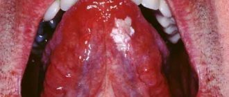
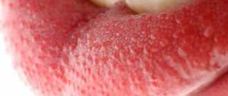
How to distinguish viral stomatitis from bacterial one? It is extremely difficult to do this at home, so it is better to consult a doctor. The main differential feature is the localization of the rash. With viral herpetic stomatitis, vesicles with transparent contents first appear on the tongue (its tip, along the side surfaces and under it), and then can even spread to the pharynx and tonsils.
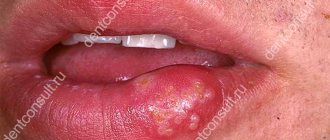
For bacterial stomatitis, the location of the rash on the gums and those areas where the skin borders the mucous membrane (for example, on the red border of the lips) is more common. Also, with a disease caused by streptococci, “jams” are often observed - pustules on the corners of the mouth, which quickly begin to bleed, become covered with a crust, crack and cause constant discomfort while eating and talking.
Types of diagnostics
An experienced doctor can identify herpetic stomatitis in adults during an initial examination, relying on only two methods.
- Clinical picture.
Based on the totality of the patient’s specific complaints and distinctive external signs, the dentist will not only assess the severity of the disease, but also differentiate it from ordinary stomatitis, candidiasis, etc. - Immunofluorescence.
Express microscopy method, the most accurate for diagnosing acute herpetic stomatitis.
Periodontal disease
Dental periodontal disease is a serious disease in which the last stage of gum inflammation occurs. This is often the cause of the development of infectious diseases, gastritis, stomach ulcers or cirrhosis of the liver. Even more often, the patient’s teeth simply fall out, and he cannot lead his usual lifestyle or eat his favorite food.
How to recognize periodontal disease
The signs of this dental disease are unclear and blurred. The patient is most often concerned about:
- exposure of the necks of the teeth;
- presence of tartar;
- burning gums;
- discomfort when eating.
There are 3 stages of periodontal disease:
- Easy. The patient has no complaints; very rarely there is a reaction to cold or hot food. The presence of periodontal disease can be determined during a dental examination. The mild stage of the disease is best treated.
- Average. The roots of the teeth are exposed by an average of 4-6 mm. The patient begins to experience a burning sensation in the mouth, and there is an acute reaction to eating hot, cold or sour foods.
- Heavy. The roots of the teeth are exposed by 8-10 mm. Chewing food causes severe pain.
Treatment methods
Diagnostics
Before starting treatment for periodontal disease, the dentist conducts an initial examination, during which he determines the extent of damage to the teeth and gums: which teeth can be restored and which will have to be removed. This is necessary in order to draw up an algorithm for further actions. The patient is then sent to the diagnostic room to take targeted and panoramic X-rays. Using them, the periodontist determines the depth of the pockets and the condition of the bone tissue.
Removing plaque and tartar
Inflammation of the gums, which is always observed with periodontal disease, mainly occurs due to soft plaque, subgingival and supragingival calculus. The main reason for their appearance is poor oral hygiene. Therefore, the specialist’s task is not only to treat the disease, but also to teach the patient proper hygiene.
General and local therapy
To increase immunity, the patient is prescribed a complex of vitamins and anti-inflammatory drugs. If the inflammation is minor, the dentist will prescribe a course of local therapy, which can be done independently at home.
Splinting teeth
An increase in tooth mobility indicates that the jawbone and soft tissue around them have begun to rapidly deteriorate. To prevent the teeth from changing position and falling out (for example, they may fan out), they are held together with fiberglass tape and filling material. This is also necessary before surgical treatment.
Surgical operations
If periodontal pockets reach 5-10 mm, it is impossible to prevent the progression of the disease without surgical intervention. First, the pockets are cleaned of granulations and food deposits. This procedure is called curettage. It comes in two types - open and closed.
Closed is carried out with special instruments, curettes. It is carried out only for periodontal disease at the initial stage (pockets reach 3 mm), when there is slight inflammation of the gums.
Open curettage is necessary in advanced stages of periodontal disease. With its help, all granulations and food deposits are completely removed. This operation is more difficult to perform. To completely clean out the pockets, incisions are made in the gum. Flaps of the mucous membrane are peeled away from the bone and the root surface is cleaned with curettes and an ultrasonic scaler. To restore bone tissue, the periodontist implants synthetic bone.
Next, the patient undergoes flap surgery to prevent receding gums. The doctor removes a 1.5 mm marginal strip of gum, since after prolonged inflammation the gum changes in such a way that it can no longer adhere normally to the tooth. After this, the mucous membrane flaps are pulled to the neck of the tooth.
Timely diagnosis and choosing the right treatment will help stop periodontal disease and maintain healthy teeth!
Causes of herpes stomatitis
There are two types of herpetic stomatitis: acute and chronic. Acute herpetic stomatitis, according to Dr. Komarovsky, occurs only in children under 3 years of age, when for the first time the child’s body, already deprived of antibodies to the herpes virus received from the mother, is first exposed to a viral attack from the outside. Moreover, the source of infection, as a rule, is the parents themselves - carriers of the virus, who kiss the baby or lick his pacifier or feeding spoon.
Recurrent or chronic stomatitis is already the lot of adults. The disease is recurrent in nature as soon as the body’s immune forces are weakened. In this case, primary infection can occur either through airborne droplets (sneezing), or through household contact (for example, through the use of the same dishes with a virus carrier) or hematogenous (through blood during injections, etc.). The incubation period of the disease can last up to two weeks depending on the state of the immune system.
Attention!
The pathogenesis of the disease in dentistry is still unknown. But if the body is weakened, any injury to the palate or gums can provoke activation of the herpes virus types 1 and 2!
Types of inflammatory diseases of the oral cavity
These diseases are classified depending on what causes the inflammation, as well as on the location of its source in the mouth.
Thus, general lesions of the mucous membrane are called stomatitis [1]. If the mucous membrane of only the tongue, lip, palate, gums or alveolar process becomes inflamed, they speak of glossitis , cheilitis, palatinitis, gingivitis or periostitis, respectively.
Depending on the reasons that caused the inflammatory process, these diseases are divided into:
- infectious;
- traumatic;
- symptomatic;
- specific.
If left untreated, inflammation can occur with complications and have negative consequences both for the teeth, gums, mouth and pharynx, and for the general health of a person.
Treatment of herpetic stomatitis
How to treat herpes stomatitis? Unfortunately, the herpes virus, once entered into a person’s blood, remains in a “dormant” state for the rest of his life. But treatment of viral stomatitis is possible provided that you do not delay visiting the dentist when the first signs of the disease are detected. During the period of therapy, in order to avoid infecting loved ones, you should eat and drink from separate containers and avoid kissing. The patient is recommended a diet that excludes spicy, smoked, sour and salty foods, which can irritate the damaged oral mucosa, and an increased drinking regimen (up to 2.5 liters per day), aimed at combating the manifestations of general intoxication of the body.
Drug therapy
Rinse
To stop the spread of infection throughout the oral cavity, as well as to prevent the occurrence of sore throat, anti-inflammatory drugs such as Stomatidine and Miramistin are prescribed. The greatest effect is achieved by repeating the rinsing procedure every 3 hours, strictly adhering to the instructions for medicinal solutions.
Antiviral drugs
To suppress the reproduction of the herpes virus, the patient is advised to take “Immudon” and “Acyclovir” orally according to the scheme, and for external use - “Viferon” ointment or “Silicea” gel.
Vitamin therapy
Tablet vitamin complexes such as “Complivit” help improve immunity.
Attention!
Before use, consultation with a specialist is recommended!
Treatment at home with folk remedies
On the Internet you can often come across the question: “How to treat herpetic stomatitis with folk remedies?” Let’s make a reservation right away: when treating this disease, you cannot rely solely on traditional medicine methods. Therapy must be comprehensive, and only a doctor can choose it correctly.
An effective folk remedy for rinsing the mouth is propolis tincture diluted with boiled water 1:3. In case of pronounced “jams” in the corners of the mouth and painful ulcers inside, applications with natural sea buckthorn oil have an analgesic and wound-healing effect.
Syphilis
Syphilis is an infectious pathology caused by Treponema pallidum. Mouth ulcers are a typical symptom of syphilis that appears throughout the entire period of the disease. Moreover, the ulcers themselves undergo a certain development process:
- The ulcers are round in shape and have dense edges. They are painless and have a white coating.
- Over time, the ulcers begin to bleed slightly.
- After about 1-3 months, the ulcers heal, and in their place strong scars form, around which dense bluish edges are concentrated.
After defeating syphilis, dense scars still remain in the oral cavity. They are the most eloquent sign of a recent illness.
Treatment of syphilis is carried out in a closed venereology clinic. During remission, the oral cavity is treated with anti-inflammatory and antibacterial drugs.
Prevention measures
- Strengthening the immune system.
Avoid excessive exercise and stress. Take a multivitamin in the fall and spring. Regular exercise and hardening increase the vitality of the body. - Healthy lifestyle.
Get rid of bad habits: scientists have proven that excessive smoking and alcoholic beverages can become a catalyst for the activation of the herpes virus in the body. - Timely treatment of chronic diseases.
Do not forget that simple stomatitis, if you do not seek medical help in a timely manner, can develop into herpetic stomatitis. Chronic caries and acute respiratory viral diseases suffered “on the legs” also seriously undermine the body’s immune defense. - Maintain personal hygiene.
According to statistics, it is poor oral hygiene that most often opens the way for herpes infection. - Avoid oral trauma.
Even a minor microcrack from a prick with a fish bone or careless use of a toothpick can become an “entry gate” for the herpes virus.
Attention!
With reduced immunity, herpes stomatitis becomes chronic and relapsing: the disease can recur 2-6 months after recovery.
Ulcers as local manifestations of general diseases on the oral mucosa
- Oral tuberculosis
is usually a secondary manifestation of pulmonary tuberculosis. It occurs as a result of penetration of tuberculosis bacteria into the oral mucosa through damaged epithelium. The membranes of the cheeks, tongue, and floor of the mouth are affected. First, typical tuberculous tubercles are formed, and then, after their disintegration, small ulcers are formed, which increase in size over time. The ulcer itself is not deep, with a loose bottom, which is covered with easily bleeding granulations (young tissue), uneven edges are observed, soft to the touch. With this disease, there is a sharp pain in the ulcer. In addition to the local manifestation in the form of an ulcer, there is a general deterioration in the well-being of patients: emaciation is noted, the amount of plaque on the tongue increases, sweating and body temperature increase. General treatment of tuberculosis of the oral mucosa is carried out in specialized anti-tuberculosis institutions. As for local treatment, in this case, sanitation of the oral cavity is carried out during the period of remission (weakening of the disease), treatment of the mucous membrane with antiseptic and anti-inflammatory agents. - Syphilis
is a chronic infectious disease caused by the so-called Treponema pallidum. All periods of development of this disease (in addition to the incubation period, which lasts 21 - 24 days) are characterized by the presence of ulcers in the oral cavity. At the initial stage of development of the disease, the presence of a painless ulcer is observed, which has a round or oval shape with raised, smooth edges and a cartilage-like specific infiltrate. The bottom of the ulcer is bright red, shiny or covered with a grayish-dark coating. The ulcers heal in 3 to 12 weeks with or without scar formation. Even with tertiary syphilis, there is no sharp pain, as, for example, with a tuberculous ulcer. The ulcer is surrounded by a powerful infiltrate, which is a dense bluish-red ridge that rises above the level of the mucosa. Its edges are smooth, bright red, covered with granulations, and bleed easily. After the ulcer heals, a retracted star-shaped scar forms. This process lasts 3 - 4 months. After the ulcers heal, scars remain, which are a sign of previous syphilis. General treatment of syphilis is carried out in a venereology hospital, local treatment is carried out during the period of remission or recovery (sanitation of the oral cavity, elimination of local traumatic factors). - Acute necrotizing gingivostomatitis
is a viral infectious disease. Most often, ulcers are localized on the mucous membranes of the gums, cheeks, soft palate, arches and tonsils. Favorable factors for the development of this disease are a decrease in the overall resistance of the body, a violation of the integrity of the oral mucosa, and a deficiency of vitamins in the body. There are also cases of this disease occurring against the background of cooling or overwork. It can also be a complication of viral infections, as well as allergic stomatitis. Usually young people (under 30 years of age) are affected, more often men.
Among the symptoms it is necessary to highlight: pain when eating; extremely unpleasant odor from the mouth; increased salivation; elevated body temperature. The gum mucosa becomes swollen, painful, and bleeds when touched. The epithelium of the gingival margin and gingival papillae becomes cloudy. The surface of the gingival margin is covered with a grayish-yellow coating, which is easily removed. The ulcers have soft, uneven edges and are covered with a dirty green coating (with a foul odor), which is easily removed. In this case, a loose bleeding bottom is detected. The surrounding tissues are swollen.
Treatment of necrotizing gingivostomatitis is carried out in accordance with the general condition of the body, taking into account its location and severity of the lesion. In moderate and severe stages of the disease, broad-spectrum antibiotics are prescribed, as well as drugs that prevent or reduce the manifestation of allergies. At any stage of development of the disease, vitamins C and P, high-calorie foods, juices are prescribed, and in some cases, according to indications, taking cardiac medications.
Local treatment is carried out under anesthesia (removal of necrotic tissue). The oral cavity is treated with warm solutions of antiseptics and anti-inflammatory drugs. The ulcer is also sprinkled with white clay powder. After acute inflammation has been relieved, professional oral hygiene is carried out.
Mouth ulcers can also form due to HIV (gum ulceration occurs in approximately 30% of people infected with HIV). How to cure mouth ulcers in this case? Treatment is specific and carried out by infectious disease doctors. Dental care is provided in all dental institutions with careful adherence to safety precautions.
Prevention of all of the above oral diseases consists of eliminating the causes of their occurrence. For example, to prevent infectious diseases that manifest themselves on the oral mucosa, such as syphilis, measures are necessary that prevent infection from entering the body. In other cases, wellness measures are very important, which include systematic independent and professional oral hygiene, which is offered by almost all dental clinics in Moscow.