Types of inflammatory diseases of the oral cavity
These diseases are classified depending on what causes the inflammation, as well as on the location of its source in the mouth.
Thus, general lesions of the mucous membrane are called stomatitis [1]. If the mucous membrane of only the tongue, lip, palate, gums or alveolar process becomes inflamed, they speak of glossitis , cheilitis, palatinitis, gingivitis or periostitis, respectively.
Depending on the reasons that caused the inflammatory process, these diseases are divided into:
- infectious;
- traumatic;
- symptomatic;
- specific.
If left untreated, inflammation can occur with complications and have negative consequences both for the teeth, gums, mouth and pharynx, and for the general health of a person.
Causes of development of inflammatory diseases
The human oral cavity is inhabited by many beneficial, opportunistic and pathogenic microorganisms, the totality of which is called microflora [2]. Aerobic and anaerobic bacteria, various types of fungi, viruses, and protozoa were found in it [3]. If a person is healthy, the composition of the microflora is balanced, due to which the health of the entire oral cavity is maintained. However, under the influence of various unfavorable factors, general and local immunity can decrease, and the balance of microflora can be disrupted due to the excessive development of certain pathogenic microorganisms
Often these reasons act in a complex manner - against the background of a chronic or acute disease, the patient’s immune defense decreases, which leads to intensive reproduction of a certain strain of microorganisms, its predominance in the intraoral flora and the start of the inflammatory process. For example, candidal stomatitis is caused by fungi of the genus Candida. The causative agent of herpetic stomatitis is the herpes simplex virus.
Risk factors for oral inflammation [1]:
- systemic diseases (diabetes, chronic diseases of the gastrointestinal tract, endocrine and other disorders);
- weakened immunity (for example, due to HIV, unfavorable environmental conditions, hypo- and avitaminosis, poor nutrition);
- past infectious diseases, especially in children (ARVI, influenza, chicken pox) [4];
- long-term use of potent medications, in particular antibiotics;
- unsuccessful prosthetics;
- prolonged stress;
- lack or insufficient oral hygiene ;
- bad habits (smoking), etc.
Thus, it is believed that one of the key reasons for the development of aphthous stomatitis is chronic diseases of the gastrointestinal tract. Oral candidiasis can develop due to long-term use of antibiotics, and leukoplakia can develop in response to constant irritation of the mucous membranes of the lips and oral cavity, for example, with a poorly fitted denture [1].
Skin rashes in children: rashes, exanthemas, enanthemas
Rashes on the skin (exanthema, exanthema ) and mucous membranes (enanthema, enanthema ) can occur not only with viral and bacterial infections, but also with diseases of a non-infectious nature.
It is important to decide whether these changes represent a primary injury to the child's skin or whether the clinical signs have changed due to secondary factors (infection, trauma, or treatment). Examination by a pediatric dermatologist Moscow - Markushka clinic.
Elements of rashes in a child, children. Primary and secondary elements
There are primary and secondary elements of rashes. Primary elements are classified as roseola, spot, papule, nodule, wheal, vesicle, vesicle, hemorrhage. Secondary elements include pigmentation and depigmentation, scale, crust, erosion, crack, abrasion, ulcer, scar, cicatricial atrophy, lichenification, vegetation.
Primary elements of rashes in a child, children: roseola, spot, erythema, hemorrhage, pinpoint hemorrhages - petechiae, papule, tubercle, node, blister, vesicle, bubble
Roseola is a pale pink or red speck ranging in size from 1 to 5 mm. The shape is round or irregular, the edges are clear or blurred, does not protrude above the skin level, disappears when the skin is pressed and stretched. Roseola occurs in many infectious diseases, especially typhoid fever. Multiple roseola 1-2 mm in size are usually described as a pinpoint rash (with scarlet fever), in the process of resolution they become covered with scales or disappear without a trace.
The spot (makula) has the same color as roseola, size - from 5 to 20 mm, does not protrude above the skin level. The shape is most often incorrect. The spot disappears when pressure is applied to the skin and appears again after the pressure is removed. Multiple spots ranging in size from 5 to 10 mm are described as a small-spotted rash (for example, with rubella - a child is vaccinated against rubella at the Markushka children's clinic). Spots 10-20 mm in size form a large-spotted rash (for example, with measles, allergies - examined by a pediatric allergist in Moscow, Markushka clinic).
Erythema is large areas of hyperemic skin that are red, purple-red, or purple in color. It occurs as a result of the fusion of large spots formed by the dilation of blood vessels not only of the papillary layer of the skin, but also of the subpapillary vascular plexus. Spots larger than 20 mm that tend to coalesce should be considered erythema. Erythema is most typical for erysipelas, thermal, and ultraviolet burns .
Hemorrhage (haemorrhagia) is bleeding into the skin as a result of destruction of skin vessels. It looks like dots or spots of various sizes and shapes and does not disappear when the skin is stretched. The color is initially red, purple or violet, then, as the hemorrhage resolves, it becomes yellow-green and finally yellow (formation of hemosiderin during the breakdown of red blood cells). Color changes are clearly visible in larger hemorrhages.
Pinpoint hemorrhages are called petechiae (petechia). Multiple round hemorrhages measuring 2 to 5 mm are described as purpura . Irregular hemorrhages measuring more than 5 mm are called ecchymoses . Hemorrhages may overlap with other elements of the rash. In such cases, they speak of petechial transformation of roseolas, spots, papules. As a rule, this is observed in severe cases of the disease. Hemorrhagic rashes are detected with typhus (often in combination with roseola - roseola-petechial rash), hemorrhagic fevers, and sepsis. Hemorrhagic elements of irregular shape on a dense basis (stellate rash) are characteristic of meningococcemia and pneumococcal sepsis. Minor hemorrhages can also have a non-infectious origin (capillary toxicosis, toxic-allergic vasculitis, vitamin deficiency C, etc.).
Papule (papula) is an element of the rash that rises above the level of the skin, which is often determined by touch. It has a flat or dome-shaped surface, size - from 1 to 20 mm. The shape and color are the same as those of roseolas and spots. Papules often leave behind pigmentation and flaking of the skin. Papules that merge with each other form plaques, and when the latter merge, areas appear that are located on large areas of the skin, the size of a palm or more. Often, during a routine clinical examination of a child, it is very difficult or even completely impossible to distinguish roseola from papules. On the other hand, the same sick child can simultaneously have roseola, papules (typhoid fever, paratyphoid fever, infectious mononucleosis), papules and spots (measles - child measles vaccination, children's medical).
A tubercle (tuberkulum) is a limited, dense, cavityless formation protruding above the surface of the skin with a diameter of 1-2 to 5-10 mm. The tubercles are formed as a result of the accumulation of a specific inflammatory infiltrate in the dermis. Clinically, the tubercle is similar to a papule, but differs from it in that when palpating the tubercle, a dense infiltrate in the skin is always clearly visible. In addition, tubercles, unlike papules, undergo necrosis during reverse development, often form ulcers and leave behind a scar or cicatricial atrophy of the skin. The tubercles are most typical of cutaneous leishmaniasis, leprosy and tuberculous skin lesions, tertiary and late congenital syphilis.
A node (nodus) is a cavityless, limited compaction that goes deep into the skin, often standing above the skin level. The size of the knots ranges from a hazelnut to a chicken egg and more. They are formed as a result of the accumulation of cellular infiltrate in the subcutaneous tissue and the dermis itself. Inflammatory nodules have a soft or doughy consistency, unclear boundaries, and the skin over them is red. Nodules that appear as a result of specific inflammation (colliquatic tuberculosis, syphilitic gumma) have a dense consistency, are sharply demarcated from the surrounding tissues, and are prone to decay and ulceration with subsequent scarring.
A blister (urtica) is an acutely inflammatory, cavity-free element slightly elevated above the skin level, measuring from 2-3 to 10-15 cm or more, has a round or oval shape, and is often accompanied by itching. Color - from white to pale pink or light red. The blister usually forms quickly and disappears quickly, leaving no trace behind. It occurs as a result of limited acute inflammatory swelling of the papillary layer of the skin and simultaneous expansion of the capillaries. The appearance of urticarial elements is characteristic of allergic reactions of various origins (drug, food, cold allergies), including those of an infectious nature. Sometimes occurs in the pre-icteric period of hepatitis B (vaccination of a child against hepatitis in the Markushka children's clinic).
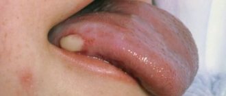
A vesicle (vesicula) is a cavity element measuring from 1 to 5 mm, representing a detachment of the epidermis. Usually the bubbles are filled with transparent, cloudy or bloody contents, they can shrink and give a transparent or brown crust. If the cover of the bubble is opened, then erosion is formed - a wet surface of pink or red color limited by the size of the bubble. The bubbles do not leave any scars on the skin. If a large number of leukocytes accumulate in a vesicle, it turns into an abscess - a pustule. Inflammatory changes are noted at the base and around the vesicle. Pustules are divided into single-chamber (chickenpox) and multi-chamber (natural smallpox). A group of blisters located on inflamed skin is called herpes. Vesicles are characteristic of herpes and enterovirus infections, chickenpox and natural pox. Chickenpox vaccination - Markushka Children's Clinic.
Bubble (bulla) is a cavity element with a diameter of up to 3-5 cm, located in the upper layers of the epidermis and under the epidermis. The contents of the blisters can be serous, bloody, or purulent. They can collapse, forming a crust, or open up, forming an erosive surface that turns into unstable pigmentation. The bubble occurs more often against the background of an erythematous spot, less often - against the background of unchanged skin (neonatal pemphigoid). The elements can be located both inside the epidermis, in the styloid layer (pemphigus vulgaris), and under the epidermis (multiform exudative erythema, dermatosis herpetiformis). It is observed with bullous form of erysipelas, sometimes with chicken pox, thermal burns.
Secondary elements of rashes in a child, children: hyperpigmentation, depigmentation, scales, erosion, abrasion, ulcer, cracks, tears, crust, scar, lichenification, vegetation
Secondary morphological elements are formed as a result of the evolution of the primary elements of the rash .
Hyperpigmentation (hyperpigmentatio) is a change in skin color as a result of an increase in melanin in it or the deposition of hemosiderin of primary elements.
Depigmentation (depigmentatio) occurs as a result of a decrease in the melanin content in the skin, observed after the disappearance of a nodule, tubercle - resolution of spotty-flaky (pityriasis versicolor, eczematoids) and papular (psoriasis) elements.
Scale (sguama) is an accumulation of rejected cells of the stratum corneum, sometimes the underlying layers of the epidermis. Scales occur on primary morphological elements - papules (psoriasis, syphilis), tubercles, after the resolution of blisters (eczema), etc.
Erosion (erosio) is a skin defect within the epidermis as a result of the opening of a vesicle, blister, or abscess, repeating their shape and size. When vesicles and pustules merge, erosions have scalloped edges. Erosion can also occur as a result of maceration of the skin in the area of folds or during maceration of other elements of the rash, most often papules. When erosion heals, there is no scar left; usually there is only temporary pigmentation.
An abrasion (excoriatio) is a violation of the integrity of the skin that occurs as a result of scratching, scratching, or other damage. Abrasions can be superficial - within the epidermis, sometimes involving the papillary dermis, and heal without a scar. Deeper abrasions, involving the deeper layers of the dermis, leave behind a scar. Abrasions are characterized by a tendency to become infected.
An ulcer (ulcus) is a deep skin defect that reaches the dermis, subcutaneous fat, fascia, muscles, and bones. It occurs as a result of the breakdown of the tissue of the primary element (tubercle, node, ecthyma). Its size is from 1 mm to the size of a coin or palm and more; the shape can be round, oval, linear, oblong, irregular. The surrounding tissue is either inflamed (edema, hyperemia) or infiltrated. Ulcers always heal with the formation of scars.
Cracks, tears (fissura, rhagades) - linear damage to the skin in the form of its rupture, resulting from excessive dryness due to loss of elasticity due to inflammatory infiltration or overstretching of the skin. Cracks can be located within the epidermis and dermis. They are usually localized in the corners of the mouth, interdigital folds, on the palms, soles, above the joints, and in the anus. A superficial crack after healing leaves no traces. After healing of deep cracks, linear scars remain.
A crust (crusla) is formed on the skin as a result of drying of the discharge of a weeping surface (vesicle, vesicle, abscess, ulcer, erosion). The crusts can have different colors (with serous exudate, transparent with a yellowish tint; with purulent exudate, yellow, greenish or brown; with hemorrhagic exudate, brown or black) and shape (layered, oyster-like, etc.).
A scar (cicatrix) is the formation of connective tissue at the site of a deep defect. Occurs after healing of deep skin defects at the site of ulcerated tubercles, deep pustules, nodes, deep burns, wounds. Scar formation is accompanied by the death of sebaceous and sweat glands, hair follicles, blood vessels and elastic fibers, and the disappearance of the skin pattern. Typically, scars are located below the skin level or are at its level, less often they rise above the skin level - hypertrophic scars.
Lichenification (lichenificatio) is a focus of increased skin pattern, accompanied by thickening and compaction, hyperpigmentation, and dryness. Foci of lichenification are most often localized in the neck, elbow and popliteal folds, wrist and ankle joints, inguinal folds, scrotum and occur in chronic dermatoses accompanied by itching (eczema, neurodermatitis).
Vegetation (vegetatio) is a papillary thickening of the skin that occurs as a result of the growth of the styloid layer of the epidermis and papillomatosis of the dermis during a long-term inflammatory process. More often it forms in the area of papular elements and ulcers. Vegetations can erode, bleed, and are prone to secondary infection.
Symptoms of inflammatory diseases
Inflammation in the oral cavity is manifested by both general and local symptoms [1]. Even before the onset of local manifestations, general malaise, pain, irritation, rashes in the mouth, increased body temperature, and decreased appetite are noted [1]. In addition, inflammation in the mouth itself can act as a symptom of another disease or general pathology [1].
Symptoms of certain inflammatory diseases
Stomatitis is a wide group of diseases that can be caused by individual pathogens, for example, fungi (candidal stomatitis) or the herpes simplex virus (herpes stomatitis), and systemic diseases, for example, chronic diseases of the gastrointestinal tract (aphthous stomatitis) [2] .
Most often, stomatitis appears as rashes (ulcers, blisters or other forms) on the oral mucosa. The nature of the rash is a striking distinctive feature that plays an important role in diagnosis. For example, with aphthous stomatitis, the ulcers never appear on the outer surface of the lips, as is the case with lesions caused by the herpes simplex virus [2].
Herpetic stomatitis, as a rule, accompanies a general infection of the body and is characterized by rashes on the inner surface of the cheeks, tongue, palate, and lips [2].
With insufficient oral care, bacterial microflora may develop and cause deeper lesions of the mucous membrane.
If the necessary treatment of acute herpetic stomatitis is not carried out, a recurrent form occurs, which is accompanied by regular rashes on the oral mucosa of vesicles and aphthae "Microbiology, virology and immunology of the oral cavity", ed. Doctor of Medicine V. N. Tsareva.
Catarrhal stomatitis occurs quite often and develops due to the lack or poor hygiene and the presence of chronic foci of infection in the oral cavity. Symptoms of this type of stomatitis are swelling of the mucous membrane, the appearance of plaque on it, first white, then brown, and bad breath [1].
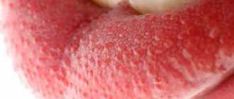
Candidiasis (candidal stomatitis, thrush) is caused by fungi of the genus Candida, which are normally always present in the mouth in small quantities [2]. Factors contributing to the sharp growth of fungi are long-term use of antibiotics and some other drugs, hypovitaminosis, endocrine and other disorders.
A striking symptom of candidiasis is the appearance of a white or yellowish-white coating on the tongue and the mucous membrane of the cheeks due to the development of inflammation in the mouth. The plaque is easily removed, revealing reddened, inflamed and eroded areas of the oral mucosa underneath.
Leukoplakia refers to chronic inflammatory diseases. It develops in response to constant irritation of the oral mucosa, for example, from a part of a denture, a sharp edge or chipped tooth, hot drinks or food, alcoholic beverages, or smoking [1]. Leukoplakia manifests itself in the form of whitish thickenings, usually in the cheek area along the line of closure of the teeth, in the corners of the mouth, on the back and on the lateral surfaces of the tongue.
Change in structure with fluid accumulation
With such lesions, the structure of the mucous tissue changes so that a cavity filled with liquid is formed inside.
Bubbles and bubbles. They are formed in the epithelial layer or under it, filled with serous or hemorrhagic contents, and can be grouped. The cavity is closed by a thin layer of epithelium, which can break through. Bubbles can group and break quickly. Bubbles form and last longer. Both types of lesions provoke the formation of healing ulcers on the surface of the mucous membranes. They arise due to damage by viruses, traumatic injuries, and disturbances in tissue nutrition.
Ulcers. They can form from blisters or on unchanged mucosa. The cavity is filled with purulent exudate (whitish, yellowish, greenish contents with a pungent odor). They can be deep or superficial, often painful. Indicate an inflammatory process, appear after traumatic damage to the mucous membrane, due to infectious, viral diseases.
Cysts. A formation with dense walls that form a cavity. It is filled with transparent contents (can become purulent, serous, bloody). Appear due to blockage of gland ducts on the mucous membranes or as a symptom of periodontal diseases.
How to get rid of stomatitis and other inflammatory diseases of the oral cavity
Treatment of stomatitis and other inflammatory diseases is usually comprehensive, aimed at alleviating the patient’s condition, eliminating the root cause and risk factors.
Often, inflammation of the oral cavity is painful, accompanied by itching, irritation, and swelling of the mucous membrane. Because of this, patients have difficulty eating. To alleviate the condition, the affected areas are treated with various anesthetic and antiseptic rinses, powders, and solutions. This also prevents possible complications of diseases, for example, the transition of aphthae or foci of inflammation into ulcers, which require more time to heal.
Another important area of treatment is the elimination of local causes of inflammation of the oral mucosa. These include local foci of infection, for example, caries , dental plaque (tartar), traumatic chipped teeth, protruding parts of fillings or dentures [1]. For this purpose, appropriate treatment and professional hygiene are carried out.
And finally, a significant part of therapy is devoted to the diagnosis and elimination of a general disease that can provoke inflammation of the oral cavity (for example, endocrine disorders or chronic diseases of the gastrointestinal tract) [1]. In special cases, antiviral drugs [5], antimycotics [6] and antibiotics [1] are used.
Prevention of stomatitis and other inflammatory diseases includes [7]:
- treatment of major systemic diseases;
- strengthening general immunity: adherence to the principles of a healthy lifestyle, nutrition, giving up bad habits, eliminating hypo- and avitaminosis;
- strengthening local immunity: regular careful hygiene, avoiding injury and irritation of the mucous membrane in the mouth.
Regular individual and professional oral hygiene is a key way to prevent intraoral inflammation. It must include:
- brushing your teeth twice a day, after breakfast and before bed, with a soft-bristled brush and toothpaste;
- the right choice of toothpaste: if you are prone to inflammation of the mouth and gums, as prescribed by a specialist, you should give preference to pastes with anti-inflammatory and antiseptic properties;
- use of antibacterial rinses with anti-inflammatory effects;
- cleaning interdental spaces with dental floss or tape;
- regular visits to the dentist (once every 6 months) for a preventive examination and professional removal of dental plaque.