We are used to the fact that we need to go to the dentist if we have problems with our teeth or gums. What to do if you have inflammation of the tongue or glossitis? Dentists identify more than 10 types and subtypes of infection. The infection can be caused by both fungus and bacteria. Viral infection is less common. Inflammation can be caused by a lack of certain vitamins and minerals, or hormonal changes. There are many reasons for the disease, so it is important to identify what exactly caused the inflammatory process.
Causes of glossitis
- improper oral hygiene;
- bacterial infection;
- fungal infection;
- heavy metal poisoning;
- bad habits (smoking, alcoholism);
- congenital pathologies of the tongue (folded tongue);
- allergic reaction;
- tongue injury (often caused by malocclusion);
- oral infections;
- lack of iron in the body;
- burn of the mucous membrane (hot food or drinks);
- infectious diseases (AIDS, tuberculosis, scarlet fever, measles).
Manifestations of diseases of internal organs on the mucous membrane of the mouth, tongue and lips
Diseases of the mucous membrane (MU) of the mouth, tongue and lips are one of the important and complex areas of practical medicine. Patients often find themselves in difficult clinical situations when they receive untimely and inadequate care. The fact that the human body is a complex, hierarchical, dynamically self-governing formation, the stability of which is due to the simultaneous functioning of a number of organs and systems, began to escape the attention of the doctor.
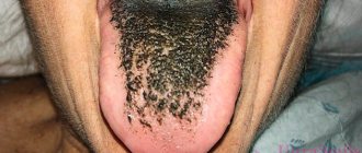
The existing relationships between lesions of the mucous membrane of the mouth, lips, tongue and systemic pathology should alert both patients and dental and somatic doctors. In the medical literature, a large group of tongue diseases are presented that occur separately, without affecting other parts of the oral cavity: desquamative glossitis (“geographical” tongue), black (“hairy”) tongue, folded tongue, rhomboid glossitis [1].
Changes in the condition of the mucous membrane of the mouth and tongue can occur and be detected both before other clinical manifestations of systemic diseases, and simultaneously with them. In many systemic (somatic, general) diseases, the oral mucosa reacts with the appearance of various types of disorders: disorders of tissue trophism, bleeding, swelling, dyskeratosis . Some manifestations of pathology in the mucosa of the mouth and tongue clearly indicate one or another type of organ or systemic disorder and are of great diagnostic value. However, in most cases, despite the different etiology and pathogenesis, the manifestations of systemic diseases on the oral mucosa are not specific in nature and are characterized by similar, sometimes outwardly identical clinical signs, which creates difficulties in their recognition.
Often occurring lesions of the oral mucosa due to diseases of the gastrointestinal tract, cardiovascular system, endocrine pathology, deficiency of vitamins (especially group B), macro- and microelements can attract the attention of specialists in various fields. Since changes in the mucous membranes of the mouth and tongue can occur and be detected earlier than other clinical manifestations of systemic diseases or simultaneously with them, patients themselves often turn to a dentist. In turn, gastroenterologists, endocrinologists, cardiologists, and hematologists can involve a dentist for consultation and joint supervision of patients with lesions of the oral mucosa, lips and tongue.
In this regard, we consider it necessary to share our clinical observations. This message may be useful to specialists whose professional interests relate to the diagnosis of these diseases and the treatment of such patients. Our previous publications were devoted to these issues [2–10]. The latest report [11] provided data on cancer screening for diseases of the oral mucosa, tongue and lips.
During our many years of clinical practice, changes in the CO of the mouth, tongue and lips were observed in the pathology of various organs and systems of the body, and metabolic disorders. Lesions of the mucous membranes of the mouth, lips and tongue are most often found in diseases of the gastrointestinal tract. In this case, ulcerative, aphthous and necrotizing stomatitis develop. The best studied in this regard are language changes characterized by a number of features:
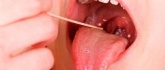
- coated tongue (Fig. 1), which is detected most often (with exacerbation of gastritis, gastroesophageal reflux disease, peptic ulcer, pancreatitis, enteritis and colitis, the amount of plaque increases);
- swelling of the tongue, diagnosed by tooth marks on its lateral surfaces, usually associated with enteritis and colitis;
- changes in the papillae of the tongue, manifested in the form of hyperplastic and atrophic glossitis;
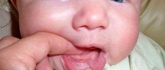
- desquamation of the epithelium (sometimes in this case they talk about desquamative glossitis (Fig. 2), which some authors regard as an “independent disease of the tongue”);
- paresthesia and disorders of taste sensitivity, often accompanying not only diseases of the digestive system, but also developing as a result of an imbalance of vitamins and pathology of the autonomic nervous system.
Rice.
1. Coated tongue in a patient with chronic pancreatitis in the acute stage. Rice. 2. Desquamative glossitis in an 82-year-old patient suffering from peptic ulcer localized in the pylorus, who underwent radiation therapy for lung cancer and surgery for cancer of the rectosigmoid angle. Damage to the mucous membrane of the mouth, lips and tongue is observed with a deficiency of vitamins and microelements. The most common deficiencies in clinical practice are B vitamins and iron.
Pain, numbness, burning in the tongue, in the gums and lips are observed with hypovitaminosis B12, which is accompanied by neurological disorders and changes in hematopoiesis. Both with iron deficiency anemia and with latent iron deficiency, changes in the CO of the mouth, tongue and lips are caused by manifestations of tissue iron deficiency (sideropenia) . Patients (usually young girls and women) complain of perversion of taste sensitivity, paresthesia, burning of the tongue, pain and swelling in the tongue, sometimes difficulty swallowing dry and solid food, and choking. With severe sideropenia, the tongue, due to atrophy of the filiform and mushroom-shaped papillae, becomes smooth (“polished”, “varnished”), angular cheilitis develops, folds on the back of the tongue and trophic changes in the mouth are sometimes observed. It should be remembered that the appearance of symptoms of sideropenia often precedes a decrease in hemoglobin levels and can persist even after correction of anemia if iron therapy is insufficient.
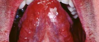
Pain, numbness and burning in the tongue, in the gums, lips are observed with hypovitaminosis B12, which is accompanied by neurological disorders and changes in hematopoiesis (B12-deficiency anemia), so the mucus of the mouth is pale. Bright red areas of inflammation appear on the tongue, reacting to the intake of acidic foods and medications. These lesions are often localized along the edges and at the tip of the tongue, sometimes involving the entire tongue (“scalded” tongue) (the so-called Hunter-Meller glossitis). Often ulcerations and petechiae develop on the tongue. Such changes can spread to the gums, mucous membranes of the cheeks, soft palate, and less commonly to the mucous membranes of the pharynx and esophagus. Subsequently, the inflammatory phenomena subside, and the papillae of the tongue atrophy. The tongue becomes smooth and shiny (“varnished” tongue; Fig. 3).
Rice. 3. “Lacquered” tongue in a patient with B12-deficiency anemia.
With relapses of the disease, along with atrophic changes, hyperplasia of the papillae at the root of the tongue develops (hypertrophic papillitis). At the same time, leukokeratosis of the tongue and lip mucosa may be observed.
Vitamin B1 deficiency is accompanied by hyperplasia of the fungiform papillae of the tongue, paresthesia and allergic reactions of the oral mucosa. Hypovitaminosis B2 is manifested by a peculiar change in the skin, red border of the lips, mucous membrane in the corners of the mouth (angular stomatitis), oozing, maceration of the epithelium. A superficial form of desquamative glossitis is noted (triad: dermatitis, cheilitis, glossitis). With hypovitaminosis B6, symptoms of a disorder of the nervous system (polyneuritis) and the gastrointestinal tract, angular stomatitis, cheilitis, and glossitis are observed.
Lesions of the oral mucosa in diseases of the cardiovascular system are observed in more than half of patients with this pathology, in which the following are detected:
- swelling and cyanosis of the mouth and lips; in case of myocardial infarction, swelling of the tongue may be accompanied by the appearance of erosions, ulcers and “cracks”;
- vesical-vascular syndrome - the appearance (usually in older women suffering from arterial hypertension) of dense blisters (after opening, erosions form) with hemorrhagic contents on the mucous membrane of the soft palate, the lateral surfaces of the tongue, and the cheeks. It is necessary to differentiate this syndrome from pemphigus and erythema multiforme;
- ulcerative-necrotic lesions of the mouth with the formation of trophic ulcers in the absence of a pronounced inflammatory reaction in the surrounding tissues. We observed a patient with necrosis and sequestration of the body and branch of the mandible. These lesions must be differentiated from: traumatic ulcers, malignant tumors, Vincent's ulcerative necrotizing stomatitis, necrotic lesions of the mouth due to blood diseases.
The most common endocrine pathology in dentist practice is diabetes mellitus. There is a direct relationship between the severity of inflammatory changes in the oral mucosa and the course of the disease, its duration and the age of the patient. With a short duration of diabetes, the oral mucosa becomes hyperemic, swollen, and bleeds. With increasing duration of the disease, hyperkeratosis of filiform papillae and hyperplasia of fungiform papillae most often develop. The tongue is coated; hyperemic mushroom-shaped papillae in the form of reddish dots rise along the entire back of it.
The dentist is responsible for diagnosing the early manifestations of “general” diseases, conducting a thorough examination of the patient through the efforts of somatic doctors. Folding and an increase in the size of the tongue are often noted; a combination of tongue folding with hyperkeratosis of the filiform papillae or, conversely, with their desquamation (diffuse or focal) and dryness of the mucous membrane of the tongue is possible. There is a “geographical” language. Teeth imprints are identified on the lateral surface of the tongue. With decompensation of diabetes, decubital ulcers are possible and in almost all patients changes in the lips are detected: dry mouth and red border of the lips in combination with cracks, jams, crusts, bright hyperemia, especially pronounced in the Klein zone, angular cheilitis. In the diabetes compensation phase, dry mouth and angular cheilitis disappear.
Changes in the CO of the tongue are more stable, remaining in the phase of decompensation and compensation. Oral candidiasis often develops.
Basics of medical tactics for lesions of the mouth, lips and tongue:
- For rational treatment of diseases of the mouth, lips and tongue, a thorough examination of the patient and contact between the dentist and other specialists is required, first of all, with the therapist, as well as specialized specialists - gastroenterologist, endocrinologist, hematologist, cardiologist.
- An axiom for the dentist should be the elimination of all unfavorable irritating factors in the oral cavity in the patient, which can support and provoke the development of the pathological process. It is unacceptable to use the so-called cauterizing agents and long-term use of the same mouthwashes.
- Treatment of diseases of the oral mucosa must be carried out in compliance with the principles of bioethics; these diseases must be considered from the standpoint of the state of the whole organism, therefore, in most cases, one cannot limit oneself only to local effects on the lesions of the mucous membrane carried out by the dentist.
- Treatment should begin only after at least a preliminary diagnosis has been established and meet the following requirements:
- be comprehensive;
- provide a pathogenetic approach;
- do not violate the anatomical and physiological properties of oral mucosa;
- eliminate the pain factor;
- help optimize the epithelization of lesions;
- provide for the active involvement of the patient in performing medical procedures at home.
In some diseases, changes in the color and general appearance of the oral mucosa and surface of the tongue do not have independent diagnostic significance. However, in combination with other symptoms, the appearance of the lips, tongue, and oral mucosa can help clarify the diagnosis. The prognostic significance of changes in organs and tissues of the oral cavity is great. The dentist is responsible for recognizing and diagnosing early manifestations of “general” diseases and conducting a thorough examination of the patient through the efforts of somatic doctors. We are confident that timely and correct assessment of the described conditions in the practice of doctors of other specialties is very necessary and advisable.
Literature
- Danilevsky N. F., Leontyev V. K., Nesin A. F., Rakhniy Zh. I. Diseases of the oral mucosa. - M.: OJSC "Dentistry". — 271 p.
- Tsepov L. M., Mikheeva E. A., Nesterova M. M. Erosive and ulcerative lesions of the mucous membrane of the mouth, tongue and lips. Tactics of a dentist // Dental South. - 2009. - No. 8 (68). — P. 10-13.
- Tsepov L.M., Nikolaev A.I., Petrova E.V. et al. Symmetrical recurrent decubital ulcers of the mucous membrane of the lower lip in a patient with facial paraspasm // Dental South. - 2007. - No. 7 (56). — P. 28-29.
- Tsepov L. M., Tsepova E. L. Diagnostic value of changes in the lips, oral mucosa and tongue in various diseases and pathological conditions // Dental South. - 2010. - No. 6. - P. 14-16.
A complete list of references is in the editorial office.
The most common types of glossitis
The most common occurrences in dental practice are:
- acute catarrhal glossitis;
- tongue abscess;
- desquamative glossitis.
Acute catarrhal glossitis is the most common type of inflammation. Inflammation can be caused by microbes or mechanical damage to the tongue. The predominant symptoms are pain, redness and swelling.
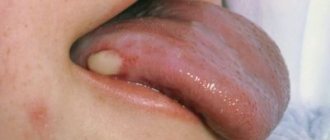
A tongue abscess is the appearance of an abscess in the tongue. The abscess can be superficial, under the mucous membrane, or maybe in the thickness of the tongue. Abscesses in the thickness of the tongue, in addition to pain in the tongue, can cause a disturbance in the general condition. A person develops a fever, a headache, and weakness. Most often occurs due to injury to the tongue.
Desquamative glossitis, also known as “geographic tongue,” most often appears in children. It appears in the form of various spots on the tongue, which look like a white coating, alternating with areas of pink mucous membrane. There are no changes other than appearance. Scientists have identified a clear reason for it. The main factors are believed to be bacteria, allergic reactions and hormonal imbalances.
White spots on a child's tongue
The easiest way to notice the formation of white areas in the oral cavity of children, which can signal the following problems:
- 1 Candidiasis is an inflammation caused by excessive growth of fungi in the mouth. The spots look like cottage cheese flakes. The problem occurs when the body's defenses are reduced.
- 2 Stomatitis is a common inflammation of the oral cavity in children, accompanied by painful white spots. The cause of infection is bacteria1.
- 3 Leukoplakia is a consequence of frequent minor injuries accompanied by infection.
- 4 Diseases of the digestive system: the tongue is often called an indicator of the functioning of the gastrointestinal tract, so the presence of white areas in combination with symptoms of digestive dysfunction is a reason to worry about the health of the stomach and intestines2.
Symptoms of glossitis
- increased salivation;
- swelling and redness;
- pain and burning when eating;
- plaque on the tongue in the form of spots;
- bad breath;
- papillomas or warts on the tongue;
- speech disorder;
- foreign body sensation.
Prevention of glossitis - high-quality oral hygiene and no bad habits. It is important to undergo timely preventive examinations and also eat well. All these factors actively contribute to the development of the disease and bring a number of problems.
What does the tongue look like normally?
In the absence of health problems, the child’s tongue looks moist, evenly pink, without areas of redness or swelling1. In some cases, parents may see plaque due to the following reasons:
- Insufficient and irregular oral hygiene.
- Eating food with coloring substances (lollipops, chocolate, persimmons, beets, carrots, ice cream, etc.).
- Using seasonings that have a coloring effect (curry) in cooking.
- Taking medications with a coloring effect (iron preparations, activated carbon, iodine sprays, solutions with furacillin and tannins).
- Excessive amounts of fatty foods in the diet.
Whitish spots on the tongue of an infant may appear due to the curdling of mother's milk in the mouth or minor regurgitation.
Treatment of glossitis
A specialist must make an accurate diagnosis and identify the cause of the disease. If you suspect that you have glossitis , and all symptoms indicate this, contact your dental clinic. This is the only way to create the right treatment plan and provide timely assistance. Quite often in such cases, doctors prescribe antibiotics, anti-inflammatory drugs and rinsing the mouth with special antiseptic solutions. In advanced stages, glossitis is treated surgically. Deep abscesses must be opened in the maxillofacial department. Under no circumstances should you take medications without a doctor’s recommendation.
You can cure glossitis, caries or any other diseases of the oral cavity right in your sleep. Family Dentistry Center "Medexpert" provides dental treatment under medicinal sedation. Thanks to this approach, the patient falls into a healthy sleep, ceases to feel pain and discomfort, while the vital functions of the body remain unchanged. Sedation is widely used in pediatric dentistry and even helps fight dental phobia. Dental treatment can be comfortable and painless - tested for yourself.
Red spots on a child's tongue
The most common type of spots is red, signaling an inflammatory process4. Reasons for such color changes to occur:
- Allergy . When an allergen enters the mouth, such as food or medicine. Sometimes children put animal fur or plant flowers into their mouths, which can also cause spots.
- Herpes . It is quite rare, but a child can develop a herpes infection in the oral cavity. It indicates a weakened immune system. The baby will be bothered by pain and itching. The temperature may rise.
- Scarlet fever . This childhood infection is not identified by spots on the tongue, but they are a specific sign. Key symptoms: high fever, severe pain and redness of the throat, cough, rash all over the body in the form of small dots.
When to see a doctor
Visit your dentist if the spots don't go away within a week. White spots, cracks, ulcers and leukoplakia can cause oral cancer. In this case, the sooner a dangerous disease is recognized, the more favorable the outcome awaits the patient [6].
“According to WHO, in the Russian Federation alone in 2015, 589,341 cases of malignant neoplasms were identified (including 270,046 and 319,335 in male and female patients, respectively).
The increase in this indicator compared to 2014 was 4.0%, and these numbers are growing” Mikhalchenko D.V., Associate Professor, Head of the Department of Volgograd State Medical University [6]
More rare situations
Yellow spots on a child’s tongue may indicate diseases of the digestive system1. Usually the problem manifests itself in the formation of a dense coating on the base of the tongue, which is difficult to remove. The cause should be sought with the help of a gastroenterologist.
Blue areas are extremely rare in childhood, as they signal problems with the cardiovascular system. Also, dark blue spots may just be hemangiomas that do not require any correction.
Bald spots are areas where taste buds are missing3. The child does not feel them, and they cannot be treated in any way, since the death of the papillae is irreversible. The main task of parents is to determine the reason for the formation of such areas.