Full text of the article:
Working with diseases of the internal organs is especially difficult because they cannot be seen. Previously, doctors had to treat patients literally “blindly,” since it was possible to examine a person’s internal organs only during surgery. Nowadays, doctors don’t have to pick up a scalpel; various types of scans help make a diagnosis. However, patients are wary of this type of examination. This is due to the high cost of some procedures and the fear of radiation. Let's try to figure out what the features of certain scans are and when to resort to them.
X-ray
The oldest and most common method of visualizing the human body. X-rays are used everywhere, from surgery to dentistry. The method is simple and clear: a person is irradiated with special rays that easily pass through soft tissues and linger in hard ones. Thanks to this principle, an image is transmitted to a photographic film or sensor located on the side opposite the ray source, and radiography or fluoroscopy is available to the doctor.
Main advantages
such an examination: speed and cost. Almost all hospitals are equipped with X-ray machines; the procedure is quick and inexpensive.
Main disadvantages:
exposure and image quality. When performing radiography, the patient is irradiated, and the picture is two-dimensional. The doctor can hardly see the internal organs individually, since their shadows overlap each other. It is also impossible to see the cartilage tissue and brain in detail. The cartilage practically does not block the rays, the brain is securely closed by the skull. X-rays are not suitable for their examination.
It will be most effective to carry out radiography for damage to bones, joints and teeth.
Radiography
Some of these rays are absorbed by tissues and only the remaining rays reach the X-ray film, which illuminate it, creating a specific image that has diagnostic value.
In clinical practice, there are two most common types of X-ray examinations:
- Plain radiography – performed without the use of contrast agents.
- Contrast radiography - the study is supplemented by the introduction of medications into the human body, which gives an additional image on the x-ray.
Types and indications for radiography
GVM International performs a list of different types of radiography, each of which has specific indications:
- Mammography
- Plain radiography of the abdominal cavity
- Plain X-ray of the chest cavity
- X-ray of the stomach
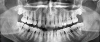
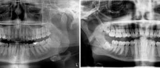
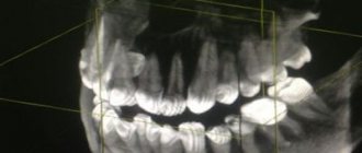
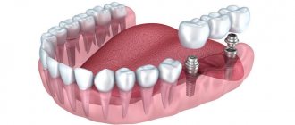
- X-ray of teeth
- X-ray of the intestine with barium passage
- X-ray of bones, joints and spine
- X-ray of the nasal sinuses
- Urography
Preparation and methodology
Some types of radiography require special preparation before they are performed:
- X-ray of the stomach and passage of barium through the intestines - studies are carried out on an empty stomach.
- Mammography – performed from 6 to 12 days of the menstrual cycle.
- Irrigoscopy requires quite a long preparation. Three days before the procedure, any foods that stimulate gas formation are excluded from the patient’s diet. The night before and immediately before the diagnostic procedure itself, a cleansing enema is performed.
- Urography requires personal supervision by a radiologist before execution, since the patient must be administered intravenous contrast 15 minutes before the examination.
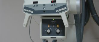
The X-ray diagnostic technique is quite simple. The area of the body to be examined is placed directly in front of the device screen and a emitting lamp is directed at it. After positioning the patient, the examination itself is performed, which only takes a few seconds. Depending on the type of x-ray, the number of shots and the intervals between them may vary.
Contraindications for carrying out
There are no absolute contraindications to X-rays, but they should be limited to pregnant women and children under 16 years of age. For these categories of patients, x-rays are prescribed only in extreme cases.
Preparing the patient for various types of x-ray examination and magnetic resonance imaging
Methods and means of radiation diagnostics
FLUOROGRAPHY - A method of x-ray examination - photographing organs of the human body from an x-ray screen; mass medical examination based on this method.
RADIOGRAPHY is a method of x-ray examination in which an image of an object is obtained on x-ray film by directly exposing it to a radiation beam.
Types of radiography:
- normal (information is stored on film);
- digital (information is stored on a digital medium).
Digital radiography is more convenient, since the digitized image can be transmitted over the Internet or recorded on a disk or flash drive.
LINEAR TOMOGRAPHY (classical tomography) is a method of x-ray examination with which you can take a picture of a layer lying at a certain depth of the object under study. This type of research is based on the movement of two of three components (X-ray tube, X-ray film, object of study).
MAGNETIC RESONANCE TOMOGRAPHY (MRI, MRT, MRI) is a tomographic method for studying internal organs and tissues using the physical phenomenon of nuclear magnetic resonance. The method is based on measuring the electromagnetic response of atomic nuclei, most often the nuclei of hydrogen atoms, namely on their excitation by a certain combination of electromagnetic waves in a constant magnetic field of high intensity.
Algorithms for preparing patients for X-ray examinations
Fluorography, x -ray of the chest organs
1. Before the examination, the patient must remove clothing from the upper part of the body (up to the waist) and remove jewelry located at the examination level.
2. The patient must inform the medical staff about previous diseases, operations on the chest organs, the presence of foreign bodies in the area of study, and warn about the presence of pregnancy.
X-ray examination of the stomach and duodenum
1. The study is performed on an empty stomach; before this, you should not eat or drink for 8-10 hours.
2. To obtain objective data, it is recommended to follow a diet for three days before the procedure. Foods that cause flatulence (legumes, brown bread, fatty, fried, smoked foods, fruits, vegetables) should be excluded. Preference is given to lean boiled meat (chicken, beef), lean fish, white stale bread, water-based porridge, and eggs.
3. If you are constipated, you should have a cleansing enema on the eve of the examination.
4. Immediately before the procedure, you must remove any jewelry and remove removable dentures.
Irrigoscopy (x-ray examination of the colon)
Purpose: diagnosis of diseases of the large intestine: determination of the shape, position, condition of the mucous membrane, tone and peristalsis of the sections of the large intestine.
Preparation for the procedure:
1. 3 days before the test, exclude gas-forming foods from the diet: legumes, rye bread, cabbage, milk, yeast products, brown bread, fruit juices.
2. Take activated carbon 1-2 tablets. – 3 times a day for three days.
3. Take 30-60 ml of castor oil at 12-13 o’clock the day before the test.
4. Give 2 cleansing enemas - in the evening (at 20 and 21 o'clock) on the eve of the study and 2 in the morning, with an interval of 1 hour.
5. Do not eat, do not drink, do not smoke, do not take medications IMPORTANT! Only hypoglycemic agents and anticoagulants ! Warn the doctor.
6. Come for the study on an empty stomach.
Intravenous (excretory) urography (x-ray examination of the kidneys and urinary tract)
Purpose: diagnosis of kidney and urinary tract diseases
Contraindications to the study: pregnancy (X-rays can negatively affect the development of the fetus), X-ray examination with a barium suspension within the last four days, the patient’s inability to remain still for even a short period of time, obesity (images with excess body weight are uninformative and unclear) .
- Exclude gas-forming foods (vegetables, fruits, dairy, yeast products, brown bread, fruit juices) from the diet for three days before the test.
- For flatulence, take activated charcoal as prescribed by your doctor.
- Avoid eating 18-20 hours before the test.
- Ensure that you take a laxative as prescribed by your doctor the day before lunch, and limit fluid intake from the afternoon before the test day.
- Do not eat, take medications, smoke, do injections or other procedures in the morning before the test.
- Empty your bladder immediately before the test.
ARTICLES ON THE TOPIC
- Early diagnosis of breast cancer is a chance for every woman.
- Ductography in the diagnosis of breast cancer.
Fluorography
Another type of examination that all residents of our country regularly undergo. Fluorography was “invented” almost a hundred years ago. This is a kind of accelerated radiography. Scientists proposed photographing a screen with an image obtained from radiography. This made the procedure faster and more widespread. Screening tests began to be administered to everyone in order to detect latent pulmonary tuberculosis.
Main plus
The procedure is fast,
the main disadvantage
is the image quality. The patient also receives a dose of radiation, and the doctor has a rather blurry picture, so fluorography is recommended to be supplemented with questionnaires and laboratory tests for the presence of tuberculosis.
Expert opinions
Ilya Gipp, Ph.D., Head of MRI-guided therapy:
— Many of these devices can be used for treatment. For example, a special installation is attached to an MRI machine. It focuses ultrasound waves inside the body, increasing the temperature in a targeted manner, and burns out tumors - for example, uterine fibroids.
Kirill Shalyaev, director of the largest Dutch manufacturer of medical equipment:
- What seemed impossible yesterday is reality today. Previously, during CT scans, a drug was administered to slow down the heart. The newest CT scanners make 4 rotations per second, so there is no need to slow down the heart.
| What radiation doses do we receive* | ||
| Action | Dose in mSv** | Over what period of time will we receive this radiation in nature? |
| X-ray of a hand | 0,001 | Less than 1 day |
| X-ray of a hand using the very first machine in 1896. | 1,5 | 5 months |
| Fluorography | 0,06 | 30 days |
| Mammography | 0,6 | 2 months |
| Mammography with MicroDose characteristic | 0,03 | 3 days |
| Whole body CT scan | 10 | 3 years |
| Live in a brick or concrete house for a year | 0,08 | 40 days |
| Annual norm from all natural radiation sources | 2,4 | 1 year |
| Dose received by liquidators of the Chernobyl accident | 200 | 60 years |
| Acute radiation sickness | 1000 | 300 years |
| Epicenter of a nuclear explosion, death on the spot | 50 000 | 15 thousand years |
| *According to Philips **Microsievert (mSv) is a unit of measurement of ionizing radiation. One sievert is the amount of energy absorbed by a kilogram of biological tissue. | ||
Mammography
A separate type of radiography designed to diagnose breast diseases, which is why women undergo mammography. There is no consensus on the recommended age for the procedure. Mammography helps ensure the absence of a malignant tumor with an accuracy of 89%. It is believed that women should be screened regularly starting at age 39, although some cancer societies recommend screening at a younger age.
Mammography is prescribed to diagnose breast cancer, the procedure is quick
, this is a plus, but the patient
is irradiated
, and the risk of an incorrect diagnosis remains, this is a minus. Mammography can be digital or film; digital mammography provides a clearer image.
How many times can an x-ray be taken?
If we are talking about analog devices, then experts recommend a break between irradiations of 3 weeks and take one photo per visit.
. However, it happens that it is necessary to increase the number of studies, then they are carried out every couple of days, reducing the negative impact as much as possible. Several x-rays on an analog device in one day can have a bad effect on your health.
The invention of digital equipment has made it possible to greatly reduce risks and allow for more frequent x-ray examinations. There is no longer any need to make compromises between harm and health benefits; doctors prescribe as many procedures as necessary to effectively monitor the progress of treatment.
Computed tomography (CT)
Computed tomography is also carried out on the principle of radiography, but as a result the doctor receives not a flat two-dimensional picture, but a three-dimensional image. This is achieved by simultaneously taking a large number of images, which are assembled into a single image. CT scanner sensors are highly sensitive and distinguish a huge number of shades, so the doctor can examine in detail all the bones and organs of the patient. The image quality can be further improved by injecting the patient with a special substance, the so-called “contrast”. The contrast helps to distinguish healthy tissues from altered ones and detect abnormal structures in the body, and also makes it possible to study the condition of blood vessels in detail. CT with contrast is not prescribed in every case; often a simple computed tomography is sufficient.
CT scan is done quickly
, with its help to screen for lung cancer. You can also use computed tomography directly during surgery.
Disadvantages of CT
can be considered a high radiation dose to the patient. Therefore, CT is not prescribed to pregnant women, children and overweight patients (more than 200 kilograms).
Ultrasound examination (ultrasound)
X-ray is not the only way to “look inside” the human body; another technology is ultrasound. Some animals, such as bats, use sound waves to orient themselves in space. People have also learned to use waves to solve some problems, including in medicine. A picture of the internal organs can be obtained by sending a sound wave into the human body and monitoring its return. The computer helps process the results and present them in the form of a three-dimensional picture.
The main advantage of this research method is safety.
. Ultrasound can be performed even on pregnant women; in addition, ultrasound devices are mobile and can easily be placed in the patient’s room to monitor the condition of organs and blood flow in real time.
However, ultrasound cannot provide a high-definition image, so the use of this method of research is limited, for example, gastrointestinal diseases cannot be diagnosed using ultrasound.
Magnetic resonance imaging (MRI)
The principle of MRI is based on the property of atomic nuclei to respond to a strong magnetic field. The calculation is based on the reaction of hydrogen nuclei, of which there are many in the composition of water molecules, and the human body, as is known, consists of 60% water. When entering a magnetic field, the nuclei of atoms are oriented along it; they can be excited and the energy that they will give off when the influence weakens can be recorded, i.e. “relaxation”. Computer analysis allows you to convert the information received and determine the location, density and structure of tissues in the body.
MRI allows you to “see” cartilage, soft tissue and the human brain without causing harmful effects
, so the procedure can be performed by anyone and as many times as desired.
However, the examination takes a long time
, and closed-type tomographs can cause attacks of claustrophobia. True, there are open-type devices. MRI procedures should not be performed on people who have electrical devices (such as pacemakers) or metal implants implanted in their bodies.
MRI will be effective in studying tumors, brain and vascular abnormalities.
Scintigraphy, SPECT, PET
Perhaps these are one of the rarest procedures on our list. These examination methods are based on radiation diagnostics, but it is used in reverse. The patient is not irradiated from the outside, but a special radioactive drug is injected into him to make him “glow from the inside.” First, scientists invented and tested scintigraphy. With its help, it was possible to obtain two-dimensional images. Then research went further and single photon emission computed tomography ( SPECT)
), followed by positron emission tomography (
PET
). The difference between these methods is rather technical; they use different radiopharmaceuticals and different types of detectors that record radiation from the patient’s body.
The question arises: “Why such difficulties?” The fact is that thanks to these procedures, you can see formations in the pictures that are not visible in pictures obtained by external irradiation. Metastases and tumors can appear inside bones or organs and remain silent for a long time. The radiopharmaceutical is injected into the body and accumulates in the tissues, which allows it to “illuminate” certain areas.
Main disadvantage
of this examination method is the cost. The radiopharmaceutical is developed individually for each patient; in addition, the patient receives radiation exposure, and the procedure itself is more complex than those we described earlier. However, in some cases it cannot be avoided, for example, in cases of oncological and neurological diseases, diagnosis of heart and thyroid diseases.
What is the difference between X-ray and digital X-ray?
The principle of digital diagnostics in medicine is completely similar to the method that is used for research in industry, construction and even, for example, at customs, in veterinary medicine. Modern X-rays in any advanced clinic or hospital are a special method of radiation diagnostics, during which all images are processed digitally.
Radiography of this type has many advantages, thanks to which it is successfully displacing analogue techniques from absolutely all areas of medicine. Even hospitals are striving to acquire such equipment, not to mention advanced paid medical centers, which have long forgotten about the old analog equipment.
The essence of the digital radiography method in medicine
The main and, perhaps, the most fundamental change affected the very method of capturing the image. If previously a special material with a special photosensitive coating was needed, now such a need has disappeared completely. The image is immediately generated in electronic form and is ready for viewing and processing.
During the shooting process, a special system of converters and detectors is used, which allows you to display the image immediately on a computer monitor. The sensitivity of flat panel detectors is several times higher than that of film, so a specialist can reduce exposure time and the radiation dose itself, and still get an image of exceptional quality.
Hybrid Imaging Techniques
Probably, science would not be science if it did not constantly move forward and try to create something new from the old. So, doctors began to combine different scanning methods to obtain even more detailed and high-quality images. PET and SPECT are combined with CT, MRI complements PET. Such experiments are not cheap, but can sometimes help make decisions about further treatment for the patient.