The dentofacial system is an important component of the human masticatory apparatus. It meets morphological and functional characteristics, such as number, size, shape, color, structure of dental units. There are a number of genetically determined or external factors that lead to disruption of these characteristics. Dental anomalies can have various consequences: some lead to periodontal pathologies, various forms of caries, others seriously limit chewing function, cause injury to the mucous membrane, and others cause aesthetic problems.
Improper growth of teeth results in speech and bite defects and breathing problems. As a result, the patient’s general health seriously suffers, and psychological problems arise.
Reasons causing anomalies
Anomalies of the dental system are very diverse and include both anomalies of dental units and incorrect formation of the dentition, as well as abnormalities in the development of the jaws. The most frequently diagnosed abnormalities of dental units. They can be caused by a wide variety of reasons, including both exogenous (caused by external factors) and endogenous (formed as a result of the physiological state of the patient).
Endogenous causes
Endogenous anomalies are formed as a result of genetic and endocrine disorders.
- Many structural features of the dental system, leading to improper development of teeth, are genetically determined. This can occur through direct inheritance (abnormal number and shape of teeth, edentia, diastema), through inheritance of a mismatch in the size of the jaw bones, through inheritance of a mismatch in the size of the jaw and teeth (crowding of teeth or sparse arrangement of teeth). Dental anomalies of this type include disorders associated with hereditary and congenital pathologies (clefts of the soft and hard palate, cleft lip, Down syndrome, Vaanderburg syndrome, Seckel syndrome, Shershevsky-Turner syndrome).
- Another part of the endogenous causes of dental anomalies are problems of the endocrine system, such as hypothyroidism (in later stages), hyperparathyroidism, hypocortisolism. Their frequent symptoms may be delays in the eruption and replacement of teeth, and disturbances in the formation of the enamel layer.
Exogenous causes
Exogenous (external) causes of dental anomalies are, for the most part, external unfavorable factors that affect the baby’s body during the formation of tooth germs. There are prenatal (prenatal), postnatal (postpartum) and intranatal (associated with childbirth) periods.
- An example of a prenatal factor is a pregnant woman living in poor environmental conditions. Various developmental disorders of the fetus can also occur as a result of the mother’s poor lifestyle.
- Factors in the intranatal period may include complicated labor, birth injuries, consequences of asphyxia and oligohydramnios.
- In the postnatal period, dental anomalies develop as a complication of pathologies in early childhood. These include childhood infections, rickets, hypovitaminosis, and micronutrient deficiency.
All these reasons are of a common nature. Local factors that affect dental development include everything related to improper oral care, feeding errors or bad habits. Among them are feeding too soft food, prolonged sucking of a pacifier or finger in early childhood.
Childhood injuries and caries with complications lead to dental anomalies.
The death of tooth germs or the development of supernumerary teeth can occur due to osteomyelitis.
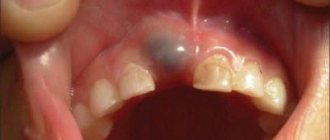
What is the danger of a wedge-shaped defect?
The wedge-shaped defect is interesting because today most dentists (at least in our country) have no idea about the cause of this disease.
Out of the goodness of their hearts, they, of course, try to alleviate the physical and moral suffering of patients by hiding wedge-shaped defects behind fillings, veneers or crowns. But problems with these structures arise much faster than with exactly the same ones, but installed for some other reason. So, if on the same day, by the same doctor, two separate fillings were built on the same tooth from the same material (and the best one at that): one covered a wedge-shaped defect, and the second covered a carious cavity of approximately the same size. size, then the second has a much better chance of celebrating its tenth anniversary. This causes justifiable bewilderment of the patient and no less justified anger of the dentist. The search for those to blame begins...
And for a long time, erosion and abrasion were considered the culprits of this disgrace. Abrasion is the mechanical abrasion of a tooth by solid foreign bodies, and erosion by liquid or gaseous ones. Therefore, a hard toothbrush in the first case and acidic foods (citrus fruits, carbonated water, juices) in the second were declared the main enemies.
Both theories are interesting, not without arguments and have numerous supporters. We also agree that both processes contribute to the progression of the wedge-shaped defect, that is, they are aggravating factors. But they cannot be the root cause. If only because wedge-shaped defects are found not only in people, but also in animals. You can’t suspect horses of using too hard brushes or cats of abusing Coca-Cola. Any solid body, no matter how strong it may seem, is subject to abrasion under the influence of regular mechanical loads. Tooth enamel is no exception. Although this is the hardest tissue of the human body, it can nevertheless be completely worn out as a result of daily use. In some advanced cases, only the roots may remain from the teeth.
In addition to direct abrasion of the rubbing surfaces of the teeth themselves, mechanical stress leads to damage to the enamel at the neck of the tooth (in the vast majority of cases, on the outside). That is, to the formation of a wedge-shaped defect. The fact is that a tooth is not an absolutely rigid structure. It tries to absorb the rather significant load that falls on it when chewing and swallowing food (up to 100 kilogram-force or 1000 newtons), partly due to micro-bending relative to its vertical axis. Simply put, the tooth bends, and the maximum stress, according to computer modeling data, occurs precisely at the neck of the tooth. The enamel in this place has a minimum thickness. In addition, the tooth in this part experiences tension, while in other places it experiences compression. And the tensile strength of enamel is 40 times less than the compressive strength.
As a result, cracking and subsequent peeling of the enamel occurs with the formation of a V-shaped defect. Just like how the paint flies off a metal rod or the chocolate icing on a frosted cheesecake cracks when you try to bend them. This is the mechanism of development of the disease. But this is not yet the reason, since all people’s teeth are subject to abrasion, and not everyone has a wedge-shaped defect. The primary source of this misfortune is initially the inharmonious closure of the dentition - a violation of occlusion or, in Russian speaking, a malocclusion.
In fact, a correct bite is an extreme rarity, and the majority of the world's population lives their lives happily without realizing that they have such a problem. An insignificant proportion of 99.9% of people with non-ideal occlusion consult an orthodontist. And even then, the first and almost always the only goal of those who dare to undergo orthodontic treatment is aesthetics. Only a few remember that teeth should not only “stand straight,” but also function correctly.
This is how the picture of the disease forms into a single whole: the patient has minor deviations in bite (or significant ones, but left without treatment) => the load when chewing food is distributed inharmoniously (some teeth work less, others overwork for them) => hard-working teeth wear out faster fellow quitters => as a result, flat areas of rubbing are formed on them => during chewing, instead of cutting the food bolus, it is crushed => in order to crush the food, the jaws have to develop greater force => the enamel cannot withstand the load, cracks and flies out => a wedge-shaped defect.
Anomalies in the number of teeth
All anomalies in the number of teeth are divided into three types:
- hyperodontia (excess of dental units above normal);
- edentia (complete absence of teeth);
- hypodontia (insufficient number of teeth).
Hyperodontia
Hyperodontia is a disorder of the formation of the dentition, which is expressed in the presence of supernumerary teeth (over 32 dental units in an adult). The cause of the disease may be a violation of the mechanism of formation of tooth germs in childhood or infancy. Hyperodontia mainly affects the incisors of the upper jaw. Other teeth are involved in the pathological process less frequently. Extra dental units grow small and have a cone shape.
The disease can be true or false.
- With true hyperodontia, the formation of extra dental units occurs in the human jaw.
- False hyperodontia occurs when the loss of primary teeth is delayed, which interferes with the normal growth of permanent teeth.
With polyodontia, teeth may erupt away from the dentition or next to a correctly positioned dental unit, which leads to its gradual displacement. The dentition can be seriously deformed, which leads to serious diction problems.
If supernumerary teeth interfere with the development of normal teeth, they must be removed. Broken teeth are corrected by installing braces.
In those rare cases when a row violation does not occur, the supernumerary tooth is preserved. Prosthetics are used to correct the shape.
Hypodentia
Hypodentia is a deficiency of dental units. This disorder occurs most often due to the death of tooth germs in the fetus. A small number of teeth causes a shift in the midline, the formation of wide gaps between teeth (diastema and three), and shortening of the dentition. All this adversely affects the bite. Possible ways of correction are prosthetics.
Edentia
Adentia is a complete or partial absence of teeth. With edentia, the continuity of the dentition is disrupted, which leads to difficulties in chewing and speaking. The reason is a violation of the development or death of tooth germs under the influence of genetic factors or under the influence of unfavorable external factors during the formation of the dental plate in the fetus (the formation of the germs of permanent teeth in the fetus occurs after the 17th week of intrauterine development). The disorder is corrected using prosthetics or dental implantation.
Human wisdom teeth
A “wisdom tooth” is the third outer molar with three to five roots. In structure it is no different from its “neighbors”. To the question “How many wisdom teeth does a person have?” cannot be answered unambiguously. They erupt around the age of twenty, one on each side of both jaws. However, there are people without wisdom teeth. This is a variant of the norm, since in the process of human evolution the need for the “eight” disappeared, and the structure of the jaws underwent corresponding changes. Today, third molars are considered a vestigial organ.
Anomalies of magnitude
There are two forms of this type of deviation, including macrodentia (increase in the size of dental units) and microdentia (small teeth).
Macrodentia
With macrodentia, the size of dental crowns is significantly increased. The cause of the disorder is, in most cases, dysfunction of the endocrine system, which is characterized by the fusion of several rudiments together. The disease is localized mainly in the area of the upper incisors. Large teeth interfere with the process of eruption and growth of neighboring dental units, which leads to their abnormal arrangement and crowding. Giant teeth are found on or outside the dentition line. This is a serious cosmetic defect that causes serious functional impairment and psychological disorders. Pathologically enlarged teeth are removed, and the growth of adjacent teeth is corrected using prosthetics or braces.
Microdentia
Microdentia – teeth that are too small. The disorder mainly affects the upper lateral incisors, but may affect the incisors of the lower or both jaws. The cause of the anomaly has not been studied, but it is reliably known that it develops in genetically predisposed patients. With the problem of small teeth, patients develop too large interdental spaces, which significantly disrupts the aesthetics of the dentition. To correct the pathology, incorrectly growing teeth are removed or covered with crowns.
When is orthodontic treatment necessary?
Orthodontic treatment of patients with splintered teeth is carried out in cases where their presence has provoked the development of malocclusion and crowding. And also when there is not enough space on the jaw to create a beautiful and proportional restoration. Orthodontics in this situation allows you to expand the space in the area of defective elements, create gaps in order to subsequently carry out high-quality and functional prosthetics, or artistic restoration.
On a note! If there are ugly or irregularly shaped teeth, then patients most often turn to an orthodontist, although this doctor cannot influence this defect in any way. But in some clinical situations (crowding, lack of space on the jaw), it creates conditions for subsequent beautifully shaped restorations. If you are going to install veneers, lumineers or crowns, then an orthopedist will help with this, if you are carrying out an artistic restoration, a dentist-therapist.
For pathologies such as overnumeration and crowding, the doctor may first recommend the removal of irregularly shaped incisors.
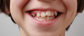
Shape anomalies
There are pathologies in the development of dental units, due to which they acquire an unnatural shape. These disorders are named after the scientists who first described them. The following types of dental shape anomalies are found: spiny teeth, Pflueger teeth, Fournier teeth, Hutchinson teeth.
- Spiked or awl-shaped teeth. With this pathology, the teeth acquire a cone-shaped shape. Wide at the base, they gradually narrow and, towards the cutting edge, become sharpened into a spike shape. This problem is combined with microdentia. There are irregularities and stains on the surface of the teeth. The disease affects the front and lateral incisors. The disease occurs in childhood and is caused by genetic factors in combination with external factors. Among them are vitamin D deficiency and endocrine system problems.
- Hutchinson's teeth are characterized by a modified crown shape of the incisors. Externally, they have the shape of a barrel, since the neck is significantly thickened. The cutting edge of the teeth acquires an arched notch. The enamel layer also suffers, which is present only on the sides and absent in the center.
- Fournier's teeth are a form of systemic hypoplasia of dental units associated with metabolic disorders at the stage of intrauterine development of the fetus. The barrel-shaped shape of the teeth is preserved, but the notch of the incisal edge is absent. The color of the enamel is not disturbed, but the enamel layer is underdeveloped, which can be seen during microscopic examination.
- Pflueger's teeth are a disorder that affects permanent dental units. Their crowns take on a conical shape. Thickening develops in the cervical region, and the chewing surface is significantly underdeveloped. The chewing function of the teeth is completely preserved.
- Turner anomaly. With this pathology, there is no enamel on the teeth. They have an abnormally lumpy surface, and replacement tissue, dentin, forms in the exposed areas.
Systemic hypoplasia with a violation of the shape of the teeth has three degrees of development. The third degree is the most dangerous, in which the crown is severely deformed and the enamel layer decreases. In this case, the defective teeth are removed and replaced with dentures. It is also possible to restore affected teeth using reflective components.
Types of pathology
Doctors distinguish several types of pathology.
Spike teeth
The tenon-like shape is characteristic of the lateral incisors located on the upper, and less commonly, the lower jaw. Such elements have a sharply narrowed cone-shaped shape. As for the length of the coronal part, it can be either shortened or, conversely, elongated. With this type of pathology, in a number of clinical situations large interdental spaces in the smile zone are noticeable, but sometimes they are absent, and the contact between adjacent units is very dense.
A child often develops a spiky tooth due to genetic disorders, DNA gene mutations, and developmental defects such as ectodermal dysplasia and cleft palate. In some cases, the development of the disease occurs against the background of congenital partial adentia or pathology of the development of tooth buds.
Hutchinson's teeth
The central incisors are barrel-shaped, and in the area of the cutting edge they have a characteristic semicircular notch. With this type of anomaly, the matter is not always limited to one problem: patients with a similar defect suffer from systemic enamel hypoplasia.
The defect develops if the child’s mother or the baby himself has had infectious diseases, rubella or syphilis.
Fournier's teeth
Fournier's central incisors differ from Hutchinson's incisors only in that they do not have a characteristic notch on the cutting edge. The reasons for their formation are infections and congenital syphilis.
Pflueger's teeth
This anomaly most often develops on premolar teeth. It is characterized by underdevelopment of the masticatory tubercles. The enamel in this pathology has various microdefects; it is covered with spots, tubercles, and grooves. The disease is also characterized by the fact that without treatment, hard tissues quickly wear out; they are very sensitive to various types of influences and temperature changes.
On a note! Anomalies of Hutchinson, Fournier and Pfluger are closely related to systemic hypoplasia caused by insufficient development of enamel. Therefore, in patients with this diagnosis, not only the aesthetics of the smile suffers. They experience pain and discomfort; hard tissues are very sensitive to spicy, cold and hot foods, to mechanical stress, and are susceptible to fragility and abrasion, as well as the development of caries.
Anomalies of hard tissue structure
An altered shape, abnormal size or color of enamel is formed against the background of an abnormality in the structure of the hard tissues of the dental unit. Among such anomalies are:
- Hypoplasia (underdevelopment of tissue). The initial degree of the disorder is manifested by the presence of chalky spots on the enamel and areas where tissue deficiency is observed. Subsequently, all kinds of pits, grooves, and recesses appear on the surface of the enamel. The defect affects all teeth in the dentition.
- Hyperplasia (excessive tissue formation). Pathology also affects all teeth at the same time. It is characterized by areas where tissue growth is observed - tubercles, sagging enamel. The consequence of the pathology is a change in the shape and size of the teeth, a violation of the occlusion (the line of contact of the upper and lower jaws).
- Anomalies of amelogenesis (enamel formation) are expressed in the presence of brown or yellow spots on the surface of the teeth. Areas where the natural composition of the enamel is disrupted become especially sensitive. Microdentia develops against the background of pathology. The cause of the development of pathology is a deficiency of microelements that take part in the formation of dental tissue. Treatment consists of replenishing this deficiency. Additionally, local remineralizing therapy and physiotherapy are performed.
- Disturbance of dentinogenesis consists of dysfunction of the mechanism of dentin formation. The main symptoms of the disease are as follows: teeth become yellow-brown or grayish in color. The fragile dentin in these areas quickly wears off, causing tooth decay and then tooth loss. The disease occurs in genetically predisposed patients. The problem can only be solved by replacing the units destroyed by the disease with prosthetics.
Why wedges form on teeth and what to do about it
The defect can develop on both the upper and lower jaw, on one or more teeth. Most often, canines and premolars are affected - namely, they experience a large chewing load. The disease develops gradually and unnoticed. Over time, the defect deepens into the dentin, and the person feels pain. If the problem continues to be ignored, the base of the tooth becomes so thin that it can break.
A wedge-shaped defect appears in people of different ages, including teenagers. However, the older the person, the greater the risk of developing pathology.
The moment of appearance of the wedge-shaped defect cannot be tracked independently. But if you undergo regular examinations with a dentist, treatment will begin on time and will be completed with minimal time and money.
At first glance, a wedge-shaped defect can be confused with cervical caries. The affected teeth are marked with triangular-shaped spots and depressions, up to 4 mm in depth in the acute stage of development.
As for where wedges on teeth come from, there are three generally accepted theories:
- Chemical. The culprit is aggressive acids from food, drinks and resulting from an imbalance in the acid balance of the oral cavity.
- Mechanical. The defect is caused by an external load.
- Physico-mechanical. Pathology develops in response to improper chewing load.
Color anomalies
Healthy, young teeth look snow-white due to a thick dentin layer and high-quality enamel, which has sufficient characteristics of whiteness, shine and transparency. Lifestyle, age, genetic factors, and ecology can slightly change the color of teeth from bluish to yellow. Small deviations are not considered pathology, although they indicate a change in the quality of the enamel.
Various pathological processes occurring in the body have a more significant impact on the condition of the enamel. The cause may be carious lesions, the use of medications, or a lack of certain microelements in the body. Teeth can take on a gray, pinkish, brown, purple and even black tint.
When starting to treat a patient, the dentist excludes the development of chronic pathologies. After a course of therapy and stable remission is achieved, other causes of discoloration of the enamel are eliminated. The final stage of treatment is a course of teeth whitening.
Specialists at the Consilium Dent choose the enamel lightening technique together with the patient. The selection criteria are age, condition of the enamel, and the wishes of the patient.
Surgical methods for correcting bites
If the pathology of the bite is associated with improper development of the jaws, then often in such cases they resort to surgical methods.
Underbite in a French Bulldog dog
| Rice. 1. | Rice. 2. | Rice. 3. | Rice. 4. |
The x-ray shows that underbite is the cause of the short lower jaw (Fig. 1,2.). In this case, only surgical correction of the bite is possible. After the surgical operation, the lower jaw was lengthened (Fig. 3,4.).
Position anomalies
Incorrect position of teeth always develops as a consequence of another pathology and forms an incorrect bite. May affect one or both jaws. Incorrect position of teeth makes chewing food and oral hygiene difficult. This can lead to digestive problems and the development of cavities. There are several options for the pathological position of teeth within or outside the dentition:
- External or vestibular position means that the tooth is located outside the dentition, closer to the vestibule of the mouth. This position is typical for canines and incisors and develops as a serious cosmetic defect.
- Oral or internal position. The teeth are deviated inward closer to the tongue. The disorder is typical for canines, incisors and premolars. May cause tongue injuries and dysfunction of the temporomandibular joint.
- Mesial and distal position. Dental units are displaced forward or backward along the dentition, which leads to its shortening.
- Supra and infraocclusion are high or low positions of teeth relative to the occlusal curve. The cause of the disorder is underdevelopment of the alveolar process. It appears due to the presence of some obstacle that interferes with the normal formation of the tooth germ. Effective therapy is surgery.
- Tortoanomaly. The tooth is rotated around a vertical axis. The disorder is typical for incisors, canines, and premolars. The anomaly can cause injury to adjacent teeth. Dental units are rotated into the correct position using orthodontic structures.
- Transposition. The teeth change their location with each other. The disease affects canines that exchange places with premolars or lateral incisors. The disorder develops at the stage of tooth germ formation. The therapy is as follows: problematic teeth are removed followed by prosthetics.
- Crowding of teeth occurs due to lack of space when tooth germs are located too closely. Crowded teeth are closely adjacent to each other and rotate around their axis. Occurs in combination with macrodentia or with an undeveloped basal part. To eliminate the violation, some dental units are removed.
- Diastemas and tremas are wide spaces between teeth. The disorder develops as a consequence of an abnormal shape and size. The problem is treated with orthodontic methods or by installing veneers, which helps restore the aesthetics of the smile.
Misaligned teeth cannot always be cured using braces. Severe pathology, which is characterized by a skeletal disorder of the maxillofacial region, is treated with surgical reconstruction. The pathological fragment is transferred to the desired position and then secured. A fragment containing part of the dentition or the entire dentition can be used.
Treatment methods
You need to understand that if teeth have grown in an irregular shape, the defect is irreversible. Therefore, all the methods that modern dentistry offers will be aimed at visually concealing and eliminating the defect, restoring the correct anatomical shape for the normal functionality of the maxillofacial system, improving the appearance and beauty of the smile.
Composite restoration
Artistic restoration, namely changing the shape of a tooth and its color using a light-curing composite and photopolymers, is the least invasive technique that quickly allows you to get results in just 1 visit to the clinic. Composite restorations are good because their cost is low, and the patient, if necessary, can remove filling materials in the dentist’s office or replace them with new ones, renew his smile, or choose a different treatment method at any time.
Important! Irregularly shaped teeth are a consequence of internal pathologies and diseases of the body. Therefore, patients often require complex treatment: not only a visit to the dentist, but also constant monitoring by specialized specialists (endocrinologist, infectious disease specialist).
Installation of veneers or lumineers
Lumineers and veneers for spiky teeth are a good way not only to comprehensively transform your smile for a long time (hide irregular shapes, unsightly shades, gaps, crevices, cracks and fillings), but also protect hard tissues from food temperature changes, bacteria and premature destruction. The method is suitable only for adults when the milk bite has completely changed to a permanent one.
Prosthetics with crowns
This method is radical, since the installation of crowns is preceded by large-scale preparation of hard tissues. However, patients whose tenon-shaped teeth have begun to deteriorate severely due to hypoplasia or carious diseases, or when the defects are not amenable to other, less radical methods of restoration, cannot do without it.
Prosthetics with crowns can be performed on both adults and children. For young patients, temporary plastic solutions are used, as well as strip-crowns made of acrylic and light-curing composites, which allow preserving the anterior elements of the row (in this case, the incisors) with various malformations and do not interfere with the correct growth and formation of a permanent bite.
If installing a crown is not possible, then doctors perform removal. Next, the defect is eliminated using implantation, bridge-like fixed or removable dentures.
Diagnostics
An experienced dentist diagnoses dental abnormalities after an external examination and questioning of the patient. To clarify the diagnosis and identify the causes of the disease, a more detailed examination of the dental system is carried out:
- The construction of diagnostic plaster models and odontometric measurements make it possible to accurately identify changes in the size of dental units and their characteristics, which indicate the presence of pathologies.
- The color and transparency of the enamel are determined using a photographic method.
- Computed tomography or teleradiography makes it possible to obtain information about the condition of abnormal dental units in order to determine further treatment tactics.
- Panoramic radiography of the jaws is an important diagnostic method
- Electromyography of the jaws helps to assess the functional state of the facial muscular system.
To carry out differential diagnosis, the patient is referred for consultation to specialized specialists: endocrinologist, otolaryngologist, geneticist. Based on the diagnostic results, the attending physician determines treatment tactics. Depending on the type of disorder and the complexity of the disease, the patient may need the help of a dentist, surgeon, orthopedist, or implantologist.
Dimensions of human teeth
The upper central incisors are twice as wide as their antagonists. The remaining dental units of the same name have approximately equal parameters. The size is determined using special tables with the optimal size and permissible deviations. Experienced doctors calculate proportions by dividing the length of a person’s teeth by the width. A result of about 0.75 millimeters is considered close to ideal. For more detailed diagnostics, other professional formulas and techniques are used.
Size deviations from the norm occur due to improper formation of the jaw, fusion of tooth buds, or genetic predisposition. Teeth that are too large are called macrodentia, and abnormally small teeth are called microdentia. Pathologies are accompanied by problems with bite and chewing functions, but can be successfully corrected by a dentist.
Interesting fact!
The longest tooth in the world belongs to an Indian teenager. The size of its crown is almost four centimeters. About a year ago, the tooth was removed, and the young man was included in the Guinness Book of Records.
Prognosis and prevention
Consilium Dent clinic uses advanced methods of therapeutic, surgical, and orthopedic treatment of dental anomalies in patients. By skillfully combining them, experienced specialists are able to restore the aesthetics of a smile and ensure the normal functioning of the dental system, even if the degree of deviation is significant.
Prevention of anomalies in the formation and development of dental units begins during the period of intrauterine development of the fetus and is carried out throughout a person’s life. The main stages are as follows: monitoring the successful course of the prenatal period, caring for harmonious postnatal development, organizing a correct lifestyle while the baby is growing up, caring for dental health and general health throughout life. By conducting an annual preventive examination, the dentist identifies dental anomalies at an early stage, when the pathological process is still reversible.
Expert of the article you are reading:
Is it possible to fix a hole between teeth at home?
It is impossible to remove diastema at home. To carry out effective correction, specialists use a whole range of diagnostic measures, as well as devices that are manufactured specifically for the patient. It is naive to believe that the problem can be eliminated simply by tying the two front teeth with floss. Moreover, such manipulations are dangerous, as they can damage the gums or the teeth themselves.
Sometimes you can hear stories about how a child's teeth were bandaged and the cleft disappeared. But, in this case, we are talking about a false diastema, which goes away on its own without any intervention.