A dental visiograph is equipment with which you can quickly obtain accurate images of teeth to monitor treatment or evaluate tissue. Unlike an x-ray, the doctor immediately receives the image on the monitor, meaning little time is spent on the examination. The picture is clear; to improve the quality, special tools are used to increase picture brightness, clarity and contrast.
The radiovisiograph is compact in size and easy to use. To perform the procedure, a small sensor is placed in the Patient’s oral cavity, which transmits the image to the screen. No special preparation is needed; the patient does not feel pain or discomfort.
The device includes the following components:
- a base in the form of a high-frequency device, with the help of which particles acquire the desired direction of movement;
- wireless or wired sensor (depending on model);
- analog-digital system for converting images into electronic form;
- laptop or PC screen for viewing and image correction.
The radiation exposure during the procedure is minimal - 2-3 μSv, which is much less than during a standard examination. The resulting images can be stored electronically, accessing them if necessary to monitor treatment or the achieved result. The resulting visiograms can be used in the following cases:
- identification of foci of infectious lesions;
- search for pathologies invisible using other research methods;
- assessment of the course of the disease, including caries, periodontitis or pulpitis.
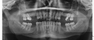
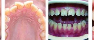
Diagnostics also makes it possible to detect foreign bodies in the tooth canal and cavities remaining after filling. Cysts, granulomas, and inflammations are painted black in the resulting picture; they stand out against the background of light healthy tissue. This improves the quality of the analysis and allows the doctor to immediately localize the pathology and the depth of the lesion. The ease of use of the device makes it easier to control therapy, for example, when filling cavities or dental canals.
Visiography of a tooth is
Dental visiography is the most informative method for diagnosing pathologies of the teeth and jaw, carried out using a special device that operates on the basis of radio frequency radiation.
The procedure is absolutely safe and not contraindicated even in childhood. The radiation exposure to the body is minimal (10 times lower than with classic x-rays). The procedure is also called “radiovisiography”. The main difference between the procedure and classical x-rays is the ability to instantly obtain an image of the area under study not on film, but on a monitor screen. In addition, experts confirm the high quality of the image and the possibility of obtaining a targeted image.
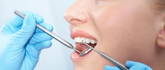
A dental radiovisiograph is used to identify any pathological processes in the teeth or jaw, including surrounding soft tissues and temporomandibular joints. As a result of diagnosis, a specialist can determine hidden processes (including hidden caries), inflammation, maxillofacial injuries, pathologies of the maxillary sinuses, and the condition of the jaw bones. Visiography is also used in dentistry to monitor the progress of treatment and its results.
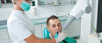
What does a visiograph look like and when is it used?
Dental radiovisiographs are small in size. To take an image, a small sensor of the device is placed in the patient’s oral cavity and transmits the result of the image directly to the computer.
The visiograph consists of several components:
- The basis is the high-frequency apparatus in the photo above, which sets the desired direction of particle movement;
- Small wired or wireless sensor;
- Analogue-digital system providing image digitization;
- A PC or laptop that allows you to view, store, and send images via email.
The wired visiograph is equipped with a special sensor that is applied to the tooth. Every radiologist should know how to take targeted photographs of teeth using such equipment. Using a wired connection, the image is transferred to the computer screen. A wireless radiovisiograph has a scanner capable of reading an image and displaying it on the screen.
The radiation dose during the examination is small and amounts to 2-3 μSv. Compared to this device, fluorography creates an exposure of 500-800 μSv.
The resulting image can be stored in an archive and on a hard drive. A visiograph belongs to the category of diagnostic and treatment dental equipment and allows you to obtain a clear image of a diseased tooth.
What does the photo show?
Clinic Dr. Martin successfully uses digital radiovisiography for diagnostic purposes when indicated for therapeutic or surgical treatment.
Computer radiovisiography is increasingly used in endodontics, which deals with the treatment of dentin and pulp. The visiograph images clearly determine the shape and length of the root canals, which makes it possible to flawlessly perform mechanical treatment and filling.
Targeted images of a visiograph allow you to obtain accurate information about the condition of soft and bone tissues for high-quality implantation. Thanks to a detailed study using computer technology, the orthodontist can select the correct size and shape of the implant and monitor the result of its installation, as well as the healing process of the pin.
Dental radiography is performed in dentistry according to indications. The patient is referred by a specialist for diagnostics before planned implantation, installation of braces or any other design/products. The visiograph is able to identify any orthodontic products in the dental system. They are identified in the image in white. Foci of inflammation (granulomas, cysts, etc.) are displayed in dark color. The pathological zone of connective or bone tissue is highlighted in gray shades in the images.
Indications and contraindications for the procedure
List of main indications for use of the equipment:
- Prevention of pulpitis, caries, periodontitis;
- Viewing the condition of a filled tooth, decayed gums, damaged bone tissue, or installed implant.
Radiovisiographic examination allows complete control of the treatment process. It will help avoid many complications. This method is safer than x-ray examination. It does not pose a health hazard.
Radiovisiography can be performed during pregnancy and lactation.
The list of relative contraindications includes:
- Mental illnesses;
- Neurological disorders.
Advantages
Classic X-rays involve exposure to a significant dose of radiation to the body, so the procedure is allowed only a limited number of times during the year, observing time intervals. In addition, the image on such a device is printed on film, which does not exclude distortion of the image.
The operating principle of the radiovisiograph offers significant advantages:
- low level of radiation exposure;
- displaying the image on a computer monitor immediately after the start of the procedure;
- storing data in a computer database;
- transfer of data to a specialist or patient in electronic format, including sending an image using modern methods;
- high quality image with the ability to zoom in and view the desired area in detail;
- availability of using special filters without introducing a contrast agent into the body;
- the ability to take a targeted shot;
- Absolute safety for the patient’s body with no side effects.
Diagnostics using a visiograph is many times faster than when examined using standard X-ray equipment. The photo is taken in a few minutes. And immediately after the examination is completed, the images will be ready.
Difference from X-ray and computed tomography
Computed tomography allows you to obtain a three-dimensional image of the jaw to examine it in all planes. A visiographic device helps to examine each problematic tooth individually. A similar examination is carried out before the therapeutic procedure, and the method is also used to monitor the treatment process.
X-ray can only create flat images. Even interproximal radiography is not always able to determine the presence of tumors in the oral cavity. Using a radiovisiograph, you can see a tumor in the initial stage of its formation.
Radiovisiography is safer than radiography; the level of dangerous examination in the first case is relatively low. The signal from the visiograph passes to a special CCD sensor or microcircuit that is more sensitive than x-ray film.
Example of resulting images
How is the diagnosis carried out?
The visiograph is a modern diagnostic device consisting of three main parts: a high-precision sensor, an analog-to-digital converter and a cord connecting the parts of the device. The operation of the visiograph is ensured by a whole complex of parts, including x-ray equipment with a special guide (tube). The device itself has an external resemblance to a tomograph.
The sensor is a silicone chip (digital sensor) that records all the signals that come to it. It is the sensor that transmits the signal to the device, which is formatted into an image with 16-bit quality. The analog-to-digital converter is a monoblock equipped with a USB port for connecting to a computer and a special port for connecting to a sensor. The data received by the sensor passes through the connecting cable to the computer. All information is converted using a built-in program and displayed on a computer monitor.
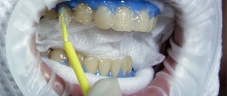
The diagnostic procedure takes on average 3–5 minutes. The patient sits down on a chair near the visiograph, the specialist adjusts the direction of the X-ray tube in the desired direction and places the sensor against the area being examined. Then the device turns on and a photo is taken.
Frequent breakdowns of radiovisiographs
| Damage to the sensor layer for recording radiation from the visiograph sensor |
| Malfunction of the radiovisiograph matrix |
| Damage to the electronic board |
| Damage to the connecting cable or connector for connecting the sensor to the PC (USB) |
| Software malfunctions, visualization errors |
| Intermediate unit faulty. |
| Damage to the body of the visiograph sensor, depressurization |
| Damage to controls (select models) |
| Loss of image clarity, noise, banding, artifacts (for no apparent reason) |
Diagnostics at the clinic Dr. Martin
Visiography is recognized as the best method of modern diagnostics in dentistry when it is necessary to obtain a targeted image that allows one to examine in detail a separate area of the dentofacial structure and joints of the lower jaw.
Clinic Dr. Martin offers dental imaging at an affordable price. Diagnostics can be either a stage of treatment by our specialists or a separately provided service. The cost for the procedure and the image is indicated in the price list on the clinic’s website.
You can make an appointment with a specialist, a consultation or an examination by calling the contact number, or using the website service by ordering a call back from a consultant (“Make an Appointment”).
How is a radiovisiograph repaired?
1. Consultation.
2. Diagnostics of the visiograph sensor.
3. Coordination of work.
4. Repair of the dental radiovisiograph sensor.
5. Connection and checking modes.
6. Transfer of equipment.
Disadvantages of radiovisiographs - is the radiation dose dangerous?
It must be said that radiovisiography has only one drawback
It consists in a lower (1.5-2 times) spatial resolution than that of film.
The sensor cannot as clearly convey the difference between structures that differ markedly in density.
True, this minus is compensated by another opportunity - to adjust the brightness and contrast of a digital image.
IMPORTANT: Are you an expectant mother? Do not forget to warn your doctor about this before treatment.
Is the radiation dose dangerous?
Here are the doses dental patients “receive” in studies:
- 1 image of a tooth: on the lower jaw - 2 μSv, and on the upper jaw - 3 μSv.
- 1 panoramic (digital) image - 15-20 µSv.
COMPARED WITH AN X-RAY : 1 fluorogram will “give out” 800 μSv.
Workflow efficiency
When developing its products, Gendex has never forgotten about the efficiency of clinical workflows. Likewise, the GXS-700 intraoral visiograph is designed for easy integration into your clinic management system. The system has additional access to patient contact information and scheduling of doctor visits.
The GXS-700 intraoral imaging device can be easily transferred from one dental unit to another, providing fast and competent patient care.
The unique Always Ready feature automatically detects the presence of radiation and begins recording images, so there is no need to activate the system before shooting.
The high-speed USB 2.0 port allows you to quickly and easily change sensors, which significantly improves the efficiency of the shooting process. Digital images are displayed without any time delay, providing the dentist with immediate diagnostic information. This allows you to avoid wasting valuable time in emergency situations, as happens, for example, when processing film. This ensures fast and uninterrupted medical care for the patient.
Specifications
| Size 1 sensor | Size 2 sensor | |
| External dimensions (mm) | 25 x 37 | 31 x 42 |
| Pixel size | 19.5 µm | 19.5 µm |
| Sensory technology | Improved version of CMOS | Improved version of CMOS |
| Scintillator technology | Csl scintillator | Csl scintillator |
| Image format | 1026 x 1539 pixels | 1346 x 1850 pixels |
| Number of pixels | 1.6 megapixels | 2.5 megapixels |
| Image Resolution | Theoretical: 25.6 l/mm Visible: 20+ l/mm | Theoretical: 25.6 l/mm Visible: 20+ l/mm |
| USB connector | Hi-Speed USB 2.0 Port | Hi-Speed USB 2.0 Port |
| Length of cable | 2.7 m | 2.7 m |
| Guarantee | 2 years | 2 years |
What is a dental visiograph? – photo, description and characteristics
A visiograph is equipment for digital x-ray diagnostics; during the process, the patient is exposed to significantly less radiation compared to a conventional x-ray, and the image comes out almost instantly. The official name is radiovisiograph.
Some confuse a visiograph and an x-ray due to the similarity of purpose, but this is wrong. The patient is prescribed an image in the following cases:
- to identify cysts and measure the root canal;
- for a complete examination of the oral cavity, when diseases are invisible to the naked eye;
- to diagnose the condition of the filling along the length of the apex;
- to detect various pathologies.
Call us or 8 (926) 100-10-10