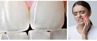
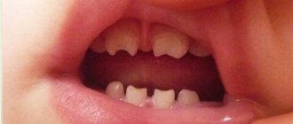
Increased sensitivity of teeth (hyperesthesia) is a short-term occurrence of pain under the influence of temperature, chemical or mechanical irritating factors. Usually occurs when drinking cold water, sour, sweet, salty, or touching with a toothbrush. The intensity of pain varies from mild to unbearable.
According to WHO, every second person in the world suffers from hypersensitivity. In Russia it is about the same: 45–65% of adults aged 20–55 years. More often women make complaints.
A little anatomy
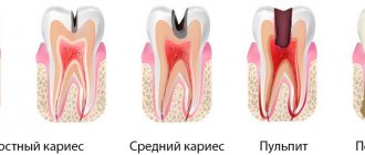
A tooth consists of a crown and root part, connected by a neck. The coronal part is covered with enamel, the root part is covered with cement. Beneath the enamel and cement there is dentin, a hard tissue. Inside there is soft tissue - the pulp; blood vessels and nerves pass through it.
Dentin is not sensitive, but consists of many tubules in which fluid circulates. The irritant causes fluid movement, which is detected by the nerve endings of the pulp. A person feels their reaction as pain.
Pathogenesis and epidemiology
The mechanism of development of the symptom is due to developing dystrophic and degenerative processes in the nervous tissue as a consequence of metabolic disorders. Developing autonomic disorders are caused by conduction disturbances along the reflex arc of the receptors. With an infectious etiology, the structure of the nerve sheaths and nerve fibers are disrupted, which reduces muscle sensitivity.
In terms of distribution, the symptom is characterized by development after:
Cutaneous hyperesthesia
- infectious diseases – 63%;
- meningitis – 16%;
- allergies – 12%;
- parasitosis – 14%;
- shock, intoxication – 98%;
- dental diseases – 67%.
The symptom is also caused by the disease and has its own epidemiological characteristics.
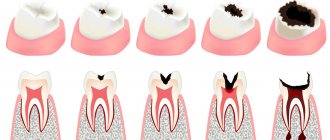
Tooth sensitivity: causes
- Demineralization of enamel. It becomes more loose due to the leaching of calcium, phosphorus, and other trace elements.
- Thinning of enamel. As a result of increased abrasion due to malocclusion and the occurrence of wedge-shaped defects.
- Untreated caries or violation of the marginal seal of the filling.
- Exposure of roots as a result of injury, metabolic-dystrophic process or inflammation of the gums.
- Changes in the pH of saliva due to the consumption of certain drinks, foods, and medications. A pH of less than 5.5 is considered dangerous.
- Some diseases accompanied by gastroesophageal reflux and endocrine disorders.
- Vitamin deficiency, exposure to radiation, work in hazardous industries, living in a region with an unfavorable environmental situation.
- Smoking.
More often than not, several reasons are discovered at once. The enamel becomes thinner, loses strength, and cannot protect dentin from irritants. The result is pain.
Provoking factors
Hyperesthesia does not appear immediately. There are several factors that you need to pay attention to in order to eliminate them in time. Don't wait for discomfort to appear. It is better to initially develop healthy habits that will help you maintain your health so that you never experience acute dental pain. Sensitivity increases when any of the factors listed below are present.
- Insufficient oral hygiene. Soft plaque is an accumulation of microbes that eat food microparticles stuck in crevices, releasing organic acids that dissolve enamel minerals. Externally, the dental unit looks intact, but the density of the enamel is significantly reduced. Its demineralization occurs. The first alarm bell is increased sensitivity, then caries develops.
- Consumption of certain foods. Juices, wine, sweet soda, fruits, candies, and other sweets contain phosphoric and other acids that negatively affect the strength of enamel.
- Constant use of aggressive whitening pastes containing abrasive and chemical components.
- Ultrasonic cleaning. Under a dense coating, the enamel becomes thinner and becomes loose. After professional cleaning, it is exposed, its sensitivity increases sharply. Usually, dentists, taking this point into account, use strengthening pastes at the end of the procedure for the preventive treatment of tooth sensitivity.
Types of hypersensitivity
If sensitivity is increased on one or more teeth, it is called limited. If for everyone - generalized.
Table 1. Types of hyperesthesia
| № | View | Reaction |
| 1. | Light | for cold, hot |
| 2. | Average | as with 1st degree plus for sour, sweet, salty |
| 3. | Expressed | as in grade 2 plus mechanical irritants (when brushing teeth, eating) |
What is dental hyperesthesia
Let's say you drank hot coffee and started eating ice cream. I immediately felt a “ache” in my teeth. The feeling is not pleasant. This is how hard dental tissues reacted to a sharp change in temperature, showing excessive sensitivity. Dentists call this phenomenon hyperesthesia. A similar reaction can occur to other chemical, mechanical and temperature irritants: spicy, sweet, sour, exposure to cold, brushing teeth, chewing. The pain can be sharp or nagging, long-lasting or short-lived.
Dental hyperesthesia is a painful reaction of teeth to chemical, temperature and physical stimuli. Pain can be localized in one or several dental units, or spread to all elements of the dentition (generalized hyperesthesia). It has been proven that emerging sensitivity (in the absence of caries) precedes non-carious lesions of the dental element (elements) and is an early diagnostic symptom of this clinical situation. The pathology is mainly observed in people aged 30 to 60 years.
Sensitivity of teeth. Stages of treatment
- Eliminate the cause of hyperesthesia: get rid of plaque, deposits, stones, caries, wedge-shaped defects.
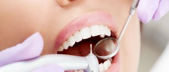
- Carry out professional oral hygiene.
- Strengthen the enamel with calcium and fluoride.
- Teach the patient how to use a toothbrush and floss correctly.
- Choose suitable dental care products: toothpaste, mouthwash.
Table 2. Tooth sensitivity: causes and how to treat
| № | Cause | What to do |
| 1. | Soft coating. | Careful hygiene with home remedies. |
| 2. | Hard coating. | Professional hygiene in dentistry. |
| 3. | Caries in the white spot stage. | Deep fluoridation, remineralization. |
| 4. | Caries, pulpitis, periodontitis. | Dental treatment. |
| 5. | Exposure of the cervical part, wedge-shaped defect. | |
| 6. | Malocclusion. | Orthodontic therapy. |
Summary
- If hypersensitivity occurs, avoid drinks and foods containing acids (fruits and fruit juices, wine, etc.).
- If you have previously used whitening toothpastes, stop using them, because... with a high probability, hypersensitivity is caused precisely by their use.
- Consult your dentist about the cause of sensitivity - it may be caused by caries or exposed tooth necks. Remember that caries in the form of a white spot is a reversible form of caries, and if you undergo remineralization therapy on time, you will not only get rid of increased sensitivity, but also prevent the destruction of this area of enamel.
- If there are dental deposits, they must be removed, followed by a course of remineralization and/or deep fluoridation.
- Maintain proper oral hygiene - cariogenic microflora in the cavity, feeding on food debris, produces acids that wash calcium from the enamel and lead to the appearance of both hypersensitivity and dental caries. Check out the link above for comprehensive hygiene tips, as well as videos on proper brushing and flossing techniques.
Sources:
1. Higher professional education of the author in dentistry, 2. Personal experience as a dental therapist, 3. National Library of Medicine (USA), 4. The European Academy of Paediatric Dentistry (USA), 5. “Therapeutic dentistry. Textbook" (Borovsky E.).
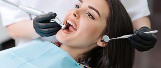
Elimination of tooth sensitivity
Using ultrasound, the doctor removes soft and hard deposits from the teeth. After removing plaque, teeth become more sensitive for a short time, so remineralization or deep fluoridation is immediately carried out. During remineralization, the enamel is treated with active compounds of calcium and phosphates. Deep fluoridation – coating with sodium fluoride. Both procedures significantly strengthen the enamel's resistance to irritants.
The doctor uses agents that reduce the movement of fluid in the dentinal tubules. It “seals” them using desensitizers or reduces their volume through remineralization. Protecting exposed dentin reduces the force of transmission of the irritant impulse from the enamel to the nerve.
Basic points of proper hygiene
Use a synthetic, medium-hard or soft brush. Change it every three months.
The toothpaste should be suitable for very sensitive teeth. Typically, such products contain hydroxyapatite, strontium chloride, fluorides, potassium nitrate, or a combination of calcium carbonate and arginine.
Take enough paste. For children under 3 years old - the size of a grain of rice, from 3 to 14 years old - the size of a pea, for adults you need to squeeze out about one centimeter.
Brush your teeth for 2 minutes: 30 seconds on each surface, top and bottom. Monitor time using an hourglass or mobile phone timer. Electric toothbrushes emit a short beep every 30 seconds and a long beep every 2 minutes from the start of brushing.
Cleaning sequence
- Using sweeping movements, clean the outer and then the inner surfaces of the bottom row. Move from molars to incisors. Then do the same on the top row. Hold the brush at a 45-degree angle and do not use a sawing motion. This leads to damage to the enamel.
- Brush chewing surfaces with small circular movements.
- Close your jaws and walk along your gums in a circular motion.
- Brush your tongue using a leisurely four to five strokes from root to tip.
- Treat the interdental spaces with dental floss.
- Rinse your mouth.
It is more effective to clean with an electric brush or irrigator. The quality of hygiene increases 3–4 times. Hyperesthesia can be dealt with faster.
Is it possible to strengthen sensitive teeth at home?
Yes. If you can’t get to the dentist, you can try to help yourself. There are several remedies that can solve the problem of hyperesthesia at home.
Table 3. Popular products for home use
| № | Name | Mechanism of action | Age category |
| 1. | ROCS Medical Minerals, GC Tooth Mousse | Remineralizing gels | Adults and children |
| 2. | Colgate Duraphat 2800 ppm | Fluoridating paste | 10–15 years |
| 3. | Colgate Duraphat 5000 ppm | From 16 years old | |
| 4. | ELMEX junior | Fluoridating paste | 6–12 years |
| 5. | ELMEX | From 13 years old | |
| 6. | LACALUT Extra Sensitive | Paste that reduces tooth sensitivity | For adults |
| 7. | Colgate Sensitive Pro-Relief | ||
| 8. | PRESIDENT Sensitive | ||
| 9. | LACALUT Sensitive, 300 ml | Rinse for sensitive enamel | From 15 years old |
Feline hyperesthesia syndrome: epilepsy as a possible etiology in two cats
Veterinary articles - translations from scientific journals / Neurology of dogs and cats: epilepsy and other diseases / Feline hyperesthesia syndrome: epilepsy as a possible etiology in two cats
Vet Rec Case Rep.
September 2021
Translation from English: veterinarian Vasiliev AV
Abridged article
Summary of the article
Feline hyperesthesia syndrome is a poorly understood pathology whose etiology is currently unknown. Epilepsy was suggested as a possible cause, but no evidence was provided. Two cats, ages 1.5 and 2 years, were assessed for neurological symptoms characterized by skin rippling, muscle spasms, licking and biting of the lumbar region, head shaking, tail swaying and suspected hallucinations, among other symptoms. Physical and neurological examination, magnetic resonance imaging of the brain, and cerebrospinal fluid analysis were unremarkable.
However, an electroencephalogram showed that polyspike-like epileptic discharges occurred in the temporoparietal region in one cat and focal sharp waves in the left frontotemporal region in the other. In both cases, the treatment duration was significantly reduced in response to anticonvulsant therapy. Based on these results, electroencephalography may be part of the diagnosis of feline hyperesthesia syndrome, and epilepsy is considered as a possible etiology for the syndrome.
Introduction
Feline hyperesthesia syndrome now includes a wide range of clinical signs that may occur spontaneously or be triggered by light touch on the lumbar region. Such signs include 1) licking/biting of the forelimbs, body, tail and perineum; 2) rippling of the skin, spasms of the lumbar muscles, involuntary movements of the ears, shaking of the tail and head; 3) excessive vocalization, aggression or increased affection and uncontrolled jumping and running; 4) mydriasis and 5) suspected hallucinations. Although feline hyperesthesia syndrome is associated with dermatological (hypersensitivity dermatitis), orthopedic (tail injury), neurological (brain and spinal disorders), muscular (myopathies) and behavioral (compulsive disorders) conditions, its etiology is unknown.
Brain pathologies such as epilepsy (idiopathic or structural) and spinal diseases (infectious myelopathies, neoplasias or intervertebral disc disease) have been proposed among the neurological causes of clinical signs consistent with feline hyperesthesia syndrome. However, several authors have suggested that the clinical signs of the syndrome hyperesthesia in cats may be a consequence of a partial complex epileptic seizure. Epilepsy occurs due to intrinsic hyperexcitability and excessive synchronous firing of abnormal neurons in the brain, which can be recorded using an electroencephalogram.
The semiology of epileptic manifestations in cats may be atypical; therefore, the use of electroencephalography may be helpful. A diagnostic approach to feline epilepsy using electroencephalography has been reported. However, the use of electroencephalography to diagnose epilepsy as a possible etiology of feline hyperesthesia syndrome has not been documented. Therefore, the purpose of this case report is to describe electroencephalographic findings in two cats with feline hyperesthesia syndrome.
Case description
Two indoor shorthair cats, a 1.5-year-old male (Case 1) and a 2.0-year-old female (Case 2), were presented for evaluation of multiple daily episodes of rippling skin with muscle spasms and spontaneous licking. /biting the lumbar region, twitching of the head with involuntary movements of the ears, licking of the forelimbs and waving of the tail. In addition, licking behavior that progressed to severe biting of the sides of the trunk, limbs, and tail over several weeks was present in Case 1 (Video 1), and possible hallucinations were described in Case 2 (Video 2).
Case 1 had previously excluded dermatologic conditions (allergic and hypersensitivity dermatitis), skeletal lesions (degenerative joint disease), and behavioral problems (compulsive disorders and disturbed behavior). However, the patient was treated with antibiotics (enrofloxacin and benzathine penicillin G), steroids (prednisolone, methylprednisolone acetate and triamcinolone acetonide), behavior modifying drugs (amitriptyline and fluoxetine), analgesics (gabapentin and tramadol) and vitamins (vitamin E), no improvement.
Differential diagnosis
The differential diagnosis was based on the chronic and transient nature of the episodes, normal physical and neurological examination findings, and pathologies that had previously been excluded (such as hypersensitivity dermatitis in case 1). Intracranial disease was suspected. Generally, congenital anomalies (eg, congenital hydrocephalus), infectious/immune-mediated meningoencephalitis (eg, toxoplasmosis and meningoencephalitis of unknown etiology) and idiopathic epilepsy were considered.
Treatment
According to the results, phenobarbital at a dose of 3 mg/kg every 12 hours was administered orally in case 1; in case 2, levetiracetam 20 mg/kg was administered orally every 8 hours. In case 2, phenobarbital was recommended, but due to the difficulties the owners had in obtaining it, levetiracetam was the preferred treatment.
Results and follow-up
The condition of both patients improved significantly: in case 1, bites in the lumbar region and tail disappeared after 3 weeks of treatment. Episodes of licking the lumbar region and forelimbs, ear movement, head bobbing, and tail wagging were significantly reduced from several times a day to one or two episodes a day 6 weeks later in case 1 (Video 3), and after a week in case 2, as expected hallucinations. One year after the start of treatment, according to a telephone survey, there were no changes in the clinical condition of both cats.
Discussion
We described two cases of feline hyperesthesia syndrome, their EEG findings, and response to treatment with anticonvulsant therapy, which raised idiopathic epilepsy as a possible explanation for the clinical signs of this syndrome. Licking/biting of the forelimbs, body, tail and perineal area, rippling of the skin and spasms of the lumbar muscles may be caused by dermatitis due to hypersensitivity not caused by fleas or food. However, not all dermatologic patterns (eg, excoriations of the head and neck or symmetrical bilateral skin lesions in the lumbar region) were present and did not meet Favreau's criteria. However, an 8-week trial was conducted using a commercial diet and the veterinarian prescribed steroid treatment with no positive response.
Lumbar hyperesthesia is associated with diseases of the spinal cord, due to degenerative myelopathy associated with feline leukemia virus, lymphosarcoma and intervertebral disc disease. But both patients were negative for feline leukemia virus, had normal cerebrospinal fluid, and no other clinical signs consistent with thoracolumbar myelopathy were observed.
Behavioral changes such as compulsive disorders or displaced behavior may explain some of the clinical features present in these patients. However, none of them had a history of conflict situations that could have triggered the symptoms. In addition, the cat in Case 1 was treated with behavior modification medications without improvement, and the cat in Case 2 had suspected hallucinations not reported in behavior disorders.
Primary myopathy/myositis with inclusion bodies in the epaxial thoracolumbar muscles, detected using electromyography and histopathology, was described in five cats with suspected feline hyperesthesia syndrome. However, the age of the cats reported here was not consistent with the age of onset of myopathy (5–8 years). Moreover, the clinical signs present in both cases (suspected hallucinations, involuntary ear movements, and head shaking) could not be explained by this myopathy. In addition, electromyography and epaxial muscle biopsy were not performed.
The MRI findings were considered incidental in Case 2 and were considered to be of non-clinical significance because the EEG showed the epileptic zone to be contralateral (left frontotemporal region) to the ventriculomegaly and there were no clinical signs consistent with hydrocephalus (altered mental states ranging from depression to hyperintensity). excitability, impaired consciousness, visual and hearing impairment, poor coordination, circling, convulsions), ventral or ventrolateral strabismus and abnormal skull shape).
Based on a review of the medical and veterinary literature, the clinical signs of feline hyperesthesia syndrome can be considered as a complex partial seizure or a partial seizure with impaired consciousness and nonmotor onset,” as currently defined by the International League Against Epilepsy in the 2017 classification of epileptic seizures.
Cats with feline hyperesthesia syndrome do not appear to fully reflect their environment and exhibit behavioral, autonomic, somatosensory, or sensory cues during episodes. In this regard, and due to the variability of clinical symptoms of feline hyperesthesia syndrome, it is proposed to group them as manifestations of partial epileptic seizure, which may help recognize feline hyperesthesia syndrome as an intracranial disorder and differentiate it from other causes associated with this syndrome (Figure 1). Epilepsy, as an etiology of feline hyperesthesia syndrome, has been documented in rare cases and cited by several authors, but no reliable evidence has been presented.
In both cases, abnormal electrophysiological brain activity was observed, and epileptiform discharges were present in cortical areas, which could explain the predominant clinical features in each case. The clinical signs present in feline hyperesthesia syndrome may resemble some of those experimentally induced by temporal lobe stimulation in cats (behavior changes, directed ear movements, and body sensations). Additionally, somatosensory signs (licking/biting) manifesting as possible paresthesia or painful dysesthesia may resemble those reported in people with parietal lobe epilepsy, consistent with the epileptic zone (parietotemporal) found in the encephalogram of Case 1.
Hallucinations, a sensory symptom associated with epilepsy, have been described in humans and dogs with fly-catching syndrome; however, only 38% of them have abnormalities associated with the occipital cortex. Additionally, hallucinations in humans may result from more widespread changes in cortical networks in the frontal and parietal regions. Thus, this may explain the differences in the epileptic zone (frontotemporal) in case 2 and the presence of sensory symptoms. Although this clinical presentation has obvious similarities to those present in humans and cats with some forms of epilepsy, not all symptoms can be explained by the epileptic zones found on the encephalogram in cats, meaning that more complex neural processes lie behind these transient episodes. brain networks.
The lack of an etiological diagnosis in feline hyperesthesia syndrome results in a long period of trial and error or multiple drug therapies until the desired clinical response is achieved. Recognition of the clinical signs of feline hyperesthesia syndrome as intracranial symptoms, exclusion of structural abnormalities, and confirmation of abnormal brain function using electroencephalography resulted in a good response to anticonvulsant therapy in both cases. Our case report strongly suggests idiopathic epilepsy as a possible etiology for this rare syndrome. However, more research is needed to confirm these findings.
Video 1. Episodes of intense licking/biting of the lumbar region and sides of the body in case 1.
Video 2. Episodes of behavioral changes with suspected hallucinations and tail licking in Case 2.
Video 3. Case 1 after 1 month of treatment. Shows a less severe episode of licking in the lumbar region, as well as improved skin and general condition.
To watch the video you need to open the article, and then at the end of the article open Supporting Information
- Feline hyperesthesia syndrome with tail self-trauma: a retrospective study of 7 cases
Figure 1 Clinical signs of feline hyperesthesia syndrome grouped for focal epileptic seizure
Subscribe to our newsletter
Tell your friends about this page!
Algorithm of actions for hyperesthesia
- Adjust your diet. Avoid completely or significantly reduce the consumption of foods and drinks containing acids and sugar. Especially fruit and berry juices, wine, candies. Eat more green vegetables, fiber-rich foods, and whole grains.
- Change your toothpaste. Never use bleach. Choose from those labeled "Sensitive".
- Check if you are practicing proper oral hygiene. If it's wrong, correct it.
- Make an appointment with your dentist to find out the type of tooth sensitivity, find out the cause and get treatment.
- Visit the dentist twice a year, even if nothing hurts.
Complications
If increased tooth sensitivity occurs, patients try to injure them as little as possible during brushing, sometimes they refuse it, so the condition of the oral cavity gradually worsens. Over time, the amount of plaque on the teeth increases and tartar forms. Hard dental deposits contribute to the development of inflammation in periodontal tissues and the occurrence of caries. Periodontal disease leads to exposure of the roots of the teeth, resulting in increased sensitivity. It turns out to be a kind of vicious circle.
Treatment of hyperesthesia in the “Family Doctor”
If hyperesthesia occurs, make an appointment with a dentist by calling the contact center in Moscow +7 (495) 775 75 66 or using the online appointment form. Our clinic has been operating for more than 26 years. Experienced doctors help patients cope with pressing problems, including hypersensitivity.
The doctor will find out the cause of tooth sensitivity and provide effective treatment. In most cases, it is enough to select suitable oral care products and carry out proper hygiene procedures. In more complex situations, full treatment is used, and if necessary, surgical treatment.
The clinic takes a gentle approach to patients. All procedures are accompanied by adequate pain relief, which eliminates the occurrence of discomfort and pain. Increased sensitivity of teeth is not just a feature of them. This is the beginning of a disease that must be cured. And it's better to do it as quickly as possible.
Diagnostics
Hyperesthesia is differentiated from other conditions that have similar symptoms:
- with inflammation of the pulp;
- with caries;
- with a cracked tooth or restoration;
- with a violation of the sealing layer.
A characteristic symptom of hypersensitivity is quickly subsiding, acute pain. To determine it, the doctor checks the condition of the nerve bundle using x-rays or electrodontometry. If the preliminary diagnosis is hypersensitivity, the dentist conducts tests to determine the degree of reaction of dental tissues to temperature, chemical, and tactile stimuli. When the degree is determined, the doctor draws up an individual treatment and prevention plan.