The dentofacial system is an important component of the human masticatory apparatus. It meets morphological and functional characteristics, such as number, size, shape, color, structure of dental units. There are a number of genetically determined or external factors that lead to disruption of these characteristics. Dental anomalies can have various consequences: some lead to periodontal pathologies, various forms of caries, others seriously limit chewing function, cause injury to the mucous membrane, and others cause aesthetic problems.
Improper growth of teeth results in speech and bite defects and breathing problems. As a result, the patient’s general health seriously suffers, and psychological problems arise.
Reasons causing anomalies
Anomalies of the dental system are very diverse and include both anomalies of dental units and incorrect formation of the dentition, as well as abnormalities in the development of the jaws. The most frequently diagnosed abnormalities of dental units. They can be caused by a wide variety of reasons, including both exogenous (caused by external factors) and endogenous (formed as a result of the physiological state of the patient).
Endogenous causes
Endogenous anomalies are formed as a result of genetic and endocrine disorders.
- Many structural features of the dental system, leading to improper development of teeth, are genetically determined. This can occur through direct inheritance (abnormal number and shape of teeth, edentia, diastema), through inheritance of a mismatch in the size of the jaw bones, through inheritance of a mismatch in the size of the jaw and teeth (crowding of teeth or sparse arrangement of teeth). Dental anomalies of this type include disorders associated with hereditary and congenital pathologies (clefts of the soft and hard palate, cleft lip, Down syndrome, Vaanderburg syndrome, Seckel syndrome, Shershevsky-Turner syndrome).
- Another part of the endogenous causes of dental anomalies are problems of the endocrine system, such as hypothyroidism (in later stages), hyperparathyroidism, hypocortisolism. Their frequent symptoms may be delays in the eruption and replacement of teeth, and disturbances in the formation of the enamel layer.
Exogenous causes
Exogenous (external) causes of dental anomalies are, for the most part, external unfavorable factors that affect the baby’s body during the formation of tooth germs. There are prenatal (prenatal), postnatal (postpartum) and intranatal (associated with childbirth) periods.
- An example of a prenatal factor is a pregnant woman living in poor environmental conditions. Various developmental disorders of the fetus can also occur as a result of the mother’s poor lifestyle.
- Factors in the intranatal period may include complicated labor, birth injuries, consequences of asphyxia and oligohydramnios.
- In the postnatal period, dental anomalies develop as a complication of pathologies in early childhood. These include childhood infections, rickets, hypovitaminosis, and micronutrient deficiency.
All these reasons are of a common nature. Local factors that affect dental development include everything related to improper oral care, feeding errors or bad habits. Among them are feeding too soft food, prolonged sucking of a pacifier or finger in early childhood.
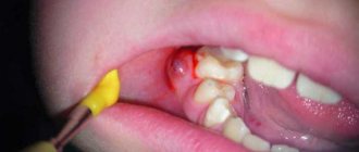
Childhood injuries and caries with complications lead to dental anomalies.
The death of tooth germs or the development of supernumerary teeth can occur due to osteomyelitis.
Dystrophies (stigmas)
The occurrence of a number of dystrophies in congenital syphilis is not associated with exposure to Treponema pallidum (the causative agent of syphilis) and does not have any diagnostic value. They develop with many infectious diseases and intoxications, for example, with parental alcoholism. Stigmas can indicate that a child may be affected by syphilis and help in making a diagnosis.
Rice. 16. Enlarged and protruding frontal and parietal tubercles without a dividing groove (“Olympic forehead”). The anomaly occurs in 36% of patients.
Rice. 17. A high hard palate (“lancet” or “Gothic”) occurs in 7% of cases.
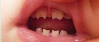
Rice. 18. Diastema (distance, gap) between the central incisors. Most often found on the upper jaw.
Rice. 19. A thickened sternal end (usually the right) of the clavicle (Ausitidian-Igumenakis symptom) occurs in patients with congenital syphilis in 25% of cases. The cause of the pathology is hyperostosis. In 13 - 20% of cases with congenital syphilis, there is an absence of the xiphoid process (Queir's axiphodia).
Rice. 20. A shortened (infantile) little finger (Dubois symptom) is recorded in 12% of cases with congenital syphilis. The little finger may be curved and turned towards the other fingers (Hissar's symptom).
Rice. 21. Stigmas indicating congenital syphilis may include spider fingers—abnormally long and narrow fingers (arachnodactyly).
Rice. 22. Girls and boys with congenital syphilis may experience hypertrichosis - growth of hair on the forehead (Tarkovsky hypertrichosis).
Anomalies in the number of teeth
All anomalies in the number of teeth are divided into three types:
- hyperodontia (excess of dental units above normal);
- edentia (complete absence of teeth);
- hypodontia (insufficient number of teeth).
Hyperodontia
Hyperodontia is a disorder of the formation of the dentition, which is expressed in the presence of supernumerary teeth (over 32 dental units in an adult). The cause of the disease may be a violation of the mechanism of formation of tooth germs in childhood or infancy. Hyperodontia mainly affects the incisors of the upper jaw. Other teeth are involved in the pathological process less frequently. Extra dental units grow small and have a cone shape.
The disease can be true or false.
- With true hyperodontia, the formation of extra dental units occurs in the human jaw.
- False hyperodontia occurs when the loss of primary teeth is delayed, which interferes with the normal growth of permanent teeth.
With polyodontia, teeth may erupt away from the dentition or next to a correctly positioned dental unit, which leads to its gradual displacement. The dentition can be seriously deformed, which leads to serious diction problems.
If supernumerary teeth interfere with the development of normal teeth, they must be removed. Broken teeth are corrected by installing braces.
In those rare cases when a row violation does not occur, the supernumerary tooth is preserved. Prosthetics are used to correct the shape.
Hypodentia
Hypodentia is a deficiency of dental units. This disorder occurs most often due to the death of tooth germs in the fetus. A small number of teeth causes a shift in the midline, the formation of wide gaps between teeth (diastema and three), and shortening of the dentition. All this adversely affects the bite. Possible ways of correction are prosthetics.
Edentia
Adentia is a complete or partial absence of teeth. With edentia, the continuity of the dentition is disrupted, which leads to difficulties in chewing and speaking. The reason is a violation of the development or death of tooth germs under the influence of genetic factors or under the influence of unfavorable external factors during the formation of the dental plate in the fetus (the formation of the germs of permanent teeth in the fetus occurs after the 17th week of intrauterine development). The disorder is corrected using prosthetics or dental implantation.
List of neurotic dental diseases
Stressful periods have a particularly detrimental effect on the state of the body. In addition, as practice shows, the quality of oral hygiene deteriorates sharply. A person simply forgets about cleaning or does not do it thoroughly enough, which increases the risk of developing caries. The parallel use of sedative medications disrupts the production of saliva and causes a feeling of dryness.
Gradually, all this is reflected in psychosomatics - muscle spasms and pain are detected. The most common occurrence is excessive clenching of the jaw.
Bruxism
A disorder characterized by uncontrollable grinding of teeth caused by emotional stress. As a rule, it makes itself felt during sleep. The causes of its appearance include stress, prolonged depression, and malfunctions of the masticatory apparatus.
Grinding
This is not so much a full-fledged disorder as a symptom. Occurs when muscle spasms occur when the patient clenches his jaw tightly. In the vast majority of cases, the person does not even know there is a problem. It is detected during routine dental examinations.
Anomalies of magnitude
There are two forms of this type of deviation, including macrodentia (increase in the size of dental units) and microdentia (small teeth).
Macrodentia
With macrodentia, the size of dental crowns is significantly increased. The cause of the disorder is, in most cases, dysfunction of the endocrine system, which is characterized by the fusion of several rudiments together. The disease is localized mainly in the area of the upper incisors. Large teeth interfere with the process of eruption and growth of neighboring dental units, which leads to their abnormal arrangement and crowding. Giant teeth are found on or outside the dentition line. This is a serious cosmetic defect that causes serious functional impairment and psychological disorders. Pathologically enlarged teeth are removed, and the growth of adjacent teeth is corrected using prosthetics or braces.
Microdentia
Microdentia – teeth that are too small. The disorder mainly affects the upper lateral incisors, but may affect the incisors of the lower or both jaws. The cause of the anomaly has not been studied, but it is reliably known that it develops in genetically predisposed patients. With the problem of small teeth, patients develop too large interdental spaces, which significantly disrupts the aesthetics of the dentition. To correct the pathology, incorrectly growing teeth are removed or covered with crowns.
Tips for parents
Many parents do not even suspect that dental hypoplasia is a very common disease among children. In order to alleviate the child’s condition, the following actions must be taken:
- Eliminate all sour and sweet foods from your diet.
- Use special toothpastes.
- For small children, purchase silicone finger brushes for oral hygiene.
- Carry out the procedure of silvering your teeth regularly.
- Monitor their condition and promptly fill teeth if necessary.
Shape anomalies
There are pathologies in the development of dental units, due to which they acquire an unnatural shape. These disorders are named after the scientists who first described them. The following types of dental shape anomalies are found: spiny teeth, Pflueger teeth, Fournier teeth, Hutchinson teeth.
- Spiked or awl-shaped teeth. With this pathology, the teeth acquire a cone-shaped shape. Wide at the base, they gradually narrow and, towards the cutting edge, become sharpened into a spike shape. This problem is combined with microdentia. There are irregularities and stains on the surface of the teeth. The disease affects the front and lateral incisors. The disease occurs in childhood and is caused by genetic factors in combination with external factors. Among them are vitamin D deficiency and endocrine system problems.
- Hutchinson's teeth are characterized by a modified crown shape of the incisors. Externally, they have the shape of a barrel, since the neck is significantly thickened. The cutting edge of the teeth acquires an arched notch. The enamel layer also suffers, which is present only on the sides and absent in the center.
- Fournier's teeth are a form of systemic hypoplasia of dental units associated with metabolic disorders at the stage of intrauterine development of the fetus. The barrel-shaped shape of the teeth is preserved, but the notch of the incisal edge is absent. The color of the enamel is not disturbed, but the enamel layer is underdeveloped, which can be seen during microscopic examination.
- Pflueger's teeth are a disorder that affects permanent dental units. Their crowns take on a conical shape. Thickening develops in the cervical region, and the chewing surface is significantly underdeveloped. The chewing function of the teeth is completely preserved.
- Turner anomaly. With this pathology, there is no enamel on the teeth. They have an abnormally lumpy surface, and replacement tissue, dentin, forms in the exposed areas.
Systemic hypoplasia with a violation of the shape of the teeth has three degrees of development. The third degree is the most dangerous, in which the crown is severely deformed and the enamel layer decreases. In this case, the defective teeth are removed and replaced with dentures. It is also possible to restore affected teeth using reflective components.
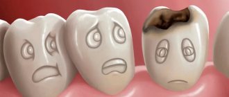
Treatment
If the hypoplasia is weak and there are stains on the teeth that are invisible to the naked eye, then treatment may not be necessary. When stains are noticeable or the process of tooth decay has begun, it is necessary to urgently consult a doctor, who will immediately take appropriate measures. No matter how sad it may sound, the disease cannot be cured completely. Dentists can correct cosmetic defects, but there is a chance that after a while you will have to contact them again.
The main treatment is teeth whitening. This helps remove stains from the enamel. However, this method is not used in severe stages of the disease. Sometimes doctors grind teeth, which helps get rid of bumps and uneven cutting edges.
Anomalies of hard tissue structure
An altered shape, abnormal size or color of enamel is formed against the background of an abnormality in the structure of the hard tissues of the dental unit. Among such anomalies are:
- Hypoplasia (underdevelopment of tissue). The initial degree of the disorder is manifested by the presence of chalky spots on the enamel and areas where tissue deficiency is observed. Subsequently, all kinds of pits, grooves, and recesses appear on the surface of the enamel. The defect affects all teeth in the dentition.
- Hyperplasia (excessive tissue formation). Pathology also affects all teeth at the same time. It is characterized by areas where tissue growth is observed - tubercles, sagging enamel. The consequence of the pathology is a change in the shape and size of the teeth, a violation of the occlusion (the line of contact of the upper and lower jaws).
- Anomalies of amelogenesis (enamel formation) are expressed in the presence of brown or yellow spots on the surface of the teeth. Areas where the natural composition of the enamel is disrupted become especially sensitive. Microdentia develops against the background of pathology. The cause of the development of pathology is a deficiency of microelements that take part in the formation of dental tissue. Treatment consists of replenishing this deficiency. Additionally, local remineralizing therapy and physiotherapy are performed.
- Disturbance of dentinogenesis consists of dysfunction of the mechanism of dentin formation. The main symptoms of the disease are as follows: teeth become yellow-brown or grayish in color. The fragile dentin in these areas quickly wears off, causing tooth decay and then tooth loss. The disease occurs in genetically predisposed patients. The problem can only be solved by replacing the units destroyed by the disease with prosthetics.
What periods would I like to highlight in his emotions:
The first period of treatment for crowded teeth with aligners is “anxiety”
After installing the attachments and giving the patient the first set of aligners, I was faced with his slight anxiety about tooth sensitivity; the patient constantly asked me if it was normal that one or the other tooth was sensitive when biting (to which I replied that this was absolutely normal and this is a period of getting used to the aligners), but after the first week and the patient got used to the aligners, his anxiety also went away. Later, when changing each set of aligners to a new one, he was already calm about the slight sensitivity of his teeth in the first 2 days of wearing the new set.
The second period of treatment for the patient is “satisfaction, sharing impressions”
Subsequently, the patient was so pleased with the chosen treatment method that he constantly told me funny / incidental stories related to removing the aligners in a restaurant or on vacation, or brushing his teeth at work after eating:
- Once
I wrapped my aligners in a napkin in the cafeteria at work and they were taken away along with the tray and thrown away, the patient panicked, called me and asked what he should do?! I suggested either looking in the trash or ordering a new set, but in the end he, a colleague and a canteen worker found them in a trash container! HOORAY) - Second story
. I was on a business trip, went to a restaurant for lunch and didn’t take the container with me, took off the mouth guards before eating, put them on a napkin next to me (for some reason I took off the mouth guards right at the table), a family with children was sitting at the next table and the children began to laugh when they saw how he took off his mouthguards, telling his parents: “Look, the guy is taking off his dentures”:
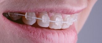
After all, aligners are generally transparent and invisible
The third period of treatment for crowded teeth is “complete satisfaction”
Complete satisfaction is also supplemented by an assessment of how others perceive him - “how cool he is.” Yes, this is cool, treatment with aligners is invisible, aesthetic, every day it adds inner self-confidence to the patient, liberates the person and reveals his potential for communication with friends. This is really cool!
Fourth period of treatment. There is no more crowding of teeth: “I will be bored without aligners”
At the end of the treatment, the patient already said that he did not know how he would live after the aligners, that he would miss them and whether it was possible to continue wearing them after treatment. Do not take this literally, the patient just had a little anxiety - the fear of losing the result he was very satisfied with!
The patient began to smile and laugh much more often!
But this is important for me, as a treating orthodontist. The patient’s smile is the most important gratitude for my work.
Color anomalies
Healthy, young teeth look snow-white due to a thick dentin layer and high-quality enamel, which has sufficient characteristics of whiteness, shine and transparency. Lifestyle, age, genetic factors, and ecology can slightly change the color of teeth from bluish to yellow. Small deviations are not considered pathology, although they indicate a change in the quality of the enamel.
Various pathological processes occurring in the body have a more significant impact on the condition of the enamel. The cause may be carious lesions, the use of medications, or a lack of certain microelements in the body. Teeth can take on a gray, pinkish, brown, purple and even black tint.
When starting to treat a patient, the dentist excludes the development of chronic pathologies. After a course of therapy and stable remission is achieved, other causes of discoloration of the enamel are eliminated. The final stage of treatment is a course of teeth whitening.
Specialists at the Consilium Dent choose the enamel lightening technique together with the patient. The selection criteria are age, condition of the enamel, and the wishes of the patient.
Basic terms in dentistry: names and causes of inflammatory diseases of the teeth and oral cavity
It plays a vital function in the digestion process: it is responsible for grinding food and influencing its subsequent breakdown by enzymes. In addition, there is a huge number of microorganisms that create natural microflora, but are considered opportunistic. This nuance determines the fact that any dental disorders progress extremely quickly.
Among the key factors in the occurrence of dental diseases are:
- poor hygiene;
- mechanical injuries and damage;
- chronic illnesses;
- metabolic disorders and abnormalities;
- the presence of thermal irritants in the body;
- bad environment;
- hereditary predisposition.
The branch of medicine that studies the emergence and development of pathologies is called etiology. It is partially correlated with diagnostic procedures, as it helps in determining pathogenesis.
Position anomalies
Incorrect position of teeth always develops as a consequence of another pathology and forms an incorrect bite. May affect one or both jaws. Incorrect position of teeth makes chewing food and oral hygiene difficult. This can lead to digestive problems and the development of cavities. There are several options for the pathological position of teeth within or outside the dentition:
- External or vestibular position means that the tooth is located outside the dentition, closer to the vestibule of the mouth. This position is typical for canines and incisors and develops as a serious cosmetic defect.
- Oral or internal position. The teeth are deviated inward closer to the tongue. The disorder is typical for canines, incisors and premolars. May cause tongue injuries and dysfunction of the temporomandibular joint.
- Mesial and distal position. Dental units are displaced forward or backward along the dentition, which leads to its shortening.
- Supra and infraocclusion are high or low positions of teeth relative to the occlusal curve. The cause of the disorder is underdevelopment of the alveolar process. It appears due to the presence of some obstacle that interferes with the normal formation of the tooth germ. Effective therapy is surgery.
- Tortoanomaly. The tooth is rotated around a vertical axis. The disorder is typical for incisors, canines, and premolars. The anomaly can cause injury to adjacent teeth. Dental units are rotated into the correct position using orthodontic structures.
- Transposition. The teeth change their location with each other. The disease affects canines that exchange places with premolars or lateral incisors. The disorder develops at the stage of tooth germ formation. The therapy is as follows: problematic teeth are removed followed by prosthetics.
- Crowding of teeth occurs due to lack of space when tooth germs are located too closely. Crowded teeth are closely adjacent to each other and rotate around their axis. Occurs in combination with macrodentia or with an undeveloped basal part. To eliminate the violation, some dental units are removed.
- Diastemas and tremas are wide spaces between teeth. The disorder develops as a consequence of an abnormal shape and size. The problem is treated with orthodontic methods or by installing veneers, which helps restore the aesthetics of the smile.
Misaligned teeth cannot always be cured using braces. Severe pathology, which is characterized by a skeletal disorder of the maxillofacial region, is treated with surgical reconstruction. The pathological fragment is transferred to the desired position and then secured. A fragment containing part of the dentition or the entire dentition can be used.
Other signs of late congenital syphilis
Lesions of the skeletal system
Osteoperiostitis and periostitis, gummous osteomyelitis and osteosclerosis are the main types of bone lesions, which occur in 40 - 50% of congenital syphilis. The lower legs (59%), nasal bones (18%), forearms (10%), skull bones (5%), and hard palate (4%) are affected.
Lesions of internal organs
Pathology of internal organs with congenital syphilis is registered in 20 - 25% of cases. The liver, spleen and kidneys are most often affected. With syphilitic damage to the heart, all its membranes, valves and vessels are affected. There is dysfunction of the thyroid, pancreas, thymus and gonads, pituitary gland and adrenal glands.
Nervous system lesions
Pathology of the nervous system with congenital syphilis occurs in 27 - 43% of cases. Of these, more than 50% are caused by damage to the brain, 32% to the spinal cord, and 11% to the tabes dorsalis. In 23% of cases, mental disability develops. With congenital syphilis, mental retardation, speech disorder, hemiplegia and hemiparesis, tabes dorsalis, and Jacksonian epilepsy are recorded. The child constantly suffers from headaches. Secondary atrophy of the optic nerves develops.
Syphilitic chorioretinitis
Syphilitic chorioretinitis leads to changes in the retina and choroid of the eye. Visual acuity is not reduced. Optic nerve atrophy leads to vision loss. With syphilis in children, a combination of chorioretinitis and damage to the optic nerve is more common.
Rice. 23. The photo shows chorioretinitis in early congenital syphilis. The disease is characterized by the “salt and pepper” symptom, which is characterized by the appearance of lumps of pigment and zones of depigmentation along the periphery of the fundus.
Diagnostics
An experienced dentist diagnoses dental abnormalities after an external examination and questioning of the patient. To clarify the diagnosis and identify the causes of the disease, a more detailed examination of the dental system is carried out:
- The construction of diagnostic plaster models and odontometric measurements make it possible to accurately identify changes in the size of dental units and their characteristics, which indicate the presence of pathologies.
- The color and transparency of the enamel are determined using a photographic method.
- Computed tomography or teleradiography makes it possible to obtain information about the condition of abnormal dental units in order to determine further treatment tactics.
- Panoramic radiography of the jaws is an important diagnostic method
- Electromyography of the jaws helps to assess the functional state of the facial muscular system.
To carry out differential diagnosis, the patient is referred for consultation to specialized specialists: endocrinologist, otolaryngologist, geneticist. Based on the diagnostic results, the attending physician determines treatment tactics. Depending on the type of disorder and the complexity of the disease, the patient may need the help of a dentist, surgeon, orthopedist, or implantologist.
Reviews
If a child is found to have Turner teeth, his parents have no reason to panic. This is a relatively mild form of hypoplasia that can be corrected with appropriate treatment.
If your child has been diagnosed with MGE, please share your personal experience with us. How severe was the destruction of the enamel, what treatment method was used, what was the result? The feedback form is at the bottom of this page.
If you find an error, please select a piece of text and press Ctrl+Enter.
Tags: enamel destruction
Did you like the article? stay tuned
Previous article
How effective are braces for diastema and are they always necessary?
Next article
What filling materials are used in pediatric dentistry?
Prognosis and prevention
Consilium Dent clinic uses advanced methods of therapeutic, surgical, and orthopedic treatment of dental anomalies in patients. By skillfully combining them, experienced specialists are able to restore the aesthetics of a smile and ensure the normal functioning of the dental system, even if the degree of deviation is significant.
Prevention of anomalies in the formation and development of dental units begins during the period of intrauterine development of the fetus and is carried out throughout a person’s life. The main stages are as follows: monitoring the successful course of the prenatal period, caring for harmonious postnatal development, organizing a correct lifestyle while the baby is growing up, caring for dental health and general health throughout life. By conducting an annual preventive examination, the dentist identifies dental anomalies at an early stage, when the pathological process is still reversible.
Expert of the article you are reading:
Nutrition
Proper and balanced nutrition plays an important role in prevention. It must be observed even at the stage of pregnancy planning. Also, the child’s nutrition needs to be monitored after birth. When doctors allow your baby to eat new foods rather than milk and formula, the main thing is to include the following in his diet:
- Milk, cheese, cottage cheese and other products that contain calcium and fluoride.
- Vitamin D. You can give your child special medications and spend more time in the sun.
- Foods rich in vitamin C. These are broccoli, oranges, tangerines, spinach.
- Products containing vitamins A and B. These include seafood, legumes, poultry and mushrooms.
Interesting moments that the patient described about the sensations on the aligners
The patient noted in the messenger that his tooth (fang) was moving and he was afraid to remove the aligner along with the fang! The movement of the canine as a result of removing crowding does occur, and as a result, tooth mobility can actually be felt. But since all dental movements during treatment with aligners are planned in advance, and the patient’s teeth are reliably protected in the aligners, all worries were removed.
As I told you earlier, once on a business trip in a restaurant, when he forgot the container and put the aligners next to him on a napkin, the children at the next table laughed that he had taken off his dentures
In fact, aligners can and should be stored in a special container while brushing your teeth or eating
Problems of treating crowded teeth with aligners
There were no problems at all with wearing the aligners. The only thing is that the activator came off once - this was after six months, but this was quickly eliminated and did not affect the course of treatment.
There was a case when a patient went on vacation and did not take his mouthguard retainer with him - his internal retainer came unstuck and the tooth moved back a little, “rotated” as orthodontists say. Now the problem is over.
Do you really need lingual braces to treat crowded teeth as your doctor advises?
Are you ready to carry this pile of iron in the form of braces - even cool lingual ones - in your mouth for the entire duration of treatment (a year or two)?
Rub your tongue against them, polish them until they shine, eliminating pieces of food? You will definitely be rubbing not covertly, but clearly, unpleasantly and constantly. Braces are uncomfortable and not aesthetically pleasing. You can have a high-quality consultation with our orthodontists and understand - what if your case of crowded teeth can be perfectly cured with aligners? If aligners can do it, then why subject yourself to self-torture with braces? So, we offer consultation on crowded teeth. Useful for you. The Star Smile company operates in more than 70 cities of Russia and will be able to offer you competent consultation with an orthodontist in your city.
Take the first step towards comfortable treatment.