The surgeon gives the patient recommendations after any operation, and invasive dental procedures are no exception. In particular, to prevent the implant, or more precisely the tissue around it, from becoming inflamed, it is necessary to strictly follow all instructions and prohibitions immediately after the intervention. It is very important to avoid hot drinks and meals in the first hours after implantation, to take antibiotics, antihistamines and non-steroidal anti-inflammatory drugs on time and in the prescribed dosage. If these conditions are not met, the risk of developing pathological reactions increases.
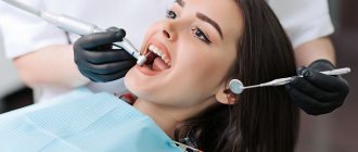
Dental implant failure: peri-implantitis and mucositis
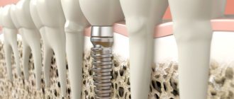
Today, implant rejection officially occurs in 2 to 5% of patients. And this process can begin either immediately or after several years of active use of the teeth. The main reason is considered to be the accumulation of pathogenic microflora, which leads to inflammation of the tissues around the installed implants. But there are two possible developments of the situation.
Peri-implantitis is a full-fledged and very severe inflammatory process that affects tissue along the entire length of the installed implant and leads to resorption (resorption) of the jaw bone. With this pathology, it is most often necessary to remove the structures in order to completely stop the development of inflammation. In any case, the doctor will diagnose and assess the condition; the more bone is preserved around the implant, the greater the chance of successful therapeutic treatment without extracting the titanium root.
But there is also such a thing as mucositis - in this case, the inflammation is localized only in the gum area, that is, it does not lead to the destruction of bone tissue around the implant body, and there is almost a 100 percent chance of saving the structure. For this purpose, drug therapy is carried out.
“Every patient must understand that the outcome of treatment is not only the responsibility of the attending physician. At the first stage - yes, the first 50% depends on the professionalism of the implantologist, on how he plans the treatment process and installs the implants. But the second 50% of success is the task of the patient himself: implants, just like living teeth, require careful care.”
Bespalov Roman Dmitrievich, maxillofacial surgeon, implantologist, work experience of more than 27 years make an appointment
Implantation without risks and complications! Our doctors are professionals who are trained in the best foreign centers. We do not save on the health of our patients!
Enroll now
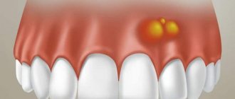
Symptoms of mucositis and peri-implantitis
With mucositis - superficial inflammation of the tissue - there is slight swelling, redness or even blue discoloration of the gums.
The mucosa may move slightly away from the crown or implant abutment. If you look at the picture, there is no bone inflammation and no bone loss. With peri-implantitis, the symptoms are more pronounced:
- redness and swelling of the gums,
- pain when pressing on the implant and its mobility,
- purulent discharge, bleeding from the area of the installed implant,
- increase in body temperature to 38 degrees.
Even with slight inflammation of the gums in the area of installed implants, you should promptly consult a doctor - the sooner a diagnosis is made and treatment is started, the greater the chance of saving the installed implant.
Stages and forms of peri-implantitis
- mild stage: loss of bone tissue around the implant is no more than 3 mm, while the mucous membrane becomes inflamed and minor pain occurs. Note that this is no longer mucositis, since bone resorption is observed. Treatment of this stage is usually conservative, with the use of antibiotics and painkillers,
- moderate stage: about half of the vertical bone tissue is lost, pus discharge appears from the area of the installed implant, bleeding and acute pain occur,
- severe stage: almost complete resorption of bone tissue, formation of deep pockets, development of infection, purulent discharge, constant bleeding.
How is diagnostics carried out?
If you find any of the listed symptoms, or the doctor himself has doubts during a routine examination, an x-ray diagnosis is required. This may be a panoramic image (OPTG), but better - a three-dimensional computed tomography, which will allow you to assess the degree of bone resorption around the entire body of the implant.
Also, the doctor must conduct a visual examination and instrumental diagnostics, or more precisely, probing. It is necessary to assess the depth of the gum pocket - normally the mucous membrane fits tightly and it is difficult to “get to” the implant itself, because it is immersed in bone tissue (if we are talking about a two-part structure). But with peri-implantitis, the gums easily peel off, and the depth of such a gum pocket can be from 5-6 mm or more.
Symptoms of inflammation
The infection, entering the gum tissue, causes inflammation, which can be recognized by the following signs.
- Acute pain that can spread throughout the entire mouth;
- redness and swelling of the gums;
- bad breath;
- temperature increase;
- enlargement of the gum pocket;
- loosening of the implant;
- bloody discharge around the installed structure.
All these signs indicate that something went wrong during the operation and implantation of the implant. In any case, you should immediately contact your doctor.
Why is peri-implantitis becoming more common? World Research Results
Due to the fact that peri-implantitis is diagnosed several times more often than 5 years ago, world implantologists are literally sounding the alarm - holding symposiums and meetings to discuss this growing problem. And they all agree that the main reason why peri-implantitis occurs is bacteria (how they get into the tissue around the implant is another matter, but this will be discussed later).
On traditional designs, which are designed for a two-stage protocol, a rough surface is provided along the entire length of the implants. This is done for their better fixation in the bone tissue - due to the presence of micropores, bone cells literally grow into the body of the implant. However, after installation of implants during the first five years1, bone resorption occurs up to 1-3 mm (this is considered a physiological norm, and manufacturers are trying to improve the design of implants to reduce this problem). As a result, the rough top of the implants becomes exposed. And if at the same time the lack of oral hygiene on the part of the patient is added to this, then a large amount of plaque accumulates on the implant. This is the main reason for the development of peri-implantitis and subsequent implant rejection.
According to studies conducted by the International Foundation of Implantologists, today it is customary to identify three main reasons for the development of peri-implantitis: the rough surface of implants in combination with poor hygiene and not entirely correct installation (above the gum level), the multicomponent nature of implants (when the connection between the implant body and the abutment is not tight, plaque accumulates ), as well as the wide diameter of the installed structures.
Naturally, we are not talking about all implants. But many manufacturers, unfortunately, do not take into account the high risks of developing peri-implantitis and produce models that significantly increase the likelihood of developing an inflammatory process. This means that their long-term service life is out of the question.
Treatment of inflammation processes after dental implantation
Treatment of post-operative gum inflammation can be conservative or surgical. It all depends on the stage and causes of the pathological process. The conservative method involves cleaning the structure under anesthesia and prescribing antibacterial therapy to the patient. The surgical method involves:
- Mandatory sanitation of the oral cavity.
- Dissection of the mucosa and removal of granulation.
- Chemical and mechanical treatment of the rod.
- Cleaning, correction of the bone bed.
- Stitching.
All procedures are performed under anesthesia, and then the patient is prescribed antibacterial and antihistamine drugs. If the implant does fail, there is still the possibility of repeat surgery. Provided that the jawbone has sufficient density and volume, re-implantation is carried out after 4-8 weeks.
The main causes of the development of mucositis and peri-implantitis
So, as noted earlier, the main reason for the development of both mucositis and peri-implantitis is the accumulation of pathogenic microflora. In simple terms, like any inflammation, these problems are caused by ordinary microorganisms: mainly spirochetes and gram-negative anaerobes. These names will mean nothing to the average person; it is much more important to understand where they come from in the oral cavity and why they begin to be active.
In general, our oral cavity contains both “good” and pathogenic microorganisms. But their number is balanced. In the absence of daily oral hygiene, with a decrease in immunity (or if it fights other diseases), the number of “harmful” microbes prevails, which provokes the development of various inflammatory processes. With mucositis it is superficial, with peri-implantitis it is deeper. But if the cause of mucositis is mainly poor oral hygiene, then there are much more prerequisites for the development of peri-implantitis:
- doctor errors: insufficient examination of the patient; installation of implants only on the basis of a panoramic image without computed tomography; penetration of microbes (including saliva) into the cavity during implant installation; errors during prosthetics - for example, overloading of the implant or injury to the mucous membrane,
- violations on the part of the patient: failure to comply with hygiene rules; refusal to treat chronic diseases (in particular, HIV infection, diabetes mellitus); use of prostheses other than for their intended purpose.
What causes gum inflammation after implantation?
Let's consider the list of reasons due to which the gums near the artificial root can become inflamed:
1. Unprofessionalism of a doctor is a common reason; not every doctor has sufficient qualifications to cope with a difficult clinical case. Sometimes errors are made due to inattention or negligence. Possible mistakes:
- incorrectly selected length and diameter of the structure;
- use of unsterile instruments;
- overheating of the bone with a bur when creating a bone bed;
- incorrect position of the rod;
- low quality of the implantation system;
- ignoring contraindications to the procedure.
2. Violation of the postoperative regimen by the patient . Even if the doctor does his job perfectly, failure to follow the recommendations can negate the results of the work. Patient errors:
- overheating or hypothermia of the body;
- increased load on the implant during engraftment;
- deliberate concealment of chronic diseases;
- insufficient oral hygiene;
- non-compliance with medication regimen;
- smoking.
3. Other reasons:
- development of an allergic reaction to the material or coating of the rod;
- structural damage;
- development of diseases of the immune, cardiovascular, circulatory systems;
- infectious diseases of the oral cavity.
What doctor mistakes lead to peri-implantitis?
At the first stage, when preparation for treatment and direct installation of implants is carried out, the positive outcome of implantation depends 90% on the doctor. After this, most of the responsibility passes to the shoulders of the patient himself.
- errors at the diagnostic stage: implantation only on the basis of OPTG (panoramic image) data without the use of three-dimensional computed tomography (CT); implantation without prior assessment of the patient’s health status,
- Chronic diseases are not excluded and are not taken into account, as well as certain conditions: pregnancy, menopause and menstruation in women, diabetes mellitus, active smoking and drinking alcohol, periodontitis and periodontal disease, ENT pathologies, poorly treated teeth in the oral cavity,
- errors at the treatment planning stage, for example, incorrectly chosen place for implants, too thin bone, too close to natural teeth,
- errors at the stage of implant installation: violation of sterility, damage to the coating of implants, saliva getting into the hole, use of non-original products, etc.,
- errors at the prosthetic stage: overloading of all or some of the implants in the complex, cement getting into the cavity, incorrect design of the prosthesis, restoration of only one jaw, etc.
Read more about all the mistakes that can be made during implantation in our separate material >>>
“The case of each patient in our clinic is reviewed by several doctors. This is necessarily a maxillofacial surgeon-implantologist, as well as an orthopedist. Already at the initial stage we evaluate every nuance, consider every detail. It is very important to us that the patient receives not only a beautiful smile, but also teeth that can be used to eat without pain.”
Chorny Stanislav Vladimirovich. orthopedic dentist, more than 20 years of experience
Non-original components mean a risk of implant rejection! We use only original implants, abutments and other superstructures that are tightly connected without joints or micro-gaps. As a whole, they passed a quality test directly at the factory - the manufacturer guarantees that they will function fully for many years.
sign up now
Diagnostics
If you find signs of a disease or a suspicion arises from a doctor during a routine examination (during visual examination or palpation), a diagnosis must be carried out:
- Computer diagnostics. Panoramic X-ray or 3D computed tomography - to assess jaw resorption.
- Periapical x-ray. Obtaining information about the condition of the artificial root and the area around it.
- Probing. To assess the depth of the periodontal pocket.
Additional studies are prescribed:
- Dentistry - a detailed examination of the mucous membrane helps to see the internal picture of the anomaly in the initial stages.
- Clinical tests, Schiller's, Russell's tests, pH-metry.
- Biochemical, bacteriological tests provide additional information about the disease.
Based on the results, a diagnosis is made and appropriate treatment is prescribed.
Who is at risk and what depends on the patient
At the stage before surgery, it is very important for you, as a patient, to follow the doctor’s recommendations and also tell him about all your health problems. We understand that if you come to the clinic, you expect to undergo implantation and restore lost teeth. But this is an expensive and complex treatment - in order not to waste money and time, and also in order not to spoil your health, you need to be extremely honest with your doctor. Today the list of contraindications to implantation is very small - over the past two years in our clinic there have been less than 10 patients whom we had to refuse treatment.
Patients with the following problems are at risk for developing peri-implantitis:
- bruxism – teeth grinding and strong clenching of the jaws leads to overload of the implants,
- long-term treatment with corticosteroids leads to poor tissue regeneration, destruction of blood vessels, increased risk of bleeding,
- previous radiation or chemotherapy suppresses the immune system and the functioning of many organs and systems,
- diabetes mellitus or osteoporosis lead to metabolic disorders,
- Alcohol abuse (especially immediately after dental implantation) leads to an increased risk of bleeding, failure to heal wounds, and inflammation of the tissue around the implant. Read our material about why alcoholic beverages must be avoided after implantation,
- refusal of hygiene or its improper implementation - including the use of dental floss, which leads to injury to the gums and exposure of the tops of the implants.
All these conditions increase the risk of developing peri-implantitis, but are not independent factors in its occurrence. This group of patients requires a more thorough diagnosis. Often, “narrow” specialists are involved, for example, oncologists, endocrinologists, who will give their opinion on the person’s state of health or prescribe appropriate therapy. In such situations, not only the doctor must weigh the pros and cons, but the patient himself must understand the risks and possible complications. If a specialist is honest and professional, he will never undertake to work with a person whose health he will harm rather than improve.
What does inflammation of soft tissues near or under the implant indicate?
Dental implantation is a complex traumatic operation, during which the integrity of soft and hard tissues is disrupted and blood vessels rupture. In the post-implantation period, swelling, bleeding, and pain are possible, which is a normal reaction of the body to injuries received.
These symptoms are not considered a pathology and go away on their own within 3-5 days. If pain and swelling persist for a longer period or tend to increase, this indicates the onset of inflammation - peri-implantitis.
Risks of implant rejection in the postoperative period
After successful dental implantation and after the implants have fully fused with the bone (that is, after six months to a year), you still shouldn’t relax. It is important to maintain good oral hygiene and maintain your health. It is clear that no one is immune from developing any disease in the future, but if you have chronic problems, you need to ensure that they do not develop into an acute stage. For example, if you have diabetes, you need to follow a diet and take all medications prescribed by your doctor, and monitor your blood sugar levels.
Immediately after installation of the implants, it is necessary to strictly follow the doctor’s recommendations in terms of therapeutic treatment. If antibiotics have been prescribed, then you must take them for a strictly specified amount of time or at least ask your doctor if you can stop taking them.
If the prosthesis was installed immediately, then you can also eat right away. But the load should be moderate - you can’t go to a restaurant on the first day and eat a hard steak, snacking on nuts and drinking a glass of whiskey.
Read our useful article about how to behave after dental implantation >>>
After 1.5 years, Alexander went to the clinic to replace the adaptive prosthesis with a permanent one made of ceramic composite. During this time, he regularly underwent preventive examinations and paid attention to oral hygiene, so there were no complications after implantation.
View review
“It’s been a year since I lost my teeth. I am happy with everything! My situation was very difficult and serious; I had to remove eight teeth at once. Thanks to the doctors for returning me to normal life. I eat, chew normally, and smile!” – our patient shared his impressions.
View review
What we guarantee
- Correct implantation, strict adherence to protocols. Our Center has a team of highly qualified doctors, employees of Moscow medical universities. Implantologists are proficient in all known protocols, and constantly undergo internships and advanced training courses in the USA, Germany, and surgical centers in Russia.
- Installation of high-quality Nobel Biocare implants. They have no restrictions on installation, are suitable for all clinical cases, the survival rate is 99.3%, and are protected from counterfeiting. Patented TI-Unite surface promotes faster healing. We provide a lifetime warranty.
- Thorough diagnosis. The center is equipped with modern diagnostic equipment, the results will be known in 15 minutes.
- Quick response to problem . You can call us at any time of the day or any day of the week if something is bothering you. In addition, we conduct preventive examinations and free CT examinations after surgery.
How is the treatment carried out and is it possible to save the implant?
Treatment of moderate and severe peri-implantitis today, unfortunately, does not give the desired results2. According to an analysis of the literature carried out by Heitz-Mayfield and Mombelli3, in almost 93% of cases, the tissue around the implants becomes infected again after treatment. That is, the risk of relapse is very, very high. Therefore, often the only correct solution is to remove the implant and carry out subsequent drug therapy.
But treatment of mucositis is quite possible. As well as the treatment of peri-implantitis if the implant is immobile and only a small amount of bone tissue is lost. In any case, the final decision is made by the doctor after conducting appropriate diagnostics.
The treatment method is only surgical, but complex, therefore it consists of several areas:
- cleaning the surface of the exposed implant,
- therapy with antibacterial drugs,
- increasing the volume of bone around the implant, correcting the position of the gums,
- the use of membranes from the patient’s own blood plasma during the surgical stage of treatment,
- prosthesis correction.
“For patients with advanced periodontitis, we recommend basal implantation, since in this case smooth implants with an antibacterial coating are used. Due to this, their contact with the mucous membrane is allowed, and even in the presence of an inflammatory process, pathogenic microorganisms will not accumulate on their surface, so the risk of peri-implantitis is minimized.”
Namdakov Nikolay Vladimirovich, maxillofacial surgeon, implantologist, orthopedist with more than 18 years of experience make an appointment
Cleansing from granulations and inflamed tissues
The very first thing to do is to clean the surface of the exposed implant from contaminants, i.e.
inflamed tissues, granulations, plaque, bacterial plaques. The surface of two-piece implants is porous, so if it is open and located above the level of the bone and gum (after all, with peri-implantitis, the bone volume is reduced), the accumulation of pathogenic microflora is guaranteed. Cleansing is carried out using ultrasound, laser or Air Flow system. Finally, treatment with antiseptics. In fact, this is the initial preparation for the main surgical intervention. The main difficulty is that many implants have an active coating, which is achieved through complex multi-stage processing in the factory. And when washing the surface of the implant, it can be damaged, which can lead to poor implantation of the structure or even complete rejection. This is also one of the reasons why treatment of peri-implantitis sometimes does not give positive results.
Antibacterial therapy
In the presence of peri-implantitis, antibiotics must be prescribed to relieve the inflammatory process. These can be general medications, but in a number of situations, an analysis of the oral microflora is first performed to assess sensitivity to various antibiotics (it is quite expensive, so there is no need to refer each patient - it is carried out according to indications). Let us remind you that taking antibiotics is not compatible with alcohol.
Bone grafting and installation of protective membranes
This is the main surgical stage, the purpose of which is to restore bone volume, as well as remove all inflamed tissue. During the operation, the gum is cut and peeled off, thereby exposing the bone defect that the doctor will work with. In fact, this stage is similar to the first, but if the initial cleaning is only superficial, now it is deeper.
After peeling off the gingival flap using a scaler (ultrasonic device) or laser, granulations and inflamed tissues (gingival and even bone) are removed, as well as a better cleaning of the implant surface. After this, bone tissue is grafted (artificial or borrowed from another area), after which protective membranes are applied, which allow the grafted material to be securely fixed. It is worth noting that the technique of performing the operation depends directly on how many bone partitions (walls) are preserved around the implant. The more there are, the higher the chance of extending the life of the artificial root.
Application of PRF membranes from blood plasma
This is the main surgical stage, the purpose of which is to restore bone volume, as well as remove all inflamed tissue. During the operation, the gum is cut and peeled off, thereby exposing the bone defect that the doctor will work with. In fact, this stage is similar to the first, but if the initial cleaning is only superficial, now it is deeper.
At the stage of surgical intervention, our clinic also uses PRF membranes - this is a platelet mass obtained from the patient’s own blood. Numerous studies and our own practice prove that the use of membranes even at the stage of implant installation is an excellent prevention of peri-implantitis. And when treating this complication, blood plasma allows you to accelerate the process of regeneration of gingival and bone tissue.
Gum plastic surgery
This is the main surgical stage, the purpose of which is to restore bone volume, as well as remove all inflamed tissue. During the operation, the gum is cut and peeled off, thereby exposing the bone defect that the doctor will work with. In fact, this stage is similar to the first, but if the initial cleaning is only superficial, now it is deeper.
Plastic surgery can be either a consequence of bone grafting or an independent procedure. In the first case, it is needed to restore the aesthetics of the mucous membrane after surgery, especially when it comes to single tooth restoration. The position of the gums also needs to be adjusted due to the increase in bone tissue. In the second case, it is carried out when exposure of the installed implant occurs due to the gums being too thin in height or width, that is, when there is little mucous membrane or it does not fit tightly enough to the crown/implant.
Correction of orthopedic construction
In some situations, overload of installed implants leads to peri-implantitis. This can happen for several reasons:
- an incorrectly designed model of the prosthesis - this is why it is so important that complex dental restoration is carried out by several doctors at once: an implantologist and an orthopedist, who will create a single, properly functioning system,
- the patient eats inappropriate foods (for example, very hard foods immediately after implant placement),
- the patient does not come for prosthesis correction. After wearing removable appliances for a long time or without teeth for a long time, a person’s bite changes. With the help of prostheses on implants, we correct it, but we do it gradually - that is why the prosthesis is regularly corrected during the first six months to a year. So that it does not cause discomfort when closing the jaws and when chewing, and also does not cause overload of the implants.
Stages
- Easy . Minor loss of bone around the implant. Inflammation of the mucous membrane appears, the pain is not pronounced, and the artificial root wobbles slightly. Painkillers, antibiotics, and in some cases surgical treatment are prescribed.
- Average . Half of the vertical bone loss. Pus appears, bleeding, acute pain, the gums peel off, the implant is unstable. Surgical treatment or removal of the implant is prescribed.
- Heavy . Complete bone resorption. Deep gum pockets, spread of infection to neighboring areas, increased purulent discharge, prolonged bleeding. The implant will have to be removed.
It is considered normal to lose bone around the implant from one to one and a half millimeters in the first year and no more than 0.2 millimeters per year thereafter. Bone loss above these levels is pathological.
Is re-implantation possible if the implant has been removed?
Classic two-stage implantation with delayed loading after removal of the implants will in most cases be impossible, since the bone tissue atrophies too much due to the inflammatory process. Naturally, in a number of situations, the option of increasing it is possible - but it will take several months for the initial rehabilitation of the tissue after the development of the inflammatory process, as well as another 3-6 months for the implanted material to engraft.
With complex implantation with immediate loading, rejection of all installed implants is extremely rare. There are cases when one of 8-10 structures does not take root, but in such a situation the load from the prosthesis is distributed evenly among the others without overloading them, and if necessary, the implant is replaced.
Treatment of complications
Depending on the stage of development of the disease, the following procedures are performed:
- surgical removal of a pus sac;
- antiseptic treatment of soft tissues;
- cleaning and removing the gum pocket;
- cleaning and disinfection of the implant;
- drug therapy.
If the inflammatory processes and suppuration progress, then perhaps the only solution will be to remove the implant.
Prevention of peri-implantitis development
Thus, the following solutions allow us to solve the problem of the development of peri-implantitis:
- thorough diagnosis of the patient’s condition and planning of the treatment process,
- the choice of solid implants in which the intraosseous part is connected to the abutment, especially in situations where tooth restoration is carried out in the presence of generalized periodontitis,
- the choice of two-piece implants with an antimicrobial coating at the upper base of the implant body, as well as the use of abutments with a smooth antimicrobial coating,
- the use of polished implants that will not accumulate plaque,
- the use of implant models that transfer less load to the alveolar bone, which is susceptible to inflammatory processes,
- strict adherence by the patient to all recommendations of his doctor.
Thus, the main recommendation for patients is to choose a professional doctor who follows modern trends in the field of implantology, improves his skills, works with high-quality brands of implants and uses safe implantation protocols.
More examples 1 According to clinical studies of implant manufacturing companies: Nobel, Straumann, Astra Tech. 2 According to studies published in the journal Perio-implant advisory. 3 Esposito M1, Grusovin MG, Worthington HV Treatment of peri-implantitis: what interventions are effective? A Cochrane systematic review.
How to improve your well-being?
The following measures will help you cope with pain after implantation.
- Compliance with the schedule of taking antibiotics, painkillers and anti-inflammatory drugs prescribed by your doctor.
- Applying ice to the cheek in the first days after surgery.
- Exclusion from the diet of solid foods that can injure the gums, as well as hot and cold drinks.
- To give up smoking.
- Limiting physical activity.
- Careful oral hygiene.
Compliance with these rules will speed up tissue restoration.
Why does a tooth hurt under a crown: reasons
Thus, if you have a toothache under the crown or the gums under the crown are inflamed, the reason almost always lies in poor-quality therapeutic preparation of the tooth for prosthetics. Of course, in most cases, dental therapists take control photographs after filling the canals, but even if they see mistakes, most doctors simply will not waste time and redo something.
Unfortunately, this approach in Russia is the norm rather than the exception. And if your tooth hurts under the crown, then the first thing you should do is an x-ray, which will show one of the main mistakes that dentists make when filling root canals. The image will show whether the tooth can be treated or whether it needs to be removed. The most common mistakes made by doctors include...
Root canals are not filled to the root apex –
Let us remind you that in most cases, teeth must be depulped before prosthetics. Depulping a tooth means that the dental pulp (neurovascular bundle) is removed from it and the root canals are filled. When filling root canals, there are certain standards, the implementation of which helps prevent subsequent inflammation in the area of the tooth roots.
In Fig. 1,2 you can see how well-filled root canals look on radiographs. However, when the doctor does not work and fills the canals not to the top of the root, conditions are created in the unsealed part of the canal for the spread of infection. Failure to fill the canal by just 1-2 mm can already cause inflammation at the apex of the tooth root (Fig. 3).
On X-ray 3 you can clearly see the unfilled part of the root canal (it is shown by a white arrow). Black arrows show the boundaries of a periodontal abscess, which on an x-ray looks like intense darkening in the area of the root apex. The reason for its formation is the development of infection in the unfilled part of the root canal. This dental disease is called periodontitis.
What does inflammation of a tooth look like under the crown (Fig. 4) –
- "Gutta-percha" is a material for canal filling,
- “periapical abscess” is a focus of purulent inflammation in the form of a purulent sac at the apex of the root (depending on the size of the purulent focus, the latter is called either a granuloma or a radicular cyst).
Poor obturation of root canals –
Inflammation can also be caused by poor obturation of the root canal with filling substances (gutta-percha and sealer). Those. the canal can even be sealed to the top of the root, but it is not sealed tightly, with many pores and empty spaces.
This could also be the reason why your tooth hurts under the crown, because... Such poor-quality canal filling also leads to the development of inflammation at the apex of the tooth roots. Poor canal obturation can also be easily determined by a targeted photograph of the tooth.
Perforation of the walls of the root canal –
Perforation is literally “a non-physiological hole.” In other words, this is a hole in the root of the tooth that is created artificially. The only physiological hole in the tooth is at the top of each root. The most common perforations that occur are:
- During instrumental treatment of root canals - using tools for mechanical expansion of the root canal, the doctor can make a number of mistakes. For example, instead of expanding the root canal along its course (from the mouth of the canal to the apex of the root), the doctor will direct the instrument perpendicularly through the canal wall, which will lead to the appearance of a “hole” in the root wall (Fig. 5, 6).
- During fixation of the pin in the root canal, doctors also very often allow perforations if the technique for fixing the pins in the root canal is not followed. Such perforations are also determined by radiographs and corresponding symptoms (Fig. 7).
Fracture of an instrument in the lumen of the root canal –
This happens quite often, but in most cases it is again due to the fault of the doctor. Below you will read about the main reasons for instrument failure in the canal. The only good reason for a breakdown that cannot be blamed on the dentist is if your root canals have a very severe curvature.
- Violation of the technique of rotating the instrument in the root canal - instruments for treating root canals are quite thin and require strict adherence to a certain technique of use. For example, most instruments cannot be rotated more than 120 degrees in the root canal. If the instrument is rotated 360 degrees in the root canal, this can naturally cause a fracture of the instrument, which is associated with the curvature of the root canals.
- Reuse of instruments—breakage of instruments can also occur due to the fact that the doctor uses “old instruments.”
Instruments for mechanical expansion of root canals are made of special metal. Any metal gets microcracks during loading, which is called “metal fatigue.” Repeated use of an instrument for root canal treatment greatly increases the risk of instrument breakage (24stoma.ru). Tools for canal treatment come in different sizes and differ in thickness. The thinnest instruments have sizes No. 6,8,10, 15. In Europe and the USA, such instruments are generally disposable and their reuse is not allowed. Instruments of other sizes can be reused after sterilization. But in Russia, in the vast majority of clinics, in order to save money, no one throws away such instruments, and they work with them “to the limit.” What affects the incidence of tool breakage. - When working in highly curved, difficult-to-pass channels - In this case, instrument failure may occur through no fault of the doctor, because
Instrumentation in such root canals is itself risky. But you cannot refuse and not do it. The presence of a foreign body (piece of instrument) in the root canal or beyond can be determined using an x-ray (Fig. 8, 9). And the problem here is that in most cases it is not possible to remove the instrument fragment from the canal. This prevents high-quality filling of the root canal, which in the vast majority of cases leads to inflammation developing, the tooth under the crown aching, or the gums under the crown hurting.
How and why rejection occurs at different times
Express rejection
Occurs with a coarse porous bone structure. This feature makes it impossible to create a reliable support for the titanium root - the bone is not able to provide the implant with the necessary stability and nutrition, and cannot provide sufficient tension to hold the implant. In our Center, the problem is solved with the help of growth stimulants (morphogenetic proteins), which are attached to the neck of the implant during installation and activate the growth and compaction of the surrounding bone mass.
Also among the common causes of early rejection:
- Excessive load on the implant with the crown at the time of engraftment during express implantation.
- Infection of jaw tissues when installing an implant immediately after tooth extraction with inflammation at the root.
The main cause of implant failure is peri-implantitis. Infectious inflammation of the tissue around the implanted rod, leading to progressive loss of the jaw bone. It develops immediately after installation, several months and even years later.
Deferred period
If rejection does not occur a month or two after implantation, then osseointegration took place without complications. However, there are cases of later rejection when a permanent crown was installed on the implant and everything looks fine. Among the reasons:
- Prosthetic errors when the angular chewing load on the titanium root is increased. It manifests itself as pain when chewing and redness of the gums.
- Inflammation of the tissues in the implant area due to a bacterial process that develops due to the accumulation of food debris between the teeth. It may be asymptomatic at first, but over time it is accompanied by bleeding gums - it looks inflamed, swollen, like periodontitis. If measures are not taken, after a year or two this condition will lead to the structure falling out.