Manifestations of distal bite in the mouth
Closing of lateral teeth during distal occlusion.
Distal occlusion is manifested in the mouth by the closure of the upper and lower teeth according to the “second class” (violation of the relationship between the projections of the antagonist teeth of the upper and lower jaw).
In the frontal segment (at the level of the front teeth) there are two closure options (the so-called first or second subclasses).
In the first case, a “sagittal gap” is observed, which reflects the discrepancy between the dentition in the sagittal plane (anterior-posterior direction). And the upper incisors are in protrusion (inclined forward). In the second option, the upper front teeth are tilted back (into retrusion). With the formation of a deep bite (when the lower teeth are not visible from under the upper ones).
Sagittal chain with distal occlusion. 2 class 1 subclass.
Distal occlusion. 2nd class 2nd subclass.
How to prevent bite pathologies
To prevent the development of anomalies, you need to follow a number of simple rules:
- During pregnancy, you need to carefully monitor your health and eat foods rich in calcium.
- After the birth of a child, do not transfer him to artificial feeding unless necessary. Bottle feeding affects the formation of the bite.
- After teething, monitor their condition and visit the dentist on time.
- Do therapeutic exercises that provide the necessary load on the facial muscles.
Prevention of malocclusion is necessary for both children and adults. Pathology can manifest at any age if appropriate factors are present. Therefore, regular examinations are necessary. If a specialist identifies a predisposition to the development of a certain disorder, he will recommend appropriate means of preventing malocclusion.
To prevent the disease, more complex measures may be needed than therapeutic exercises and compliance with a number of the rules described above. In some cases, you will need a set of devices for the prevention of malocclusions, which the doctor will select depending on the situation.
You cannot use devices to prevent malocclusions on your own, only on the recommendation of a specialist. Their unsystematic use can significantly worsen the current condition and provoke the development of pathology.
Causes of distal bite (distal occlusion)
They are almost always (in the vast majority of cases) skeletal...
- Posterior position of the lower jaw. This is the most common cause of distal occlusion
- Small (short) lower jaw.
Many people write that the cause of a distal bite may be an overdeveloped (excessively large) upper jaw. They say that this is why there is a “gap” between the front upper and lower teeth (sagittal gap)... Many years of observations and our experience have shown that this is not so. With any anomaly of occlusion, there can be no talk of any OVERdevelopment. Everything, on the contrary, is UNDERdeveloped.
The lower jaw ends up in a posterior position (the first reason) only because the lower teeth bump into the upper teeth while the upper jaw is shortened and narrowed (generally underdeveloped). Or, when the upper incisors are “wrapped” back, into retrusion (second subclass). In addition, with distal occlusion, the upper jaw is always narrower than the lower jaw. Which, by the way, is another reason for blocking the lower jaw in the posterior position. And this same “blocking” does not allow the lower jaw, which is caught in the trap, to develop normally. Here is the second reason for distal occlusion - underdevelopment of the lower jaw.
There is also a “dental” reason for distal occlusion. Or more precisely, the dental cause of closure is “second class”. This is when mesial (front) displacement of the upper lateral teeth occurs. And then, even with a normal relationship of the jaws in the sagittal plane, the closure of the lateral teeth (molars) will also be in the second class. True, this is not a true distal occlusion...
A “dental” cause of the second class may complement or may camouflage true distal occlusion. Therefore, it will not be possible to figure out what is the reason for the closure in class 2 visually (by eye). Diagnostics required.
Malocclusion, psychosocial problems
According to experts, recently correct bite has become increasingly socially important. Straight teeth and a beautiful smile have a high status regardless of social level, and society has a negative and unfriendly attitude towards malocclusion. A person’s appearance influences the opinion of a kindergarten teacher, then a school teacher or college teacher. It is often more difficult for a child with malocclusion to learn. With an incorrect bite, it is difficult to get a prestigious job that requires active communication with clients or colleagues. This attitude towards the appearance of teeth cannot but influence the adaptation of a person with malocclusion in society. In other words, if your malocclusion is having a negative impact on your relationships with the world, then the problem is much more serious and deeper than it seems at first glance.
All of the above problems are a sufficient reason to consult an orthodontist. You should not save time and effort, do not underestimate a child’s malocclusion, hoping that the problem will disappear with the change of baby teeth. If you notice any deviations, entrust the problem to a specialist. According to statistics, in cities, parents of children with malocclusion are more likely to consult an orthodontist than in villages. In many ways, visits to the orthodontist depend on family income. This fact illustrates not only the idea that only families with higher incomes can afford orthodontic treatment, but also that a correct bite is associated with a more prestigious social status.
According to statistics, teeth straightening, bite correction,
Wearing braces in adulthood is becoming more popular in society than before. Also, an increasing number of people aged 40 years and older are seeking treatment from an orthodontist as part of comprehensive dental treatment. Orthodontics is occupying an increasingly important position in dentistry. According to statistics, the majority of people who received orthodontic treatment were satisfied with the results of the treatment and noted its beneficial effects; almost all patients recognize the improvement in the dental system and general psycho-emotional state.
Diagnostics
The main goal of any diagnosis is to identify the cause of the problem. And make a diagnosis. Likewise, the diagnosis of distal occlusion is designed to identify the cause of closure in the second class. In order to separate the flies from the cutlets during treatment, so to speak. That is, dental causes of closure are in the second class from non-dental (maxillary, skeletal).
The main diagnostic methods are:
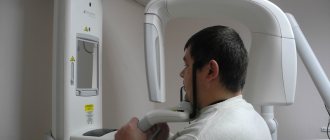
- Analysis of TRG (teleradiogram) of the skull in a lateral projection. It allows you to clearly determine all the skeletal (jaw) nuances of the problem. Both with the position of the jaws and with their sizes.
- Analysis of plaster models of jaws (for this, casts are taken). It allows you to clarify the “dental” nuances: the size of the teeth, the length and width of the dentition, the features of the relationship (including closure) of the upper and lower teeth
Analysis of TRG (teleradiogram) of the skull in a lateral projection.
Analysis of plaster models of jaws.
It is not recommended to fall below this diagnostic “bar”. Below - just by eye. But this, as we understand, is not the best option.
Diagnostics is the first and key step in the treatment of distal occlusion. Do you want to know why it’s impossible to do without diagnostics?
When will only a surgeon help?
If a person comes to an appointment not only with a greatly enlarged gap, but also with several missing molars or their strong inclination towards the throat, he is referred to a surgeon. In such situations, doctors have to disconnect part of the bone and move it to a physiological position. This is a complex operation that is performed under general anesthesia and recovery from it takes 6-8 months.
We told you what to do with non-physiological interdental distances. The main thing is to contact a competent orthodontist, be patient and set yourself up for a long journey to health. Before installing expensive structures and enrolling in surgical operations with a long rehabilitation period, it is important to undergo a full examination and start with gentle methods.
Errors in the treatment of distal occlusion
The first, main mistake is that many try to treat distal bite with one device. For example, only with braces (this is the most popular option). But braces, although a cool device, are not for skeletal problems. The level of hardware competence of the braces system does not extend further than “to place the teeth on the jaw.” And distal occlusion, as we have already said, is a purely skeletal problem. Jaws and posture are to blame. And braces are not capable of solving problems with posture or jaw problems. After all, braces cannot change the position or size of the jaw. No matter what the would-be doctors promise...
And therefore, such videos are just an advertising gimmick.
Or rather, a convenient “hook” that catches gullible patients. Convenient because it coincides with the desire to do everything quickly and efficiently... But it doesn’t coincide with reality.
And braces cannot be the main device in the treatment of distal occlusion.
Braces are the wrong choice when treating distal malocclusions. In any case, you definitely shouldn’t start with them. Braces, as mentioned and shown above, can be effectively used only at the final stage of treatment of distal occlusion. All major “feats” must be accomplished before working on the teeth. Before using braces. And they are accomplished at the level of the jaws and skeleton (posture). Otherwise, there will simply be nothing to “clog” (close) these teeth (read with braces). See “Fundamentals of the therapeutic concept of “Ortho-Arteli”.
The second common mistake in correcting distal occlusion is treatment with the removal of upper premolars (4 or 5 teeth).
And this mistake is much more serious. Because it can have the most dire consequences. See "Orthodontic treatment with tooth extraction."
After all, as we said above, the main causes of distal occlusion are skeletal. And most often the lower jaw is to blame. And if so, then a reasonable question arises: if the lower jaw is to blame, what does the upper teeth have to do with it (their removal). Where is the logic?
In general, all errors in the treatment of distal occlusion in an adult are usually associated with insufficient diagnosis. Which entails a misunderstanding of the reasons for the occurrence of distal occlusion in each specific case.
For example, the “dental” cause of distal occlusion is very often confused with the “skeletal” (jaw) cause. We remind you that in the first case you need to move the lateral teeth back. And in the second - to push the lower jaw forward and develop the upper one. And if you get confused, if you don’t figure it out... Start distalizing the upper teeth, or worse, remove them when it is necessary to advance the lower jaw... Or vice versa. It is clear that nothing good will come of it.
The third mistake is when, without understanding what’s what, what exactly is the cause of distal occlusion, but realizing, however, that the problem cannot be corrected with braces, they try to solve the problem surgically. Using orthognathic surgery. Through osteotomy of the lower and upper jaws.
This is where I would like to dwell in more detail. And explain something. Why do orthodontists send to a surgeon to “treat” a distal bite? They have already said: they understand that braces will not cope. But they don’t understand how to treat it without braces. They don’t understand because in dentistry in general, and orthodontics in particular, there are a lot of prejudices regarding the possibilities of moving the lower jaw forward and developing the upper jaw. And these, as we saw above, are the main (“key”) points in the treatment of distal occlusion.
Most orthodontists simply do not believe in this (that it is possible in principle). Because someone (even a “great one”) said two hundred years ago that this cannot be done. Time has passed. Everyone has already forgotten why it is “impossible”. But no one even bothered to check why, in fact, it was impossible...
And therefore, those doctors who blindly believe in these moss-covered “dogmas” simply do not see an alternative. And for them there are only two extreme poles in the treatment of distal occlusion: braces and surgery. That's all.
During a direct examination by a specialist, you will be able to find out your exact diagnosis, as well as receive a referral for diagnosis or a treatment plan.
And in our clinic we treat distal occlusion between these two poles. Using methods of maxillofacial orthopedics and craniodontics. Here are the ones: changing the position of the lower jaw and changing the size of the upper. And we get good results, without regard to the fact that someone once said something... We must always double-check what was said. We checked. And everything turned out to be somewhat different. Or rather, not at all the way they “frightened” us. You can also push it out. It can be developed. You just need to know how, because there are some “secrets” here. And we, at the Orto-Artel clinic, knowing these “secrets”, take advantage of these opportunities, successfully eliminating distal occlusion of any complexity.
By the way, if we are talking about surgical treatment of distal occlusion, then we need to determine when and in what cases surgery is generally appropriate. But the indication is basically the same - a small lower jaw, the so-called lower micrognathia, and the micrognathia is very pronounced.
We indicated this reason above as one of the skeletal reasons. I will add: the lower jaw should be VERY short. Literally ugly. Only then can this be resolved surgically. In all other cases, you can do without surgery. Proven and tested many times.
By the way... In our clinic, a good half of the patients have formal indications for surgical treatment of distal occlusion. But only a few got to the surgeon. Because formalities are formalities, but compromise solutions were found that satisfied everyone: both the patients and us. This is because we approach treatment not formally, but in essence. And we are guided by logic and common sense.
Well, as a special case of a short lower jaw, which cannot be corrected except by surgery, this is a small chin. If it is small and spoils the face, then even by pushing the lower jaw forward we will not get the aesthetics we want. Simply because orthodontists do not have the means and instruments (devices) to selectively influence the chin. It is impossible to stimulate the chin to grow. Only surgery.
Inferior micrognathia.
This is where the indications (and there is essentially one) for surgical intervention for distal occlusion end.
Going with a knife into the upper jaw - God forbid! Even if the upper jaw is indirectly to blame for the distal occlusion. More precisely, the upper jaw also, as a rule, suffers (dimensionally) with distal occlusion. The upper jaw is directly connected to the base of the skull. And such interventions can then backfire. Literally. And the most important thing is that there is no need to go there surgically. The upper jaw can be developed!!!
Reasons for development
The classification of dental growth anomalies, as well as types and degrees of malocclusion, allows for a combination of various factors. Both external and internal processes have an impact, from genetic predisposition to bad habits. Differentiation by age makes it possible to identify the main prerequisites characteristic of patients of certain groups.
In childhood
The reasons affecting the formation of the dentofacial apparatus in the early period include:
- Heredity - studies show that anomalies are often transmitted from parents and are of a similar nature.
- Problems with respiratory function - prolonged mouth breathing caused by ENT diseases leads to disruption of the physiological position and muscle strain, affecting the growth of elements of the jaw region.
- Bad habits typical for children - the anatomical structure is affected by constant sucking of a pacifier or fingers, attempts to gnaw hard objects, pressure with the tongue on growing units, and even curvature of posture in a sitting position.
- Insufficient presence of mineral and nutritional components in the diet both during pregnancy and in the early stages of development of the body.
- Lack of sufficient tension during artificial feeding, observed when using bottles and nipples with a wide opening.
Pathological and abnormal types of occlusion also form during the process of changing the milk set, acting as a consequence of premature loss of temporary units, the appearance of impacted and dystopic elements, as well as other negative factors.
As an adult
Patients of the older age group are less at risk of disrupting the natural anatomical state of the jaw region, however, even after its final formation, there is a possibility of the formation of anomalies. First of all, the reason is incorrectly performed prosthetics - incorrect selection of the dimensions of the replacement structures leads to displacement of the occlusion and muscle overstrain, provoking dysfunction of the temporomandibular joint. In addition, systematic abuse of bad habits, the development of pathological processes, as well as mechanical damage and injury can have a negative impact.
Anomalies of teeth position
In case of anomalies in the position of the teeth, the task of the orthodontist is to preliminary normalize the shape and size of the dentition and occlusion. For this purpose, various orthodontic structures are used - both removable and non-removable. In the distal position, the teeth are moved mesially if there is space in the dentition. The need for mesial tooth movement arises when the first molar is removed (for therapeutic indications), and in this case the second molar moves mesially.
Since this anomaly relates to the lateral teeth, in devices of any design the fulcrum is formed in the anterior or lateral part of the corresponding side, and the point of application of force is the tooth being moved. If a rubber rod is used to move a tooth in an inclined distal position, the point of application of force is the coronal part of the tooth; in the case of a corpus tooth, it is the coronal and root parts, for which a rod with a hook is used in the area of the transitional fold.
In plate devices and mouth guard plastic structures, the fulcrum is hooks welded into the base. In metal structures, hooks are also soldered in the front section on the corresponding structural elements. Primary and permanent teeth at the appropriate stage of formation can be moved in the mesial direction using hand-shaped springs (according to Kalvelis). Permanent teeth in the final stage of root formation are moved using a brace system both obliquely-rotational and corpusally. To move lateral teeth in the mesial direction, the use of a positioner is ineffective.
Treatment of mesial position of teeth is carried out individually. With early removal of the second primary molar or primary edentia of the second premolar of the upper jaw, mesial movement of the first molar is observed. In this regard, the closure of one pair of antagonist teeth is disrupted, namely the mesial-buccal cusp of the first molar of the upper jaw is located in front of the intercuspal fissure of the first molar of the lower jaw. In this case, it is possible to maintain the mesial position of the first molar and then it is advisable to move the second molar forward.
If the doctor decides to move the first molar in the distal direction in order to achieve good closure with the antagonist teeth, you can use a plate on the upper jaw with a sectoral cut, a Kalamkarov apparatus, or an Angle arch. The use of a facebow with cervical traction is especially effective. For the first molars, rings with facebow tubes are made. On the side of the distally moved first molar, a bend is made on the arch, which rests against the tube, and on the opposite side, the end of the arch does not have a stop and is freely located in the tube. In the anterior section, the facial bow is separated from the front teeth. When applying a cervical traction, the entire force of the facebow is directed toward the first molar, which should be moved distally. To move both first molars distally, the facebow has stops in front of the tubes on both sides and both teeth will move distally.
After moving the first molars in the distal direction, the integrity of the dentition is restored at the level of the second premolar using only prosthetics or with preliminary implantation. In the clinic, the mesial position of the lateral teeth is often encountered. This may be due to early removal of the primary canine, high position of the permanent canine bud, the presence of a supernumerary tooth bud, macrodentia of the lateral teeth, a change in the order of eruption of the canine and second premolar (the second premolar erupts first). In this case, the type of closure of the lateral teeth corresponds to Angle class II. To create space for the canine, the lateral teeth must be moved distally. For this you can use plate devices.
Devices 1 and 2 allow you to move the lateral group of teeth on both sides in the distal direction. In this case, the front teeth are moved in the labial direction. Plate apparatus 3 (a plate for the upper jaw with a sectoral cut) moves the lateral teeth in the distal direction, and apparatus 4 allows, using a vestibular arch with an M-shaped bend, to move the canine in the same direction (the end of the arch is welded into the distal part of the cut). Apparatuses 5 and 7 move molars in the distal direction, and apparatus 6 moves one molar.
The main problem encountered when moving a canine distally is its initial position. The choice of orthodontic apparatus and the direction of the acting force depend on the position of the crown and root parts of the tooth.
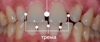
Treatment of lateral position of teeth . The most typical clinical sign of such an anomaly is the appearance of a gap between the central incisors - diastema .
The following types of diastema are distinguished: 1) symmetrical diastema, in which there is a lateral displacement of the central incisors; 2) diastema with preferential movement of the crowns of the central teeth in the lateral direction from the midline. The roots of the central incisors retain their position or move slightly in the lateral direction; 3) diastema, in which the crowns of the central teeth have shifted slightly in the lateral direction from the midline, and the roots of the central incisors have shifted significantly; 4) asymmetrical diastema, which occurs when one central incisor has moved significantly in the lateral direction, while the other central incisor has maintained its normal position.
It should be noted that the lateral displacement of the central incisors can be combined with their rotation along the axis of the tooth (tortoanomaly) and vertical displacement of the teeth (dental alveolar lengthening or shortening).
Treatment depends on the clinical picture and causes of the anomaly. If there is a supernumerary tooth germ between the roots of the central incisors, it should be removed. In case of microdentia of the central incisors, the diastema is eliminated only by prosthetics of the central incisors with solid cast or metal-ceramic structures. Such prosthetics are performed in adolescents after 14-15 years of age. In case of diastema caused by microdentia of the lateral incisors, the diastema should be eliminated, and then prosthetics of the lateral incisors should be performed with artificial crowns.
If the upper jaw develops excessively in the anterior region and a diastema occurs, one should try to delay the growth of the upper jaw using a plate with a loop for the treatment of diastema and a vestibular arch. At the same time, the loop and U-shaped bends of the vestibular arch are activated. The canine is removed and installed in place of the missing lateral incisor or moved distally. In the first option, this can be done when the root of the canine is located significantly ahead of its proper place in case of normal eruption. If the mesiodistal size of the canine allows filling the gap formed behind the central incisor, then the cusp of the canine crown can be ground down and given the shape of a lateral incisor. Moving the canine mesially is only possible if the antagonist teeth allow the canine to create normal occlusion with them; otherwise, contact with antagonist teeth (regardless of retention) will result in lateral movement of the canine.
When distal movement of the canine occurs, the gap formed in the area of the missing lateral incisor is eliminated by prosthetics. To do this, you can make a metal-ceramic structure with support on the canine and select the central incisor as the second point of support by making a cusp located on the palatal surface
If the diastema has developed due to the low attachment of the frenulum of the upper lip, they resort to plastic surgery of the low-attached frenulum. Surgical treatment should begin after the eruption of not only the central incisors, but also the lateral ones, i.e. at the age of 8-9 years. There are cases when, after the eruption of the lateral incisors, the diastema disappears on its own.
If there is a diastema caused by bad habits, it is necessary to wean children from them, and hypnosis therapy is also effective.
With a diastema formed as a result of the abnormal position of the primordia of the incisors and canines, the eruption of not only the incisors, but also the canines is required, after which the diastema may self-remove.
Treatment of symmetrical diastema is carried out with orthodontic appliances, taking into account the size of the gap between the incisors. If the diastema is 3 mm or less, a plate on the upper jaw with a loop for treating diastema or with arm-shaped springs can be used. Activation of the loop is carried out 2 times a week by squeezing the loop with crampon tongs or pliers. You can also use a plate on the upper jaw with two arm-shaped springs covering the incisors from the lateral side, and hooks open to the rear, between which a rubber ring is placed. To prevent rotation of the incisors as they move toward the midline, bend the wire along the palatal surface of the incisors.
When a diastema is combined with deep incisal occlusion or disocclusion, it is necessary to make a bite pad on top of the loop. When treating more pronounced diastema, devices are used that would facilitate the body movement of the incisors and would prevent their rotation during movement. To do this, orthodontic crowns (rings) are used on the incisors with rods soldered to their vestibular surface with hooks open to the rear, between which a rubber ring is placed. To prevent rotation of the incisors when moving them, you can solder a horizontal tube to the ring of one of the teeth, and a wire to the other, one end of which will be soldered horizontally to the crown on the vestibular side, and the other should fit into the tube. Thus, the problem of rotation is removed and tension is created for the teeth to move.
When treating diastema with predominant movement of the crowns of the central incisors, the main load of the orthodontic apparatus should be in the area of the coronal part of the incisors. To do this, use a plate on the upper jaw with a loop for the treatment of diastema, arm-shaped springs with hooks open to the back, with a rubber rod placed between them. You can make orthodontic crowns or rings for the central incisors, solder vertically directed rods with hooks open to the rear to them, and put a rubber rod between them.
In case of diastema, when the crowns of the central incisors have shifted slightly in the lateral direction from the midline, and their roots are more significant, it is necessary to create conditions for a more significant movement of the root part of the teeth compared to their crown part. In these cases, a rotational moment is created between the crown and root parts of the tooth for the correct vertical position of the incisors, and only then the diastema is eliminated. For this purpose, crowns or rings are made for the central incisors, and rods are soldered vertically on the vestibular side. The upper end of the rod should be extended and end with a hook, open back at the level of 2 tooth roots or K from the top of the tooth root. Then a stable Angle arch is applied to the dentition, to which a hook open to the rear is soldered in the canine area on the opposite side of the dentition. When an oblique rubber rod is applied, the tooth root experiences a load in the mesial direction, but the tooth does not rotate, since there is no second rod in the opposite direction. To do this, the lower hook from the bar is open forward, from it a rubber rod will go to the hook, open back, which is soldered to the Angle arch in the canine area on the same side of the dentition.
Instead of an arch, a plate on the upper jaw with Adams clasps on the first molars and button clasps located between the first and second premolar on both sides of the dentition can be used as a support. The ideal technique to correct this anomaly is braces.
When treating an asymmetrical diastema, which occurs when one central incisor is displaced laterally, only this tooth should be treated. The choice of orthodontic technique depends on the position of the central incisor, which can be different: parallel with an offset from the midline, when the root and crown of the tooth are displaced the same distance from the midline; the crown of the tooth is displaced more significantly than its root, the root of the tooth - more significantly than its crown. Lateral displacement of the central incisor can be combined with its tortoanomaly, as well as with dentoalveolar lengthening or shortening. With this form of diastema, the central incisor, which is normally located, can serve as a fulcrum when moving the abnormal incisor. To eliminate an asymmetrical diastema, a plate can be made for the upper jaw with a hand-shaped spring covering the moving incisor from the distal side. As a support, Adams clasps are used on the first molars, button clasps and a round clasp on the central incisor, located correctly. You can make an arm-shaped spring with hooks open back, and put a rubber rod between it and the second hook located on a round clasp and also open back.
For a more pronounced diastema, a crown or ring is made for the tooth being moved with a guide tube, as described above.
Very often, diastema is accompanied by protrusion of the upper front teeth. In this case, along with treatment of the diastema, the anterior portion of the upper dentition should be flattened. For this purpose, it is more correct to make a plate for the upper jaw with arm-shaped springs on 1|1 to correct the diastema and a vestibular blowout with U-shaped bends with a vinyl chloride coating.
In recent years, orthodontic devices - positioners - have been used to eliminate diastema in dental practice.
Treatment of vestibular position of teeth . Permanent teeth with formed roots are moved from the vestibular position with an Angle arch, and depending on the combination with anomalies in the size and shape of the dentition, both a stationary and a sliding arch are used. Since the bracket system is universal, it is intended to use its design features to normalize the position of permanent teeth in the vestibular position. At the appropriate stage of formation of the roots and periodontium of permanent teeth, it is possible to use a positioner.
Normalization of the position of the anterior teeth located vestibularly is carried out, as is the normalization of the position of the lateral teeth. However, the morphological, functional and topographical features of the anterior teeth determine the possibility of using devices of specific designs and different combinations of their structural elements. Thus, vestibular retracting arches are widely used in children with baby teeth and during their replacement period. Naturally, the design of the device is determined by a complex of clinical manifestations.
One of the features of normalization of labially located upper teeth is also the use of a face bow. It should be said that the use of positioners to eliminate the labial position of the anterior teeth is more effective than when moving other teeth.
Treatment of the vestibular (labial) position of the lower front teeth is carried out with a retracting arch with vinyl chloride coating in the presence of three and diastema between the teeth.
If there is protrusion of the lower front teeth and the absence of three and diastema between them, one should take the path of removing complete teeth (usually the first premolars). The choice of treatment method depends on the size of the teeth and the type of closure of the first molars and canines. The canine often occupies a vestibular position, which is called dystopia, and it is necessary to determine whether there is a place for it in the dentition. Canine dystopia can occur as a result of disturbances in the eruption of teeth and the sequence of their eruption. Thus, very often after the eruption of the first premolar of the upper jaw, the eruption of the second premolar, and not the canine, follows. In this regard, and taking into account the mesial position of the teeth when they erupt, the canine has no place in the dentition and it erupts either in the vestibular or oral direction.
Dystopia of the canine occurs with macrodentia of the upper front teeth, which take the place of the canine. It can also occur in the presence of supernumerary teeth, narrowing of the dentition, early removal of the primary canine (in this case, mesial displacement of the lateral teeth occurs). Clinically, the mesial shift of the lateral teeth can be determined by the closure of these teeth with the antagonist teeth. On this side of the dentition, the closure of the lateral teeth occurs according to Engle’s class II, and on the opposite side - according to class I.
In case of canine dystopia, it is necessary to find out whether there is a place for it in the dentition. If there is one, then there is only one task: to place the canine in the dentition. To do this, you can use a plate on the upper jaw with a vestibular arch and an M-shaped bend on the canine. When the M-shaped bend is activated (plastic is first cut out from under the canine on the palatal side), the canine experiences increased load and moves in the oral direction.
Teeth are moved from the vestibular position using a rubber rod and springs, arches, even screws. Moving with a screw involves placing it in activated form on a plate with a sectoral cut, which has clasps or a multi-link clasp on the moving teeth, as well as additional Adams or round support clasps on the opposite side. By activating the screw, i.e. returning it to its original position, the necessary movement of the teeth is achieved. When moving teeth using a rubber rod, a ring or crown with a hook, or a bracket is fixed on the tooth, which is the point of application of force, and the fulcrum is the hook in the base of the device.
If there is dystopia of the canine and there is no place for it in the dentition, a place should be created for it. If there is no space for a canine due to mesial displacement of the lateral teeth, they should be moved distally. Distal movement of teeth is possible in the absence of a wisdom tooth germ. For distal movement of teeth, a plate apparatus with a sectoral cut, a face bow, a Kalamkarov apparatus, and arm-shaped springs are used.
If there is a rudiment of a wisdom tooth, macrodentia of teeth, one should follow the path of removing a complete tooth in order to create a place for a canine. Most often, for orthodontic reasons, the first premolar is removed; in the presence of caries and destruction of the crown of the tooth, the second premolar and even the first molar can be removed. When removing a tooth, attention should be paid to the passage of the midline between the incisors, and the choice of the tooth to be removed should be such as not to aggravate the asymmetry of the position of the incisors of the upper and lower jaws.
Treatment of oral position of teeth should include normalization of the position of the tooth and its placement in the dentition. It is necessary to find out whether there is room for this tooth. If there is space, the tooth or group of teeth is moved using orthodontic appliances.
If the upper anterior teeth are in a palatal position, a plate is made for the upper jaw with a sectoral cut or protracting springs. A stable Angle arch can be made and by activating the ligatures or nuts the teeth will be moved in the labial direction. When the upper incisors are in a palatal position, Bynin and Schwartz mouthguards and a Reichenbach-Brückle plate with an inclined plane are used. The use of a positioner with a preliminary setup system is also shown.
In case of crowded position of the lower front teeth and their lingual position, which arose as a result of macrodentia, it is advisable to take the path of removing complete teeth. First you should pay attention to the passage of the midline. The tooth to be removed can be a central or lateral incisor, as well as a first or second premolar. It all depends on the lack of space in the dentition and the location of the lower incisors in relation to the midline. If the space deficit is greater than the size of the incisor, and the midline is not displaced, then the abnormally located tooth is removed. If the midline is displaced to one side or the other, then the tooth on the opposite side of the midline displacement is removed.
The question of removing the first or second premolars is decided depending on the lack of space, taking into account the violation of the closure of the lateral teeth.
It must be remembered that the removal of any incisor on the lower jaw leads to aggravation of the depth of the incisal overlap.
When the upper or lower teeth are in an oral position, the closure of the dentition is disrupted. Thus, with a palatal inclination of the upper anterior teeth, a deep incisal occlusion is formed. This is typical for class II of the 2nd subclass of Angle. Otherwise, it is distal occlusion of the dentition in combination with palatal inclination of the upper incisors. With a significant palatal position of the upper incisors, reverse incisal occlusion, or disocclusion, is formed.
In this case, it is necessary to take into account the separation of the dentition in order to eliminate blocking of the upper and lower incisors. For this purpose, plate devices are made with occlusal linings in the lateral areas of the dentition. To eliminate the pressure of the orbicularis oris muscle on the upper incisors, it is necessary to make a labial plastic bandage. You can separate the dentition using mouthguards or orthodontic crowns.
If the upper lateral teeth are in a palatal position, it is advisable to use a plate on the upper jaw with a sectoral cut and occlusal overlays on the opposite side of the dentition. When a palatal position of the upper incisors is combined with a mesial position of the lateral teeth, it is necessary to either move the lateral teeth distally or remove complete teeth (usually the first premolar - one or both sides). Thus, a place is created in the dentition for the frontal teeth, after which they are moved in the labial direction.
Very good results are achieved when treating crowded lower anterior teeth with a lip bumper. This device allows you to change the myodynamic balance between the orbicularis oris muscle and the tongue muscles. Treatment of anomalies in the vertical position of teeth involves reducing or increasing the dento-alveolar height in the corresponding section. Reducing the dento-alveolar height is achieved by creating vertical loads on the corresponding teeth to induce the process of bone resorption.
Dental alveolar elongation in the area of one tooth or a group of teeth may be associated with the absence of antagonist teeth or the presence of bad habits. Dentoalveolar elongation of the lateral teeth of the upper jaw is often observed, which leads to vertical incisal disocclusion. Dentoalveolar elongation of the lower anterior teeth leads to deep incisal disocclusion or occlusion. When dentoalveolar lengthening of the lateral teeth occurs, they should be introduced.
Treatment is carried out with a plate on the lower jaw with occlusal pads, and dentoalveolar lengthening of the lower anterior teeth is carried out with a plate on the upper jaw with a bite pad. An Andresen-Goipl monoblock and positioner are used.
When dentoalveolar lengthening of one tooth is carried out, it is implanted and then an apparatus is necessarily made for the opposite dentition with an artificial antagonist tooth.
When the tooth is suprapositioned, another task is to increase the dentoalveolar height in the corresponding section as a result of bone building. This is achieved by physiological irritation by applying a rubber ring and creating traction that transfers the load through the periodontium to the bone structures. The point of application of force is a hook on a ring fixed to the tooth being moved (crowns or braces are possible), the fulcrum is a hook on a mouthguard blocking antagonist teeth, or a hook in the design of an apparatus used in complex treatment. At the end of the change of teeth and after it, you can use a bracket system, as well as a stationary Angle arch. It should be noted that after eliminating such an anomaly, a long retention period is usually required.
Treatment of tortoanomalies involves creating a pair of forces directed in the directions opposite to the rotation of the tooth. This is achieved by creating two points of force application on the crown of the tooth being moved. The points of application of force can be hooks on rings, crowns or braces, and the points of support can be hooks on aligners blocking groups of teeth, or fixed in basic appliances. When elastic rings are applied, a pair of multidirectional forces is created, which leads to normalization of the tooth position. It is extremely important to maintain constant optimal traction. Tortoanomaly is also eliminated with the help of positioners.
At the end of the change of teeth and after it, the tortoanomaly can be eliminated using a brace system or an Angle arc, if there are other indications for their use. Treatment of tooth transposition. If such an anomaly is present in the area of the front teeth, the cosmetic and functional effect is often achieved by grinding (for example, when there is a canine in place of the incisor). Depending on the combination of clinical factors, it may be preferable to restore the optimal shape of the tooth using an orthopedic crown. In the area of the lateral teeth, grinding is usually sufficient.
Problems arise when there is transposition of the teeth and these teeth are abnormally positioned. For example, in the place of the canine there is a first premolar, the canine is vestibular at the level of the first premolar, and in the dentition there is a second premolar (in place of the first premolar), then the first and second molars. If there is a rudiment of a wisdom tooth, it is necessary to remove the vestibular canine. In the absence of a wisdom tooth rudiment, distal displacement of premolars and molars and movement of the canine in the dentition to its place are possible.
Distal movement of teeth is carried out using a plate with a sectoral cut, arm-shaped springs, a Kalamkarov apparatus, a face bow, and a positioner. It should be noted that dental anomalies lead to anomalies of the dentition and anomalies of occlusion.
Occlusion disorders: types, classification and treatment options
An incorrect bite is not as harmless as it may seem to an ignorant person, who considers the gap between the teeth to be simply an individual feature. Occlusion that is far from normal affects the beauty of the smile, disrupts the harmony of the face, and increases the load on the temporomandibular joints, which ultimately leads to health problems. This means that defects must be corrected as early as possible with orthodontic treatment.
The type of malocclusion depends on how the dentitions close; all anomalies are considered in three directions - sagittal, vertical and transversal.
Crossbite
Characterized by atypical (reverse) closure of the anterior or lateral or both the first and second teeth together with simultaneous deviation of closure in the left or right area of the bite. A double form of this bite is possible.
If professional treatment and care are not provided, patients with transverse bite experience facial distortions and serious changes in appearance, as the jaw bones develop asymmetrically. Characterized by chewing dysfunction and difficulties with pronunciation. Often patients with this bite suffer from biting the mucous membranes of the cheeks, tongue, and lips.