Dear friends, last time we talked about what wisdom teeth are like, when they need to be removed and when not. And today I will tell you in detail and in every detail how the removal of “sentenced” teeth actually takes place. With pictures. Therefore, I recommend that especially impressionable people and pregnant women press the “Ctrl +” key combination. Joke.
Where does the removal of the 8th, and, in principle, any other tooth begin?
With anesthesia.
So:
Anesthesia (pain relief)
To minimize discomfort during an injection, you need to treat the injection site with a special anesthetic gel. This is the so-called topical anesthesia. It is very often used in pediatric dentistry, but we also use it in working with adults. As practice shows, there are fewer unpleasant sensations, and the taste is pleasant... at least some kind of joy.
When removing teeth from the upper jaw, as a rule, a simple infiltration of anesthetic into the area of the tooth being removed is sufficient. It is carried out using a special syringe with specially selected anesthetics and is called infiltration .
When removing teeth on the lower jaw, infiltration anesthesia is usually not enough (with the exception of the frontal group of teeth, from canine to canine). Therefore, the anesthesia technique changes somewhat - the anesthetic, using a long but very thin needle, is applied directly to the nerve bundle responsible for the innervation of the desired areas. This anesthesia allows you to “turn off” sensitivity not only in the area of the tooth being removed, but also in the lip, chin, part of the tongue, etc.
It should be noted that during and immediately after anesthesia, a number of interesting phenomena may be observed - increased heart rate, trembling of limbs, an inexplicable feeling of anxiety. Many patients begin to panic about this. But there is no need to panic! These are side effects of most modern anesthetics and go away on their own within 10-15 minutes.
Well, the anesthesia is done! Now you need to make sure that it was carried out successfully?
Those places that should ideally be numb are listed above. Also, using a special instrument and pressing on the gum in the area of the operated tooth, we determine whether the pain still remains or no longer exists. The only thing that should be felt is the sensation of “something” touching the gum. That is, tactile sensations are still preserved, but pain is no longer present.
And then our actions differ depending on what type of wisdom tooth we are dealing with.
Exostosis, fistula or beneficial plaque: types of growths after removal
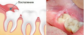
Tooth extraction is always traumatic. The doctor extracts the tooth, damaging soft and bone tissues and breaking blood vessels. After the tooth is extracted, the wound fills with blood, which coagulates into a clot. The naturally formed blood clot closes the hole, protecting it from bacteria and stimulating regeneration processes.
With the correct course of events, the hole gradually overgrows, closes, and ceases to disturb the patient. But sometimes a formation forms at the site of removal - on the hole itself or nearby. Possible options:
- white dense coating;
- cyst;
- fistula;
- exostosis.
Each formation has its own nature and reasons for its appearance. Some require treatment, others are beneficial and go away on their own.
White plaque or growth after tooth extraction
During the first hours after removal, bleeding stops in the hole and a dark blood clot appears. After a couple of days, a white coating is noticeable on the clot. Externally similar to purulent discharge, it frightens many patients. But this is the next stage of healing - fibrin protein fibers.
Fibrin performs several functions at once:
- accelerates tissue regeneration, including bone;
- helps reduce the size of the hole;
- protects the wound from bacteria and pathogenic microflora;
- helps the blood clot dissolve.
The plaque often acquires a dense, hard structure, and patients mistake the fibrinous film for a growth. White growth on the tooth socket after extraction is a natural healing process and should not be a cause for concern.
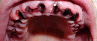
Cyst or fistula after tooth extraction
The cyst and fistula only indirectly resemble a white growth. But often patients describe the pathological condition this way. The reason is the filling of the formation with liquid, pus, which gives the lump a whitish tint.
A fistula is a channel through which purulent contents are removed from the hole. It is located near the site of the extracted tooth. The tissues around the formation are inflamed.
A cyst is a cavity or lump that occurs when tissue is damaged during surgery. It has walls and contents, and stands out strongly above the surface of the gum. Causes pain.
Both a cyst and a fistula are pathological formations that require removal. Externally, patients define them as growths in the area of tooth extraction.
If there are no visible manifestations, indirect signs of ill health will be:
- a feeling of heaviness and pressure in the area of the hole;
- soft tissue swelling;
- jaw pain;
- heat;
- discomfort when eating;
- smell from the mouth;
- burning gums;
- individual signs of intoxication of the body.
If you have unpleasant symptoms, you should consult a doctor to determine the type of pathology and its treatment. The main methods are drug therapy or surgical removal of the formation. The dentist chooses the method after assessing the clinical situation.
Exostosis - bone growth after tooth extraction
Exostosis - bumps, spines, growths on the gums that arise due to hereditary factors or after complex tooth extraction. The cause of protruding ridges is improperly fused bone, the consequences of displacement of bone tissue due to careless work of the surgeon.
The pathology looks unaesthetic, but does not cause much discomfort. If the formation is significant in size, it can put pressure on the bone and nearby teeth and become a contraindication for dental implantation.
Removal of exostosis is carried out surgically. After anesthesia, the doctor makes an incision into the soft tissue, saws off the growth, straightens the bone, and applies sutures. Healing occurs quickly and the formation does not reappear.
Other causes of gum growth
Lumps and growths on the gums appear after removal and for other reasons:
- infectious processes that began due to non-compliance with the surgeon’s recommendations after removal;
- allergic reaction to anesthetic drugs;
- mechanical damage - due to careless removal.
Sometimes a growth occurs at the site where the anesthetic injection was given. Such a hematoma resolves on its own in 2-3 days.
Other lumps require consultation with a dentist.
Impacted wisdom tooth
These are usually the hardest eights to remove out of all the others. We have already numbed the surgical field. What's next?
It's under the gum! So, we take a scalpel in our hands and make a delicate incision in the area of the tooth being removed. This creates access to the wisdom tooth being removed. It is isolated from the surrounding tissues using special instruments, and now we can visually assess its position and choose a removal technique.
If the tooth does not erupt, it means that something is preventing it. This “something” will also interfere with its removal, and this “something” could be a neighboring tooth, a bony protrusion, etc. However, you won’t also remove the seven to get to the wisdom tooth, right?
Special surgical tip for removing wisdom teeth. Rotates at the right frequency, provides the right torque, does not burn tissue or inflate emphysema. In surgery, we use only such devices.
Therefore, we divide the tooth into parts. Using a special tip with a cutter speed of 150,000 rpm - this is no longer a simple angle cutter, but not yet a turbine cutter. The latter, by the way, is highly undesirable to use for removing teeth, because at 500,000 rpm it is easy to burn everything with a hellish flame, and with air from the cooling nozzle you can also inflate emphysema over half your face. In general, for removal you need to choose the right tools; there are no trifles or compromises here and cannot be. And you should think a hundred times before removing such problematic teeth in a one-chair dental office at a rural club on the “Half-Empty Bins” collective farm.
Impacted teeth are removed mainly with an elevator, and not with forceps, as many are accustomed to thinking
So, we divide the tooth into 2-3 parts in order to remove it carefully and with little trauma to the surrounding tissues. And teeth are usually removed using an “elevator” (in the picture on the left). Forceps, which everyone associates with removal, are actually used extremely rarely.
Well, the tooth has been removed. Next, we clean the tooth socket from “sawdust” and small tooth fragments that might remain. Using a curette.
When removing wisdom teeth, no biomaterials are used; the hole is filled with a blood clot on its own, this is quite enough for normal healing.
Moreover, “pushing” biomaterials into the hole can complicate the healing process, so let the regeneration process take place naturally and simply, and not fancy, as some doctors suggest.
After removal, resorbable (absorbable) sutures are placed on the hole; most often they do not need to be removed.
The clot is in place. Next, we bring the edges of the wound together and put stitches so that food doesn’t get stuck in the wound, it doesn’t bleed too much, and it heals faster. But at the same time, the sutures should not be tight, because the wound may bleed significantly during the first 24 hours. And if you don’t create an outflow, edema often develops.
How to speed up the healing of a hole
To speed up the healing of the hole after tooth extraction, you need to follow these rules:
- Do not touch the blood clot on the socket with your tongue, much less with your hands or a toothpick, so as not to damage it. The presence of a clot is a guarantee of speedy healing of the wound.
- For three hours after visiting the dentist, you should not eat or rinse your mouth. Compliance with this point will also help maintain the integrity of the blood clot.
- · Drinks and food should not be too hot or cold for several days after removal. The food chosen is soft, without coarse inclusions, to prevent injury to the gums.
- You should not engage in heavy physical work for several days to avoid opening the wound and resuming bleeding.
- Since tobacco smoke and alcohol irritate the mucous membrane and inhibit the healing of the hole, you need to stop smoking and drinking alcohol for a while.
- To prevent infection from entering the wound and reduce inflammation after a traumatic tooth extraction, antiseptic baths are used. For them, solutions of chlorhexidine or furatsilin, infusions of sage, chamomile, and eucalyptus are used. The solutions are taken into the mouth, held for several minutes and carefully spat out.
- On the third day after removal, you can carry out antiseptic rinses with the same agents.
- Taking anti-inflammatory drugs (nimesulide, ibuprofen) will help not only eliminate pain, but also relieve the inflammatory process, which also helps accelerate socket regeneration.
- If the doctor has prescribed antibiotics, you should not refuse them, since eliminating the microbial infection shortens the healing time.
- When brushing your teeth, be careful not to touch the wound with the brush.
- To speed up healing, you can use Solcoseryl dental paste. It enhances intracellular energy exchange, due to which cell regeneration and restoration of damaged tissues are accelerated. Solcoseryl also creates conditions for the growth of granulation tissue. The paste is applied to a previously dried surface, then moistened with water. Before using the medicine, it would be more correct to ask your dentist about when to start using Solcoseryl after tooth extraction and how many times a day to apply it to the gums.
Semi-retinated tooth
In principle, the method of removing such a tooth is no different from removing a completely impacted tooth. But, as a rule, it is a little easier, because the tooth is not so deep. The main stages are essentially the same: anesthesia, creating access to the tooth (and sometimes you can do without incisions), fragmentation (dividing the tooth into parts) and, in fact, removing the teeth in parts.
After removing the lower semi-impacted tooth, sutures are placed on the socket; in the area of the upper wisdom teeth, sutures are not necessary.
Healing of the socket with gum inflammation
If tooth extraction was carried out against the background of inflammation, or the inflammatory process in the gums developed later, epithelization of the wound begins on the 10th -14th day, bone beams appear only by the 15th day. A significant part of the socket is filled with young osteoid tissue only by the end of the second month.
After a complex tooth extraction, when the gums rupture and the walls of the socket are traumatized, the edges of the gums cannot come together for a long time and the epithelization process slows down. Wound healing can only be completed after 1-1.5 months. In this case, the development of new bone tissue is delayed.
Dystopic tooth
Removing such teeth can be called a simpler case compared to others, but only if the tooth has one straight root. Then the removal can take place quite quickly. But such clinical cases are extremely rare. And, looking at the picture, we see hooks, not roots, which, with proper pressure, can simply break. There are usually 2 roots, and in this case we just need to separate one root from the other using the same tool - the “raising” tip. And carefully remove each of the roots separately. The beginning and completion of the removal of such teeth is the same as for all others.
How long does it take to remove a front tooth?
The time it takes to remove the upper teeth can vary from a few minutes to an hour. On average, the operation takes 15-20 minutes. Much here depends on the initial situation and the qualifications of the dentist.
Removing front teeth is usually simple. Some difficulties may arise when extracting the upper canine, since it has a long, massive root with an often curved apex.
During surgery to remove a front tooth, the patient sits in a chair with a slightly reclined back and headrest. The doctor is in front of the patient on the right. The procedure looks like this:
- the gum is separated from the edge of the alveolus, and the circular ligament is separated from the neck;
- the forceps are applied to the tooth, moved under the gum and fixed;
- the dentist rocks or rotates the tooth to break the fibers connecting the root to the walls of the socket;
- the front tooth is removed from the socket;
- The surgical site is inspected and broken root fragments are removed from the socket.
And it also happens...
... that wisdom teeth block the neighboring teeth and prevent them from erupting normally. In such cases, patients are referred to a surgeon by an orthodontist.
Of course, the germ of the eighth tooth needs to be removed. This is a fairly simple and relatively comfortable operation.
Look at the pictures on the right. There is a difference of three weeks between the top and bottom. It is clearly visible from them that after removing the rudiments of the eights and “unblocking”, the seventh teeth immediately began to grow.
Wisdom tooth removed. The patient is satisfied. But the fun is yet to come. Namely, the postoperative period.
The main stages of socket healing after wisdom tooth removal
Conventionally, the regeneration reactions after extraction of the “eight” can be divided into stages. Many of them remain invisible to humans, as they relate to changes occurring in the deep tissues of the gums.
- Stop bleeding. Normally, blood stops flowing within two (maximum four) hours after extraction. On the first day, the patient may still detect a small amount of bloody impurities in the saliva. There's nothing wrong with that.
- Formation of a clot. Occurs on the first day after surgery. Usually at the time of its appearance there is an increase in pain in the wound area and increased swelling. Body temperature may rise slightly. Headaches are also possible. These negative symptoms disappear in one to two days. They usually do not require additional treatment.
- Formation of a thin epithelial layer on top of a blood clot. This stage occurs on the second to fourth days after dental surgery. Then the person sees that the hole has turned white or gray. The state of health at this point usually becomes satisfactory. Toothache no longer bothers me.
- On the fifth to eighth day, the area of gray connective tissue increases rapidly . The clot is visible only in its center. The swelling goes away. If stitches were placed after removal, now is the time to remove them.
- The surface of the wound is covered with granulation tissue. This occurs two to three weeks after removal. As a result, the gums begin to look normal.
- In the period from the third or fourth week to six months, bone tissue grows in the socket.
If the removal was carried out against the background of progressive periodontal disease or periodontitis, then the patient’s recovery period increases by about ten days.
Thus, a month after extraction, a person can chew food on the affected side without restrictions . Nothing will remind him of the previous dental intervention. Since the “eight” is practically not involved in the process of grinding food (that is how far it is located in the dentition) and does not change the appearance of the smile, there is no need for prosthetics in the vacated area.