Naturally occurring mineral form of calcium apatite
| Hydroxyapatite | |
| Hydroxyapatite crystals on a matrix | |
| General | |
| Category | Phosphate mineral Apatite group |
| Formula (repeating block) | Ca5(PO4)3(OH) |
| Strunz classification | 8.BN.05 |
| Crystal system | Hexagonal |
| Crystal class | Dipyramidal (6/m) HM Symbol (6/m) |
| Space group | p 63/ m |
| Cell | a = 9.41 Å, c = 6.88 Å; Z=2 |
| Identification | |
| Mass formula | 502.31 g/mol |
| Color | Colorless, white, gray, yellow, yellowish green |
| Crystal Habit | In the form of lamellar crystals and stalagmites, concretions, from crystalline to massive crusts. |
| Split | Bad on {0001} and {1010} |
| Fracture | Conchoidal |
| Perseverance | Fragile |
| Mohs hardness scale | 5 |
| Shine | Glassy to almost resinous, earthy |
| Band | white |
| Transparency | Transparent to translucent |
| Specific gravity | 3.14–3.21 (measured), 3.16 (calculated) |
| Optical properties | Single axis (-) |
| Refractive index | pω = 1.651 pε = 1.644 |
| Birefringence | δ = 0.007 |
| Recommendations | [1][2][3] |
Hydroxyapatite Needle-shaped crystals of hydroxyapatite on stainless steel.
Scanning electron microscope picture from the University of Tartu. Nanoscale Ca-HAp coating, image taken with a scanning probe microscope. Three-dimensional visualization of half of a hydroxyapatite unit cell from X-ray crystallography. Hydroxyapatite
, also called
hydroxyapatite
(
HA
), is a naturally occurring mineral form of calcium apatite with the formula Ca5(PO4)3(OH), but is usually written Ca10(PO4)6(OH)2 to indicate that the crystalline block cell is composed of two entities.[ 4] Hydroxyapatite is the hydroxyl end member of the apatite group complex. The OH ion can be replaced by fluoride, chloride, or carbonate, producing fluorapatite or chlorapatite. It crystallizes into the hexagonal crystal system. Pure hydroxyapatite powder is white in color. However, naturally occurring apatites can also have a brown, yellow or green coloration, comparable to the discoloration of dental fluorosis.
Up to 50% by volume and up to 70% by weight, human bone is a modified form of hydroxyapatite, known as bone mineral.[5] Calcium-deficient carbonated hydroxyapatite is the main mineral that tooth enamel and dentin are composed of. Hydroxyapatite crystals also occur in small calcifications within the pineal gland and other structures, known as corpus arenacea or "brain sand".[6]
Calcium-deficient hydroxyapatite
Calcium-deficient (non-stoichiometric) hydroxyapatite, Ca10− X
(PO4)6−
X
(HPO4)
X
(OH)2−
X
(where
X
is between 0 and 1) has a Ca/P ratio of 1.67 to 1.5.
The Ca/P ratio is often used when discussing calcium phosphate phases.[9] Stoichiometric apatite Ca10(PO4)6(OH)2 has a Ca/P ratio of 10:6, usually expressed as 1.67. Non-stoichiometric phases have a hydroxyapatite structure with cation vacancies (Ca2+) and anion (OH−) vacancies. In stoichiometric hydroxyapatite, the sites occupied exclusively by phosphate anions are occupied by phosphate or hydrogen phosphate, HPO42−, anions.[9] The preparation of these calcium-deficient phases can be obtained by precipitation from a mixture of calcium nitrate and diammonium phosphate with the desired Ca/P ratio, for example to obtain a sample with a Ca/P ratio of 1.6:[10] 9.6 Ca(HET3)2 + 6 (NH4)2HPO4 → Ca9.6(PO4)5.6(HPO4)0.4(OH)1.6
When these non-stoichiometric phases are sintered, a solid phase is formed, which is a homogeneous mixture of tricalcium phosphate and hydroxyapatite, called two-phase calcium phosphate:[11]
Ca10−
X
(PO4)6−
X
(HPO4)
X
(OH)2−
X
→ (1 −
X
) Ca10(PO4)6(OH)2 + 3
X
Ca3(PO4)2
CONTENT
- 1 Chemical synthesis
- 2 hydroxyapatite with calcium deficiency
- 3 Biological function 3.1 Mantis shrimp
- 3.2 Mammal/primate/human
- 3.3 Hydroxyapatite in the remineralization of tooth enamel
- 4.1 Dentin sensitivity
Biological function
Mantis shrimp
Club appendages Odontodactylus scyllarus
(Peacock Mantis Shrimp) are made from an extremely dense form of the mineral that has a higher specific strength; this led to its investigation for possible synthesis and engineering uses.[12] Their dactyl appendages have excellent impact resistance due to the fact that the impact area is composed primarily of crystalline hydroxyapatite, which has significant hardness. A periodic layer beneath the impact layer, consisting of hydroxyapatite with lower calcium and phosphorus content (resulting in a much lower elastic modulus), inhibits crack growth by causing new cracks to change direction. This periodic layer also reduces the energy transferred through both layers due to the large difference in moduli, even reflecting some of the incident energy.[13]
Mammal/Primate/Human
Hydroxyapatite is present in bone and teeth; bone consists primarily of HA crystals interspersed with collagen matrix—65 to 70% of bone mass is HA. Similarly, HA accounts for 70 to 80% of the mass of dentin and enamel in teeth. In enamel, the HA matrix is formed by amelogenins and enamel instead of collagen.[14]
Hydroxyapatite deposits in the tendons around the joints lead to the disease. calcific tendinitis.[15]
Procedure for injecting filler
In modern cosmetology, the use of fillers is one of the safest, most effective and fastest methods for solving aesthetic and medical problems. Contour plastic surgery using these drugs can only be performed by a specialist who has been trained in a special program and has a certificate giving him the right to use this product in practice. These are the ones who work at the VIRTUSCLINIC Center for Aesthetic Cosmetology.
On average, the procedure lasts 30 minutes and is performed under local anesthesia. The doctor cleans the facial surface and determines the areas where the drug will be administered, leaving markings there in the form of dots. Some specialists take photos before and after the procedure.
The cosmetologist demonstrates the drug to the patient in its entire packaging, with the obligatory indication of its expiration date and dosage. The filler is injected using a very thin needle subcutaneously in the previously designated area. The specialist chooses the method and methods of administering the drug independently, taking into account all the features of the procedure and the patient.
The session ends with a light massage, which helps to evenly distribute the filler under the skin.
The effect of the procedure is noticeable immediately after it is performed, but the maximum result usually appears after 10–14 days.
Uses
Cosmetic
Hydroxyapatite is added to some types of cornstarch. baby powder such as aloe and vitamin E powder from Johnson's.[16] According to the website, the mineral is added as an emollient to "hydrate and soften the skin."[17]
Medical
A flexible hydrogel-HA composite in which the ratio of mineral and organic substances in the matrix approaches that of human bone.
HA is increasingly used for bone grafting materials, as well as prosthetics and dental repairs. Some implants, such as hip replacements, dental implants, and bone conduction implants, are coated with HA.[14] Since the natural dissolution rate of hydroxyapatite in vivo, about 10 wt.% per year, is significantly lower than the growth rate of newly formed bone tissue, when used as a bone replacement material, ways are being sought to increase its solubility rate and thus promote better biological activity.[18]
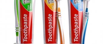
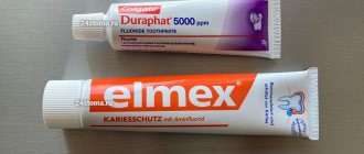
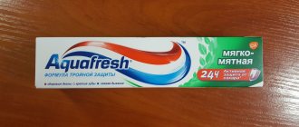
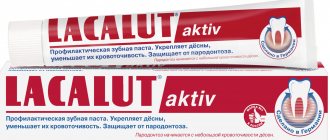
Hydroxyapatite is added to special toothpastes as an additive to prevent caries and reduce tooth sensitivity.[19]
Additive
Hydroxyapatite overgrows biomaterial
Microcrystalline hydroxyapatite (MCHA) is marketed as a "bone-building" supplement with superior absorption compared to calcium.[20]
It is a second generation calcium supplement derived from bovine bone.[20] In the 1980s, bone meal calcium supplements were found to contain heavy metals,[20] and although manufacturers claim their MCHA is free of contaminants, it is not recommended as its effects on the body have not been well tested.[20 ]
Advantages
Calcium hydroxyapatite fillers have many wonderful properties. Among them are the following:
- naturalness. The homogeneity and homogeneity of the drug, combined with the high professional qualities of the doctor, allows the patient to look as natural as possible immediately after the procedure;
- Expected Result. The structure of the gel prevents the migration of the drug through the tissues, concentrating in the place chosen by the specialist;
- no swelling;
- long lasting effect. The filler is removed from the body rather slowly, while the microspheres are replaced by their own tissues formed during this time;
- safety. The drug is biocompatible with humans, non-toxic, does not cause allergic reactions;
- easy and painless insertion procedure;
- lack of rehabilitation period;
- the skin is rejuvenated and healed naturally.
Chromatography
| This section do not quote any sources . |
The mechanism of hydroxyapatite chromatography is complex and is described as "mixed mode". It involves ionic interactions between positively charged groups on a biomolecule (often a protein) and phosphate groups in hydroxyapatite, as well as metal chelation between calcium ions of hydroxyapatite and negatively charged phosphate and/or carboxyl groups on the biomolecule. It can be difficult to predict the performance of hydroxyapatite chromatography based on the physical and chemical properties of the desired protein to be purified. Elution typically uses a buffer with increasing concentrations of phosphate and/or neutral salt.
Use in archeology
In archaeology, hydroxyapatite from human and animal remains can be analyzed to reconstruct ancient diets, migrations, and paleoclimates. The mineral fractions of bones and teeth act as a reservoir of trace elements, including carbon, oxygen and strontium. Stable isotope analysis of human and faunal hydroxyapatite can be used to determine whether the diet was predominantly terrestrial or marine (carbon, strontium);[21] the geographic origin and migratory habits of an animal or human (oxygen, strontium)[22] and to reconstruct past temperatures and climate shifts (oxygen).[23] Post-depositional bone changes may promote the degradation of bone collagen, a protein required for stable isotope analysis.[24]
Defluoridation
Hydroxyapatite is a potential adsorbent for defluoridation from drinking water, as it forms fluorapatite in a three-step process. Hydroxyapatite removes F− from water and replaces it with OH− forming fluorapatite. However, during the defluoridation process, the hydroxyapatite dissolves and increases the phosphate and ion concentration, which makes the defluoridated water undrinkable.[25] Recently, a "calcium-corrected hydroxyapatite" defluoridation technology has been proposed to overcome phosphate leaching from hydroxyapatite.[25] This method can also affect the treatment of fluorosis by delivering calcium-fortified alkaline drinking water to fluorosis-affected areas.
Chemical synthesis[edit]
Hydroxyapatite can be synthesized by several methods such as wet chemical deposition, biomimetic precipitation, sol-gel method (wet chemical deposition) or electrodeposition. [7] A suspension of hydroxyapatite nanocrystals can be prepared by wet chemical precipitation reaction following the reaction equation below: [8]
10 Ca (OH) 2 + 6 H 3 PO 4 → Ca 10 (PO 4 ) 6 (OH) 2 + 18 H 2 O
The ability to synthetically reproduce hydroxyapatite is of invaluable clinical importance, especially in dentistry. Each method produces hydroxyapatite crystals of different characteristics, such as size and shape. [9] These changes have a marked effect on the biological and mechanical properties of the compound, and therefore these hydroxyapatite products have various clinical applications. [10]
Recommendations
- Hydroxylapatite on Mindat
- Hydroxylapatite on Webmineral
- Anthony, John W.; Bidot, Richard A.; Bladh, Kenneth W.; Nichols, Monte S., ed. (2000). "Hydroxylapatite". Handbook of Mineralogy
(PDF). IV (arsenates, phosphates, vanadates). Chantilly, Virginia, USA: Mineralogical Society of America. ISBN 978-0962209734. Archived (PDF) from the original on 09/29/2018. Retrieved 2010-08-29. - Singh, Anamika; Tiwari, Atul; Bajpai, Jaya; Bajpai, Anil K. (1 January 2022), Tiwari, Atul (ed.), "3 - Polymer-based antimicrobial coatings as potential biomaterials: from performance to application", Handbook of Antimicrobial Coatings
, Elsevier, pp. 27– 61, doi:10.1016/b978-0-12-811982-2.00003-2, ISBN 978-0-12-811982-2, received 2020-11-18 - Junqueira, Luis Carlos; José Carneiro (2003). Foltin, Janet; Lebowitz, Harriet; Boyle, Peter J. (Ed.). Fundamentals of Histology, text and atlas
(10th ed.). McGraw-Hill Companies. item 144. ISBN 978-0-07-137829-1. Inorganic matter makes up about 50% of the bone's dry weight...the crystals have defects and are not identical to the hydroxyapatite found in the rock minerals. - Angerval, Lennart; Berger, Sven; Röckert, Hans (2009). "Microradiographic and X-ray diffraction studies of calcium in the pineal gland and intracranial tumors." Acta Pathologica et Microbiologica Scandinavica
.
44
(2): 113–119. Doi:10.1111/j.1699-0463.1958.tb01060.x. PMID 13594470. - Ferraz, M. P.; Monteiro, F.J.; Manuel, K. M. (2004). "Hydroxyapatite nanoparticles: a review of production methods." Journal of Applied Biomaterials and Biomechanics: JABB
.
2
(2): 74–80. PMID 20803440. - Bouyer, E.; Gitzhofer, F.; Boulos, M. I. (2000). "Morphological study of a suspension of hydroxyapatite nanocrystals." Journal of Materials Science: Materials in Medicine
.
11
(8): 523–31. Doi:10.1023/A: 1008918110156. PMID 15348004. S2CID 35199514. - ^ a b
Rey, C.;
Combs, C.; Drouet, C.; Grossin, D. (2011). "1.111 - Bioactive Ceramics: Physical Chemistry". In Ducheyne, Paul (ed.). Complex biomaterials
.
1
. Elsevier. pp. 187–281. Doi:10.1016/B978-0-08-055294-1.00178-1. ISBN 978-0-08-055294-1. - Raynaud, S.; Champion, E.; Bernache-Assollant, D.; Thomas, P. (2002). "Calcium phosphate apatites with variable atomic ratio Ca/PI. Synthesis, characterization and thermal stability of powders." Biomaterials
.
23
(4): 1065–72. Doi:10.1016/S0142-9612(01)00218-6. PMID 11791909. - Valletregui, M. (1997). "Synthesis and characterization of calcium-deficient apatite". Solid State Ionics
. 101–103: 1279–1285. Doi:10.1016/S0167-2738 (97) 00213-0. - Weaver, J.C.; Milliron, G. W.; Miserez, A.; Evans-Lutterodt, K.; Herrera, S.; Gallana, I.; Mershon, W. J.; Swanson, B.; Zavattieri, P.; Dimasi, E.; Kisailus, D. (2012). "Club Stomatopod Dactyl: A fearsome damage-resistant biological hammer." The science
.
336
(6086):1275–80. Bibcode:2012Sci...336.1275W. doi:10.1126/science.1218764. PMID 22679090. S2CID 8509385. Archived from the original 09/13/2020. Retrieved 2017-12-02. - Tanner, K. E. (2012). "Small but extremely durable." The science
.
336
(6086):1237–8. Bibcode:2012Scientific ... 336.1237T. Doi:10.1126/science.1222642. PMID 22679085. S2CID 206541609. - ^ a b
Habib, TU; Salisbury, H. G. (January 2022). "Biomaterials, hydroxyapatite." PMID 30020686. Archived from the original 2020-03-28. Retrieved 2018-08-12. Magazine citation required | log = (help) - Carcia, C.R.; Scibek, J. S. (March 2013). "Causality and treatment of calcific tendinitis and periarthritis." Current Opinion in Rheumatology
.
25
(2): 204–9. Doi:10.1097/bor.0b013e32835d4e85. PMID 23370373. S2CID 36809845. - "Archival copy." Archived from the original 09/13/2020. Retrieved 2019-07-25.CS1 maint: zipped copy as title (link)
- "Archival copy." Archived from the original on 2019-09-01. Retrieved 2019-07-25.CS1 maint: zipped copy as title (link)
- Zhu, H.; and others. (2018). "Nanostructural insight into the dissolution behavior of Sr-doped hydroxyapatite". Journal of the European Ceramic Society
.
38
(16):5554–5562. arXiv:1910.10610. Doi:10.1016/j.jeurceramsoc.2018.07.056. S2CID 105932012. - Vano, M.; Derchi, G.; Barone, A.; Pinna, R.; Usai, P.; Kovani, U (January 2022). "Reduction of dentin hypersensitivity with nanohydroxyapatite toothpaste: a double-blind randomized controlled trial." Clinical Oral Research
.
22
(1): 313–320. Doi:10.1007/s00784-017-2113-3. ISSN 1432-6981. PMID 28361171. S2CID 24712149. - ^ a b c d
Straub, D.A.
(2007). "Calcium Supplementation in Clinical Practice: A Review of Forms, Doses, and Indications." Nutrition in Clinical Practice
.
22
(3): 286–96. Doi:10.1177/0115426507022003286. PMID 17507729. - Richards, M. P.; Schulting, R.J.; Hedges, R. E. M. (2003). "Archaeology: A dramatic shift in diet at the beginning of the Neolithic" (PDF). Nature
.
425
(6956): 366. Bibcode:2003Natura.425..366R. Doi:10.1038/425366a. PMID 14508478. S2CID 4366155. Archived from the original (PDF) on 2011-03-07. Retrieved 2015-08-28. - Britton, K.; Grimes, V.; Dau, J.; Richards, M. P. (2009). "Reconstructing faunal migrations using intradental sampling and strontium and oxygen isotope analysis: a case study of modern caribou ( Rangifer tarandus granti
)".
Journal of Archaeological Science
.
36
(5):1163–1172. doi:10.1016/j.jas.2009.01.003. - Daniel Bryant, J.; Luz, B.; Froehlich, P. N. (1994). "Oxygen isotope composition of fossil horse tooth phosphate as a record of continental paleoclimate". Paleogeography, paleoclimatology, paleoecology
.
107
(3–4): 303–316. Bibcode:1994PPP ... 107..303D. Doi:10.1016/0031-0182(94)90102-3. - Van Klinken, G. J. (1999). "Bone Collagen Quality Indicators for Paleodietary and Radiocarbon Measurements." Journal of Archaeological Science
.
26
(6):687–695. doi:10.1006/jasc.1998.0385. Archived from the original on 09/13/2020. Retrieved 2017-12-02. - ^ a b
Sankannavar, Ravi;
Chaudhary, Sanjeev (2019). "A Mandatory Approach to Fluorosis Mitigation: Modifying Aqueous Calcium Solution to Suppress Hydroxyapatite Dissolution During Defluoridation." Journal of Environmental Management
.
245
: 230–237. doi:10.1016/j.jenvman.2019.05.088. PMID 31154169. Archived from the original on 05/2020/18. Retrieved 2019-06-03.