The difference between acne due to syphilis and chickenpox
Smallpox syphilide is most easily confused with chickenpox.
This disease is diagnosed mainly in childhood.
In adults it is more severe.
The difference is a pronounced intoxication syndrome.
It occurs 2-3 days before acne appears.
Severe hyperthermia, cephalalgia, myasthenia gravis, and sore throat are noted.
There are many foci of rashes, and they are located in groups.
While with syphilis there are few of them, and they are single.
Another feature of chickenpox is the extremely rapid transformation of morphological elements.
The entire cycle can take only 8 hours.
First, pink spots form.
From them papules are formed.
They turn into bubbles.
They usually contain completely clear liquid.
Unlike syphilis, in which the contents of the vesicles are often purulent.
With chickenpox, there is severe itching of the skin.
Primary syphilis
The beginning of the primary period of syphilis is characterized by the formation of primary syphiloma or chancre, which appears within 10 to 90 days (usually 3-5 weeks) from the moment of infection: The incubation period may be shortened with bipolar chancre (distanced far from each other; for example , genitals and red border of the lips). And lengthen if the patient, after infection with syphilis, uses antibiotics for intercurrent diseases.
Hard chancre (primary syphiloma) occurs at the site of the primary penetration of pale treponema through the skin and mucous membranes. The development of primary syphiloma begins with a reddish inflammatory spot, which within a few days infiltrates and takes on the appearance of a papule, followed by the appearance of erosion or ulcers. A typical chancre looks like a saucer-shaped erosion or superficial ulcer of a round or oval shape, with clear, even borders. The size of chancre can vary - from 1-3 mm to 1.5-2 or more cm (diameter on average 5-10 mm). The bottom of the chancre is smooth, shiny, “varnished” flesh-red or yellowish-pink in color. After a few days, the surface of the chancre may acquire a grayish color and a greasy appearance, depending on the coagulation of proteins and superficial tissue necrosis. Typically there is an absence of acute inflammatory elements around the chancre and very scanty serous discharge. The edges of the chancre are at the level of unaffected tissues or rise slightly above them due to pronounced infiltration at the base.
The most characteristic sign of chancre is an infiltrate of dense elastic cartilaginous consistency, palpable at the base and which determines the name of the primary syphilitic affect - “hard chancre”. The infiltrate at the base of the chancre may not be so pronounced, thin, in the form of a rounded plate, but the density of the element is preserved (leaf-shaped, lamellar infiltrate).
A feature of chancre is the absence of subjective sensations or very slight pain. When a secondary infection occurs, pronounced swelling develops around the chancre, the brightness of the element increases, the discharge becomes abundant, serous-purulent or purulent, and pain appears in the area of the chancre and regional lymph nodes.
The clinical features of chancroid largely depend on its location. The following types of chancre are distinguished:
Burn (combustiform) chancre - the edges of the erosion lose their correct outline, its bottom becomes bluish-red, granular, the compaction at the base becomes leaf-shaped.
Cockade chancre - the central zone of erosion becomes gray in color, while the peripheral zone remains flesh-red.
Chancre herpetiformis is a group of multiple hard chancres in a small area.
Most often, solitary hard chancre occurs in patients.
However, cases of the disease with two or more chancre are now increasingly being observed. Based on localization, primary syphilomas are distinguished between genital and extragenital. There are also so-called bipolar chancres with the simultaneous occurrence of primary syphilomas on the genitals and outside them.
Atypical chancre:
Indurative edema is located in areas rich in lymphatic vessels (labia majora, scrotum, foreskin). There is swelling of these areas and a pronounced compaction of the tissues, when pressure is applied to which no indentation is formed. Indurative edema can be mistaken for inflammatory phimosis in men and bartholinitis in women. The absence of acute inflammatory phenomena during indurative edema and specific lymphadenitis make it possible to establish a true diagnosis. On the fingers, hard chancre can occur in the usual clinical form (erosive or ulcerative) or occur atypically. This localization of chancre is often observed among medical personnel (gynecologists, dentists, laboratory assistants, etc.). The clinical picture of chancre-felon resembles a banal felon (club-shaped thickening of the nail phalanx of the finger, pain), however, the presence of a dense infiltrate and characteristic regional scleradenitis facilitates recognition . However, despite this, diagnosing chancre-felon can be very difficult.
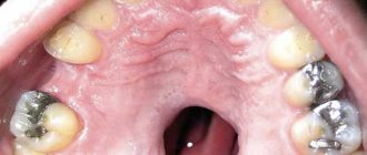
Primary syphilis of the oral mucosa. Damage to the oral mucosa in the primary period is quite common. Hard chancre can occur on any part of the red border of the lips or the oral mucosa, but is most often localized on the lips, tongue, and tonsils. A feature of hard chancre of the oral cavity is its small size and similarity to traumatic defects, which creates significant difficulties for diagnosis. Chancre of the upper and, more often, lower lip appears in the form of an ulcer or erosion, the bottom of which can often be covered with a raised brownish crust. In the corners of the mouth, usually in small folds of the skin, a slit-like chancre may be localized, resembling a crack in shape, but when the fold in which the hard chancre is located is stretched, its oval outline is determined. When hard chancre is located in the corners of the mouth, it can clinically resemble jams, which are distinguished by the absence of compaction at the base. Lip chancre often imitates impetiginous, traumatic, herpetic erosion, and with severe infiltration, epithelioma. Significant difficulties in diagnosis arise in cases where the chancre is covered with a massive brownish crust (cortical chancre). It is extremely rare to find hypertrophic hard chancre on the red border of the lips. This is a hemispherical, densely elastic formation, sometimes in the shape of a mushroom cap, rising sharply above the skin level with a diameter of up to 2-3 cm. The surface of the hypertrophic chancre is usually shiny, smooth, with scanty discharge, subjective sensations are little expressed. Regional (submandibular) lymph nodes are sharply enlarged, usually on one side, and are often painless. Diagnosis of gum chancre, located in the form of a crescent near the neck of one or several (usually two) teeth, presents great difficulties. The ulcerative form of chancre of the gums is very similar to banal ulceration and has almost no signs characteristic of primary syphiloma. Diagnosis is facilitated by the presence of regional lymphadenitis in the submandibular region.
On the tongue, the chancre is usually single, more often found in the middle third. When a hard chancre is located on the back of the tongue, due to significant infiltration at the base, the chancre sharply protrudes above the surrounding tissue, and there is flesh-red erosion on its surface. In addition to the erosive or ulcerative forms, chancre of the tongue is often presented in the form of fissure-like erosion or an ulcer with a shiny bottom. A less common sclerotic hard chancre may have the appearance of a sclerotic tip of the tongue, in which redness without sharp boundaries passes into the normal mucous membrane (chancre “without edges”). Noteworthy is the absence of inflammation around the chancre and its painlessness. Very rare and difficult to diagnose are chancre of the tonsils, which can have one of three forms: erosive, ulcerative and sore throat-like (chancre-amygdalitis). Erosive chancre of the tonsil occurs in the form of erosion of a red or opal color, round in shape, ranging in size from 2 to 10 mm, with compaction at the base, a smooth bottom and scanty discharge. Soreness, as a rule, is not noted. The tonsil around the erosion is of normal color and dense. In the ulcerative form, the tonsil is enlarged and dense. Ulcerative chancre of the tonsil is larger in size, significant depth, its bottom is covered with a grayish coating, and there is often pain when swallowing and palpation. Both types of chancre are characterized by one-sided lesions and specific scleradenitis of the cervical and submandibular lymph nodes. An atypical manifestation of chancre on the mucous membranes of the oral cavity is chancre-amygdalitis, which is characterized by enlargement and hardening of one tonsil in the absence of erosion or ulcers. When palpating the tonsil with a spatula, its elasticity is felt. An enlarged, hyperemic tonsil obscures the lumen of the pharynx and can cause a change in voice. In some cases, pain when swallowing, general malaise, and increased temperature are possible, as with a common sore throat, which makes it difficult to diagnose syphilis. Chancre-amygdalitis is characterized by specific submandibular and cervical lymphadenitis, also unilateral.
Complications of chancre. Complications of chancroid include erosive balanoposthitis, vulvovaginitis, phimosis, paraphimosis, gangrenization and phagedenism, which usually develop with the addition of a secondary infection, irrational treatment or self-medication.
Damage to the lymph nodes and blood vessels is the second most important symptom of primary syphilis. Regional scleradenitis appears 7-10 days after the onset of chancre. The lymph nodes closest to the chancroid enlarge to the size of a pea, bean or hazelnut or more,... while remaining painless. On palpation, the lymph nodes have a dense elastic consistency, are not fused to each other and the surrounding tissues, are mobile, the skin over them is not changed.
When chancre is localized on the face and mucous membranes of the oral cavity, the submandibular, anterior and posterior cervical, occipital, and preauricular lymph nodes enlarge. Recently, according to various authors, 4.4-8% of patients with primary syphilis did not have regional scleradenitis.
The third symptom of the primary period of syphilis - syphilitic lymphangitis - is not permanent and is now rare.
Differences between ecthyma in syphilis and normal
Common ecthyma has a staphylococcal or streptococcal etiology.
Rarely it is caused by gonococcus.
With simple ecthyma, a bubble or pustular element is formed.
The morphological element appears only against the background of infiltration.
It dries out quite quickly.
A crust forms.
After it leaves, an ulcer forms, the bottom of which bleeds and is covered with plaque.
Ecthyma heals in 2-3 weeks.
In some cases, it can heal without forming an ulcer.
Main localization:
- hips
- gluteal region
- shins
- hands
The main difference between ordinary ecthyma is that there is no infiltrate around the ulcer.
The crust covers the ulcerative defect completely, not partially.
Because ordinary ecthyma does not have a tendency to infiltrative growth.
Signs and symptoms of syphilis in women during the primary period of the disease
Chancroid in women has its own characteristics, which depend on their location:
- erosive chancres are more often located in the area of the labia majora, indurative edema is less often recorded;
- chancre located in the area of the urethra always has a dense base;
- in chancre located in the area of the vulvovaginal fold, the compaction at the base is not pronounced;
- primary syphilomas, located on the cervix, are erosions of round shapes, with a flat bottom, clearly defined boundaries, intense red color, scanty serous discharge;
- when lymphatic capillaries are damaged and lymph outflow in the area of the clitoris and labia is impaired, unilateral indurative edema occurs, characterized by a significant increase in the organ, which becomes dense, dark red in color, often with a bluish tint. After pressing, there is no hole left. There is no pain. The swelling subsides slowly even with treatment.
- Primary vaginal syphiloma is extremely rare;
- in the area of the nipple in women, chancre is recorded in the form of erosions, usually single, with pronounced compaction at the base, covered with a crust;
- The crescent-shaped crack has a primary defect localized at the base of the nipple.
Rice. 12. Primary syphilis in women on the labia majora (photo on the left) and cervix (photo on the right).
Rice. 13. The first signs of syphilis in women are primary ulcers on the genitals.
Rice. 14. The photo shows an atypical form of chancre in a woman - indurative edema (photo on the left) and a primary defect in the nipple area (photo on the right).
The appearance of an erosive ulcer on the genitals or inducing swelling of the labia majora is the first sign of syphilis in women.
Syphilitic rupee
One type of syphilitic ecthyma is syphilitic rupee.
It occurs in weak patients.
Characterized by the severe general condition of the patient.
Often combined with inflammatory lesions of the eyes, nasal mucosa, bones, etc.
Rupa occurs in the same way as ecthyma.
The difference is that underneath the crust, rapid tissue breakdown continues.
As a result, it is constantly increasing.
Over time, it can reach sizes up to 8 cm in diameter and up to 3 cm in depth.
When you press on the crust, pus is released.
There is an ulcer underneath.
After it heals, a large scar remains.
It may be pigmented at first and then colorless.
Rupees are usually single.
They develop slowly and last a long time.
Rupees can occur not only in the secondary period of syphilis, but also in the tertiary stage.
Regional lymphadenitis, lymphangitis and polyscleradenitis
Regional lymphadenitis
Regional lymphadenitis (scleradenitis) is, after chancre, the second most important symptom of primary syphilis. Scleradenitis develops 6 - 7 days after the initial manifestation of syphilis - chancre and often on the affected side (unilateral localization).
The lymph nodes are painless, woody in density, mobile, and not fused with the surrounding tissues. Sometimes lymph nodes appear simultaneously with primary syphiloma. Several lymph nodes may enlarge at the same time, the largest of which is located closer to the chancroid. When a secondary infection occurs, the lymph nodes become fused into conglomerates, the phenomena of periadenitis are pronounced, and sometimes fistulas are formed. Syphilitic lymphadenitis resolves slowly.
Inguinal lymphadenitis is often bilateral, it develops when hard chancre is localized on the external genitalia, ulnar and axillary lymph nodes increase when syphilomas are localized on the hands, cervical and submandibular - when localized on the lower lip, preauricular and submandibular - when localized on the upper lip, submandibular , cervical and preauricular - when localized on the tonsils, sublingual - on the tongue, preauricular - on the skin of the eyelids, axillary - when primary syphiloma is localized on the skin of the mammary gland, when chancre is localized in the rectum and on the cervix, the lymph nodes located in the small pelvis.
Rice. 24. Regional lymphadenitis is, after chancre, the second most important symptom of primary syphilis. The photo shows inguinal lymphadenitis.
Syphilitic lymphangitis
Inflammation of the lymphatic vessels (lymphangitis) is the third main symptom of primary syphilis. The causative agents of syphilis multiply in the lymphatic system and the lymphatic vessels are the first to be affected in this process. Lymphangitis begins from the site of primary syphiloma and reaches nearby lymph nodes. The inflamed vessels thicken and become like a dense elastic painful cord. Lymphangiitis, like lymphadenitis, resolves slowly.
Rice. 25. The photo shows sclerosing lymphangitis.
Diagnosis of acne with syphilis
It is impossible to determine the origin of acne based on the clinical picture alone.
Diagnostic tests are required to confirm the analysis.
To directly detect Treponema pallidum, PCR of material from the surface of acne can be performed.
When a morphological element is irritated, a discharge usually appears.
It can be collected and sent to the laboratory.
Using PCR, the genotype of Treponema pallidum is determined.
You can also take blood tests.
The doctor will determine what reactions to syphilis you should take.
A minimum of two serological tests are usually performed.
For example, cardiolipin test and ELISA or RPGA.
If the results are questionable or contradictory, additional studies may be required.
How contagious are acne with syphilis?
The infectiousness of secondary syphilides is very high.
Especially ulcers and erosions that are not covered with a crust.
These elements contain a large number of pale treponema.
They can penetrate not only into the mucous membrane, but into the skin.
Not only sexual contact, but also close household contact in a person with active rashes can lead to transmission of infection.
In addition to the direct route, there is also an indirect route of infection.
In this case, Treponema pallidum is transmitted through common objects.
For example, through clothes, towels, etc.
That is, through objects that first come into contact with the skin of a patient, and then of a healthy person.
Incubation period and pathogenesis of the disease
The entry points for syphilis pathogens are damaged mucous membranes and skin. The period of illness from the moment pathogens enter the patient’s body until the appearance of primary changes (chancroid) is called incubation. During the incubation period, bacteria multiply intensively in lymphatic vessels and nodes. Reproduction of pale treponema occurs by division at a rate of one division in 30 - 32 hours. There are no clinical manifestations of the disease during this period, serological tests remain negative. The incubation period averages 3 to 4 weeks. Sometimes the incubation period is shortened to 8 - 15 days, or extended to 190 days. In case of simultaneous infection of 2 sources, a shortening of the incubation period is recorded. When taking antibiotics after infection, its lengthening is noted.
At the end of the incubation period, primary syphiloma appears at the site of introduction of pale treponema - chancre (hard ulcer), erosive or ulcerative chancre, an increase in regional lymph nodes (regional lymphadenitis) is noted, and later the entire lymphatic system begins to respond to the infection (syphilitic lymphadenitis or polyscleradenitis). With maximum accumulation in the lymphatic system, bacteria penetrate through the thoracic lymphatic duct into the subclavian vein. Syphilitic septicemia develops. In some patients, this period is manifested by an increase in body temperature, severe headaches and muscle-joint pain, weakness, and general malaise.
Serological reactions become positive 3 to 4 weeks after infection.
The spread of pale treponema with blood throughout the body marks the development of the next stage of the disease - secondary syphilis.
- Primary affect (primary syphiloma) forms 3 to 4 weeks after the initial infection.
- Within 1 - 2 weeks, the chancre increases in size, and then after 6 - 8 weeks the ulcer scars even without treatment, erosion epithelializes after 4 - 5 weeks.
- On the 5th - 6th day after the appearance of primary syphiloma, regional lymph nodes enlarge.
- After 5 - 6 weeks, polyscleradenitis develops, which indicates the generalization of a specific process.
- Serological reactions become positive 3 to 4 weeks after infection.
- The primary period lasts about 7 weeks.
- The end of the primary period is marked from the moment of occurrence of secondary syphilides.
Rice. 2. The appearance of hard chancre in the anus and oral cavity is associated with sexual perversions.
Prevention for others with syphilis
Those around you must follow preventive measures so as not to become infected from a person who has acne due to syphilis.
Firstly, you cannot have sexual contact with him.
Even if a condom is used.
Because the source of infection is not only the genitals, but also other parts of the body.
Secondly, you should not come into close everyday contact with the person.
Thirdly, you do not need to share hygiene items with him, sleep in the same bed, or wear the same clothes.
If there were contacts before syphilis was detected, it is advisable to receive preventive treatment.
Localization
Since syphilis is transmitted primarily through sexual contact, chancroid is most often localized on the genitals. However, clinical practice shows that syphilomas are almost as often found in the mouth and anus.
This means that chancre can appear anywhere, the place of its localization can be:
- penis and scrotum
- labia and clitoris
- posterior commissure and anal area
- pubis
- oral cavity: lips, gums, tongue and throat
- inner thighs
- chest and stomach
- face
- rarely - eyelids, conjunctiva of the eyes
Chancre can be located inside the genital organs, for example, on the walls of the vagina or cervix, then syphiloma is difficult to detect.
Acne prognosis for syphilis and complications
Acne with syphilis itself will sooner or later go away on its own.
For some time the disease occurs without symptoms.
But this does not mean that the person is cured.
The rashes often recur.
And in the absence of timely treatment, the disease can progress to the tertiary stage.
She is very heavy.
Accompanied by damage to the central nervous system and internal organs.
Foci of infection can occur in almost any part of the body.
Most often affected:
- bones
- sense organs
- joints
- muscles
Gummas are formed.
Entire sections of tissue die.
They can lead to disability of the patient and often disfigure him.
They often cause organ loss.
For example, the eyes, nose or penis.
In the most unfavorable cases, syphilis ends in death.
Signs and symptoms of syphilis in men during the primary period of the disease
Hard chancre in men has its own characteristics, which depend on their location:
- small erosive chancres are often located on the head of the penis;
- ulcerative chancres are located in the head groove, they are large in size, and have a powerful infiltrate at the base;
- primary syphilomas, located on the frenulum of the penis, have an elongated shape, are easily injured and bleed during erection;
- in primary syphilomas located on the sides of the frenulum, there is no compaction at the base;
- in the region of the edge of the foreskin there are linear chancres;
- the seal at the base of the chancre, located in the area of the inner layer of the foreskin, looks like a visor;
- the hard chancre located on the crown of the glans penis resembles a swallow's nest;
- when located in the urethra, chancres have a dense consistency, are painful on palpation, always bleed, urination is also painful, scanty serous-bloody discharge is noted, in some cases, when cured, a cicatricial narrowing of the organ occurs;
- when the lymphatic capillaries are damaged and the outflow of lymph is impaired in the area of the scrotum and penis, indurative edema occurs, characterized by a sharp increase in the organ, which becomes dense; after pressing, no hole remains. There is no pain. The swelling subsides slowly even with treatment.
Rice. 10. Primary syphilomas in men - localization on the head of the penis (photo on the left) and in the area of the inner layer of the foreskin (photo on the right).
Rice. 11. Ulcerative chancre, located in the head groove, is large in size, has a powerful infiltrate at the base and is linear in direction.