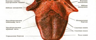
Anatomical structure of the tongue
The structure of human language corresponds to its multifunctionality, which lies in the fact that it participates in the processes:
- chewing;
- salivation;
- taste perception;
- speech.
The body of the tongue consists of striated muscle tissue, which is covered by a membrane of mucous tissue. Its surface, called the back, is conventionally divided into three parts:
- the last third, located near the pharynx, is called the root;
- the first two thirds are the body of the tongue.
A longitudinal groove runs in the middle, which is an external manifestation of the internal septum; it is, in fact, a reduced thyroglossal duct.
The mucous membrane, tightly adjacent to the muscle tissue, is covered on the outside with stratified squamous epithelium. It contains:
- salivary glands;
- taste buds;
- lymphatic ducts.
The mucous membrane of the posterior part forms three supraglottic folds, with the help of which the tongue is attached to the larynx:
- median;
- two lateral.
The tongue is abundantly covered with papillae, including:
- filamentous - act as organs of touch and, thanks to the rough surface, hold food on the tongue;
- cone-shaped – responsible for sensitivity to temperature and pain;
- mushroom-shaped - equipped with taste buds, thanks to them we distinguish many taste sensations;
- groove-shaped - located near the root, have serous glands and are also responsible for the sense of taste;
- leaf-shaped - equipped with lingual glands that secrete a mucous secretion.
The tongue is attached to the oral cavity by a fold of mucous membrane called the frenulum.
Methods of treating the disease
Treatments for this type of cancer include surgery, radiation therapy, and chemotherapy. Let's consider these methods separately.
Surgery
The main method of treatment is resection (removal) of the tumor. The principle of the surgical technique is to remove a malignant tumor from the patient. Surgery is always performed, except in cases where the tumor is inoperable.
Operation options:
- Gentle resection, in which the tongue or most of it is preserved.
- Radical removal (the operation is called glossectomy, this is the total removal of the tongue).
- Together with glossectomy or after it, reconstruction of the tongue is often performed - one-stage plastic surgery with a skin flap. This intervention allows you to preserve the functions of the removed organ.
Radiation therapy
Radiation is designed to destroy cancer cells or slow their growth. For small tumors this is the main treatment method. Radiation can also be given before surgery (to reduce the tumor) and after it (to avoid relapse). In later stages, the technique is used as palliative therapy, that is, it alleviates symptoms.
Chemotherapy
This is a systemic therapy that aims to destroy malignant cells throughout the body. Chemotherapy is given before surgery (to shrink the tumor) or after it (to avoid relapse).
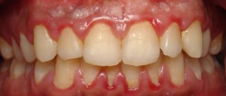
What does a healthy tongue look like?
A white-pink color of the tongue is considered normal. There are a number of other accompanying signs of his health:
- the longitudinal fold of the tongue is clearly visible;
- the papillae are clearly visible, but not hypertrophied;
- the edges are smooth.
The surface must be clean, although a slight coating on the tongue is acceptable.
For an adult, seasonal changes in the color of the tongue are possible:
- in winter, a slight yellowish coating on the tongue in adults can be considered normal if there are no other abnormalities - pain, increase in size, lack of taste;
- in summer, a light white coating, not localized, but over the entire surface, is also not considered a pathology.
A healthy child’s tongue is not much different from an adult’s tongue. One significant feature: it reacts to the slightest changes in the condition of the baby’s body - teething, the introduction of complementary foods, even a change in the brand of baby food. Therefore, plaque on a child’s tongue requires close attention.
What is plaque on the tongue?
The most numerous - filiform papillae - form a white coating on the tongue due to their structure:
- the lamina of the mucous membrane of the filiform papilla is covered with stratified squamous epithelium;
- this is a keratinizing epithelium that periodically exfoliates, covering the entire tongue with a light white coating;
- In case of any malfunctions in the human body, desquamation slows down and a layer of keratinized cells grows, which acquire different colors depending on what pathology led to the malfunction.
By the color of the plaque and where it is localized, diseases that have led to pathological changes in the tongue are judged.
Classification
Depending on the location, cancer is distinguished:
- Bodies. These include tumors of the tip, dorsum and lateral surface (most often the middle of the lateral surface). This localization occurs in 70% of cases.
- Root (accounts for 20% of all cases).
- Lower surface (sublingual area).
By growth pattern:
- Exophytic form (this includes papillary and ulcerative forms of tumors growing outward).
- Endophytic (growing inside the tissue of the tongue - infiltrative and ulcerative).
Papillary form a is a dense papillary growth of a mushroom shape. There may also be raised plaque-like growths that have clear boundaries.
Ulcerative (occurs in 50% of cases). Characterized by a superficial ulcer surrounded by a ridge. The ulcer is constantly increasing in size. At first, the ulcer does not bother the patient, but as it grows, pain and bleeding appear. The ulcer can become infected and inflamed, making diagnosis difficult.
Infiltrative form . The tumor grows into the thickness of the tissues, and they become denser. With diffuse growth, the compaction spreads to the entire tongue, which impairs its mobility.
The infiltrative-ulcerative form is characterized by thickening of the tongue with the presence of deep ulcers.
According to histological composition:
- Adenocarcinoma.
- Squamous cell carcinoma.
Classification according to the TNM system.
- Tis "cancer in place."
- T1 Tumor no more than 1 cm. Not accompanied by complaints, discovered by chance during examination.
- T2 ≤2 cm. There are ulcers and areas of compaction.
- T3 >4 cm. The tumor is equal in size to half the tongue. Metastases to the occipital lymph nodes, postauricular.
- T4 Locally advanced cancer that occupies the entire tongue and invades the tissues of the mouth and face. Metastases to internal organs (brain, liver, heart) and bones.
Symbol N - the presence of metastases in the lymph nodes:
- N0 - No lymph node involvement.
- N1 One node on the side of the tumor is affected.
- N2 Metastasis: one metastasis no more than 6 cm, several up to 6 cm on one side or both sides up to 6 cm.
- N3 Metastases larger than 6 cm.
With T1 tumors, lymph nodes are affected in 40% of cases, with T4 stage in 85%. A reliable factor for metastasis is the depth of invasion - 4 mm is considered a critical value. Most often, metastases are found in the submental, submandibular and cervical nodes (upper third).
Histopathological differentiation:
- G1 High degree.
- G2 Medium.
- G3 Low.
Why does plaque appear on the tongue?
The tongue is a muscular organ that can tell a lot about the state of the body. It is soft and easily mobile, and has a pale pink color if the person is healthy. From time to time, plaque may appear on the mucous membrane, the density of which is often seasonal. This is explained by the fact that at different times of the year the body needs certain vitamins. For example, in summer the deposits are thicker and more saturated. At this time they may acquire a yellow tint.
Bacteria constantly accumulate on the mucous membrane of the tongue. These microorganisms are the cause of plaque and bad breath.
The following factors contribute to their intensive reproduction:
- excessive alcohol consumption;
- smoking;
- poor nutrition;
- infections and inflammatory processes;
- taking medications;
- chronic diseases;
- poor oral hygiene.
Since the tip of the tongue is mobile, it is cleaned more and, accordingly, there is less plaque here. At the root, its density is higher, since in this place there is contact only with the sky. Such manifestations are also possible with dysbacteriosis, vitamin deficiency, and improper hygiene.
Reasons for appearance
There are a number of predisposing factors to the development of tongue cancer. Let's look at them in more detail:
- Tobacco and alcohol . When smoking, the risk of developing this disease is influenced by how much, often and for how long a person smokes. Those who have been diagnosed with this type of cancer need to completely stop smoking, since continued tobacco use increases the risk of developing cancer of another location. Of 10 patients with malignant neoplasms of the oral cavity, 7 are susceptible to alcohol dependence. The combination of alcohol and smoking increases the risk of oral cancer by 100 times compared to people who do not drink or smoke.
- Human papillomavirus (HPV) . The incidence of oral cancers associated with HPV has increased in the last decade. According to research, cancer associated with HPV affects mainly young patients who do not have a long history of drinking alcohol and smoking. Now many doctors associate the prevalence of HPV in the last two decades with people's passion for oral sex.
- Floor . Men suffer from this type of cancer 2 times more often than women, which may be due to frequent consumption of alcoholic beverages and smoking.
- Age . Oral cancer is more often diagnosed in patients 55-60 years old. This is due to its slow development. However, the presence of HPV changes this situation.
- Poor nutrition . According to several studies, the risk of developing such cancer increases with a deficiency of vegetables and fruits in the diet.
- Weak immune system . The risk increases in people who have congenital or acquired immunodeficiency, as well as in those who take drugs to suppress the immune system (this is necessary after an organ transplant).
- Graft versus host disease (GVHD) . This reaction is typical for the condition after stem cell transplantation: this is how the body reacts to foreign donor cells. Oral cancer can develop within 2 years.
- Lichen planus. This disease most often affects the surface of the skin, but can also appear as white spots and lines on the oral mucosa. Increases the risk slightly.
- Bowen's disease. This is an obligate precancerous condition, the tumor is located inside the epithelial layer. Sooner or later it transforms into malignant.
Types of plaque on the tongue
White plaque
A thin white coating is a common occurrence. You can especially notice it in the morning, when your teeth have not yet been brushed. Homogeneous white deposits occur in infants after feeding. This is also considered normal in older children.
The presence of other features may indicate certain ailments:
- an increase in plaque thickness is a symptom of prolonged constipation;
- elevated temperature and symptoms of intoxication - indicate infectious processes in the body;
- localization on the root of the tongue, its back – gastrointestinal diseases;
- placement on the sides of the tip of the tongue - pay attention to the kidneys.
A white coating with a cheesy structure, as well as dry mouth, indicate candidiasis (popularly called thrush). It often affects infants. The cause of the disease is weak immunity. To prevent thrush from spreading to the cheeks and gums, you should consult a doctor. He will prescribe antifungal medications. This disease is also possible in older children, but more often these are asthmatics or children with weakened immune systems. The disease may be accompanied by an unpleasant taste in the mouth.
Gray, green and brown plaque
And if the deposits are not white, but of a different color - what is it? Each shade has its own characteristics:
- Grayish
is a common symptom of gastrointestinal diseases. This could be, for example, a stomach ulcer. But a grayish-white coating is not a deviation from the norm. - Brown
. Such deposits on the root of the tongue appear in chronic alcoholism. It also occurs in smokers and with lung diseases. - Green
is a rare occurrence. Occurs with different types of glossitis. The disease can develop as a result of taking antibiotics, steroids and other substances that reduce the body's immune strength.
Please note that the tongue often changes color after eating and drinking. For example, strong tea can turn it brown.
Yellow plaque
As already mentioned, a yellowish coating appears in the summer. You need to worry if its shade becomes saturated. You should pay attention to the following signs:
- Bright yellow color - the liver and bile ducts may be affected.
- Yellowness of the lower part of the tongue is a symptom of incipient jaundice.
- A thick yellow-green coating is a sign of improper functioning of the digestive organs and stagnation of bile. These disturbances in the body may be accompanied by the formation of a red plaque.
Sometimes yellow deposits indicate an increased amount of bile.
Black plaque
Black deposits on the tongue are very rare. More often this is one of the signs of a serious illness:
- serious disruption of the pancreas, gallbladder, and gastrointestinal tract;
- high blood acidity resulting from dehydration;
- cholera.
- There is such a thing as a “villous” tongue, when the papillae on it turn black and become hard. Such manifestations can be observed in smokers and people who abuse alcohol, as well as when exposed to certain organisms and medications.
Spotted plaque
Geographic tongue, when its mucous membrane is covered with uneven red spots, scares many. This condition occurs in people of all ages. There is no danger in it, and often it goes away on its own.
Today, science does not fully know what the appearance of such spots means. Each case is individual, so it is important to monitor your condition. For example, spots may occur due to an allergic reaction. But in most cases they are then present on the skin.
Determining the disease by the color of plaque
The first diagnostician on the condition of the tongue was the Russian doctor M. A. Nechaev, who in 1833 published the book “Recognition of diseases by changes in the language” in the printing house of Kazan University. Several generations of Russian doctors were grateful to him for this unique work, which helped to carry out diagnostics without additional instruments.
Today, the technique is widely used not only among traditional healers, but also among practitioners of traditional medicine. However, the diagnosis must be confirmed after a comprehensive examination carried out in a laboratory, or using ultrasound, CT, MRI, fluoroscopy, etc.
What do you pay attention to during this diagnosis:
- plaque color;
- its consistency.
As for the color of plaque, it can be:
- white;
- grey;
- yellow;
- greenish;
- bluish;
- brown;
- even black.
And the consistency can be:
- almost transparent;
- flaky;
- viscous.
All signs are compared, and a certain diagnostic verdict is made.
Treatment
If tongue cancer is detected, there are several types of treatment:
- Surgery. The operation is used most often and allows for recovery in 80% of situations. When the tumor is removed, the tongue may be partially or completely removed. In advanced cases, part of the jaw bone is also removed, then the jaw structures can be restored using special methods.
- Radiation therapy. When fighting tongue cancer it gives good results, also up to 80% effective. Can be used either alone or in combination with surgery.
- Chemotherapy. Prescribed if metastases are detected.
Both chemotherapy and radiation therapy can be palliative - that is, they are carried out in cases where the patient can no longer be cured, but his condition can be maintained and the quality of life can be improved.
Forecasts
With timely consultation with a doctor and proper treatment, the five-year survival rate for tongue cancer is about 90%. If the cases are advanced, but without metastases, this percentage drops to 60%. When metastases have spread, the five-year survival rate is less than 35%.
Diseases of the oral cavity and plaque on the tongue
Most often, the condition of the tongue depends on the conditions in the oral cavity. The presence of plaque may be due to:
- caries;
- stomatitis with fungal and bacterial etiology;
- periodontal disease – systemic damage to periodontal tissue (gums, bone and tooth ligament);
- glossitis - inflammation of the tongue that occurs as a result of mechanical damage to the organ, or as a condition accompanying other diseases;
- gingivitis - inflammation of the gums without damage to bone tissue.
The mucous membrane of the tongue reacts very sensitively to any problems in the oral cavity caused by inflammation, caused by bacterial or fungal infections.
They are diagnosed quite easily:
- A loose white coating indicates that a yeast-like fungus of the genus Candida has settled in the mouth.
- The presence of periodontal disease and gingivitis is determined by the condition of the gums.
- The presence of caries is accompanied by an unpleasant odor and putrefactive damage to the bone tissue of the tooth.
- Glossitis is accompanied by a burning sensation, salivation, pain and inflammation.
If everything is more or less clear with these signs, then plaque caused by systemic diseases is not so easy to recognize without special knowledge.
What diseases does plaque on the tongue foreshadow?
It is believed that the nature of the disease and its location can be determined by the color of the plaque:
- White plaque is quite acceptable if it is easily removed after hygiene procedures. If it lies in a thick layer and has a cheesy consistency, then this is a sign of fungal infection, intoxication, the presence of foreign bodies in the oral cavity - implants or dentures - and the allergic reactions they cause.
- A gray coating may indicate that a course of antibiotic treatment has been carried out or there are problems with the gastrointestinal tract. Most often these are ulcerative lesions of the stomach or duodenum. A decrease in general immunity can also be the cause of plaque of this color.
- A yellow coating indicates stagnation of bile or problems with the liver. It can also be observed with kidney damage, then its localization is at the edges of the tongue. Constipation also causes such plaque, in which case bad breath also appears.
- The green color of plaque occurs from excess bilirubin during hepatitis of various etiologies. This may also be a consequence of a viral infection.
- Brown plaque can be a consequence of gastronomic preferences - among lovers of brewed coffee and strong black tea. Heavy smokers also often have a brown coated tongue. Inflammatory processes in the gastrointestinal mucosa can cause such plaque.
- A bluish coating is a consequence of problems with the cardiovascular system. This may be coronary heart disease or chronic hypotension.
- A dark, almost black coating should alert you. This is a consequence of a serious pathology in the body - oncology, severe dehydration, rare Crohn's disease or cholera infection.
For adults, a constant coating on the tongue may mean that the person is a heavy smoker. It is difficult to find among smokers those whose organs have not been damaged by nicotine tar. This means that they are no longer healthy.
Plaque in diseases of the gastrointestinal tract
The gastrointestinal tract, or digestive system, includes:
- oral cavity;
- esophagus;
- stomach;
- liver;
- gallbladder;
- pancreas;
- duodenum;
- small and large intestines;
- rectum and anal sphincter.
Any malfunctions in the organs of the digestive system cause plaque on the tongue:
- if it is concentrated in the area of the root of the tongue and has a gray tint, this means that the large intestine and rectum are affected;
- a thin yellow coating in the middle of the tongue indicates the presence of gastritis or gastroduodenitis, and a thick layer localized in the middle indicates its exacerbation;
- with cholecystitis - inflammation of the gallbladder - a yellow-brown coating appears, while the tongue itself is dry, bitterness and dryness in the mouth are felt;
- if there is a problem with the biliary tract, the plaque can take on a color from yellow to green; it is the greenish shade that indicates that not everything is in order with the biliary system;
- a bluish coating indicates an intestinal infection;
- a thick yellow coating in combination with heartburn, belching and a burning sensation indicates that pancreatitis has worsened - inflammation of the pancreas;
- a reddish-brown, and sometimes even black, coating may indicate oncological processes or abscess inflammation in the gastrointestinal tract.
In any case, plaque on the tongue is not the main sign of diseases of the digestive system. Only a doctor, having collected an anamnesis, can make the correct diagnosis.
Treatment of tongue cancer
A combination of methods is used to treat tongue cancer. The tongue is an important organ in the human body that is involved in speech and swallowing, therefore the choice of treatment method largely depends on the impact that the effect of this method can have on these processes. Also, when choosing a treatment method for tongue cancer, doctors take into account how it may affect the patient’s appearance.
Basic treatment methods for tongue cancer
In modern medicine, the following types of treatment for tongue cancer are distinguished:
- Surgery. Today, surgery is the most common treatment for tongue cancer in the early stages, leading to recovery in 80% of cases. However, there is a category of patients who, for certain reasons, cannot undergo surgery or the operation can negatively affect body functions, such as swallowing and speech. Surgical treatment of tongue cancer involves radical removal of the tumor, which includes complete glossectomy or partial hemiglossectomy of the tongue. If the cancer grows into the bone structures and soft tissues of the floor of the oral cavity, surgical intervention is accompanied by resection of the jaw bone and affected tissues. If necessary, doctors use an orthostomy and apply plastic surgery methods to restore lost structures of the maxillofacial part.
- Radiotherapy. Modern medicine has proven that the results of radiotherapy are similar to those of surgery. Clinical studies have shown that radiotherapy is effective as a single treatment or in combination with surgery, with less severe complications. Recovery is achieved in about 80% of patients.
- Radiotherapy in combination with surgery. This combination is usually prescribed in cases of large tumors. But it can also be used to treat patients with a small area of normal tissue that remains after surgery, as well as if cancer grows into the edge of the removed tissue. Also, for leukoplakia of the tongue, complex treatment is prescribed.
- Palliative radiation and chemotherapy are given to patients who have distant metastases of tongue cancer.
Removal of lymph nodes
When treating tongue root cancer in the early stages, doctors decide to perform surgery or radiotherapy on the lymph nodes. If the lymph nodes are not treated, the cancer will spread to the neck through the lymphatic system. Untreated cancer leads to recurrence of the disease. It is very important to determine the presence of a tumor in the lymph nodes of the neck. Currently, this is effectively achieved through surgical removal of the lymph nodes.
For cancerous tumors in the neck, lymph nodes are removed, which is called radical dissection. If cancer cells are found in the lymph nodes, the patient is prescribed radiotherapy to the neck. Otherwise, no further therapy is carried out.
Diagnosis and treatment of various types of tumors are carried out by specialists from the Oncology Center of the Yusupov Hospital. For this purpose, the clinic has modern high-tech equipment, and also employs leading oncologists and chemotherapists with many years of experience.
Plaque for bronchitis and pneumonia
The area of the tongue immediately following its tip is an indicator of the health or disease of the respiratory system (bronchi and lungs). Based on the condition of this area, one can judge the presence of bronchitis or pneumonia:
- Red spots indicate that pneumonia or bronchial asthma is possible.
- A light film on the front of the tongue indicates the presence of a respiratory allergy or congestion in the lungs.
Plaque on the tongue caused by inflammatory processes of the upper and lower respiratory tract is not decisive in the diagnosis of these diseases.
Tests and diagnostics
To clarify the diagnosis, patients are prescribed:
- Biopsy of a tumor formation (or scraping and impression smears from the surface of erosions).
- CT and MRI of the affected area with intravenous contrast. This study makes it possible to assess the extent of the tumor and the depth of invasion into the tongue tissue.
- CT scan of the facial bones with contrast if there is a suspicion of tumor spread to the bones of the jaw and skull.
- Ultrasound of the lymph nodes of the neck.
- Puncture of the affected cervical nodes with cytological examination.
- Ultrasound of the abdominal cavity to exclude distant metastatic process.
- Chest X-ray to exclude metastases.
- Osteoscintigraphy for metastatic lesions of skeletal bones.
Plaque due to oral chlamydia and thrush
There are two types of infectious diseases that affect the urogenital organs and the oral cavity. These are chlamydia and thrush. For candidiasis caused by a fungus of the genus Candida:
- a dirty white cheesy coating forms on the walls of the mouth and on the tongue;
- when mechanically cleaning the tongue from plaque, bloody discharge appears;
- an unpleasant putrid odor and taste appear in the mouth;
- treatment with special antifungal drugs is necessary.
Oral chlamydia shows a slightly different picture:
- thick sticky mucus forms in the nasopharynx;
- then it migrates to the upper and lower palate;
- only after this does it appear on the tongue, first in the form of spots, and later covering the entire tongue with a white pasty coating;
- At the same time, it has the smell of rotten fish.
It is diagnosed both by visual examination and by laboratory analysis of scrapings from the tongue and palate.
Diagnostics
To accurately diagnose tongue cancer, laboratory and hardware tests are required. However, first of all, the patient should consult an ENT oncologist, who will give the necessary directions. Among the diagnostic methods:
- blood for tumor markers, other laboratory blood tests;
- biopsy of the affected area followed by histological examination;
- revision (revision) of the biopsy;
- MRI of soft tissues of the neck and face using contrast;
- CT scan of the neck and head with contrast;
- PET-CT.
What else can cause plaque on the tongue?
There are many other reasons that cause plaque on the tongue:
- Chronic alcoholism leads to the development of fatty hepatosis, and later cirrhosis of the liver. As you know, a dirty yellow or even greenish coating on the tongue is characteristic of people suffering from liver diseases. In addition, alcoholics are rarely concerned about body hygiene, much less oral hygiene. This further enhances the coating and odor on the tongue of a person suffering from alcoholism.
- Plaque can occur as a side effect of taking medications, mainly antibiotics. Taken orally, they kill the beneficial microflora of the small and large intestines, causing dysbiosis, accompanied by poor digestion and absorption of food. And this, in turn, leads to the formation of plaque on the tongue.
- Intoxication of any origin necessarily causes a coating on the tongue. Thus, cancer patients after a course of chemotherapy all suffer from a dirty-brown coating on the tongue, which is caused by toxic chemotherapy, as well as tissue breakdown products destroyed by cancer cells.
- Impaired immunity, especially if failures occur in that part of the immune system that is located in the intestines, also leads to the formation of plaque, because T-lymphocytes die, settling in the form of a yellowish coating on the tongue and intestinal walls.
In these cases, consultation with a specialized specialist is necessary.
Causes of tongue cancer
In modern medicine, it is believed that the release of carcinogens during the combustion of tobacco is the main factor that influences the occurrence and development of tongue cancer. When combined with alcohol, carcinogens from tobacco smoke increase the likelihood of developing cancer among alcoholics and smokers.
Also, the development of tongue cancer can be influenced by components that are classified as occupational hazards, such as perchlorethylene, asbestos, heavy metal salts, and petroleum distillation products.
The formation of this tumor can be influenced by mechanical trauma to the mucous membrane. This type of injury can occur from sharp tooth edges, poorly fitting dentures, frequent tongue biting, poorly processed fillings, or a broken tooth.
According to research, a connection has been established between chronic persistent infection and tongue cancer, which is caused by the herpes simplex virus, HIV, or HPV. Patients who receive immunosuppressive drugs for a long time are also predisposed to tongue cancer.
If the carcinogenic factor continues to act on the organ, then changes in the mucosa will lead to oncology. Today, the following factors are classified as precancerous conditions of the tongue:
- papillomas;
- chronic tongue ulcer;
- hyperkeratic forms of lupus erythematosus;
- leukoplakia of the tongue;
- hairy leukoplakia of the tongue;
- Bowen's disease;
- lichen planus;
- systemic lupus erythematosus.
Long-term exposure to these trigger factors leads to changes in the DNA structure of tongue cells with the development of hyperplasia and dysplasia of the tongue mucosa. Most benign tumors of the tongue are susceptible to transformation into malignant tumors due to trauma to the oral cavity.
Plaque on the tongue in children
In children from birth to 5 years of age, the immune system is so imperfect that a slight coating on the tongue is considered normal. Moreover, a rare baby has avoided thrush, which affects the oral cavity and tongue from the first days of life. But you need to know and be able to differentiate plaque on a child’s tongue in order to recognize dangerous infectious and autoimmune diseases in time and seek medical help:
- Thrush is characterized by a loose, cheesy coating on the tongue and oral mucosa. Cleansing causes the baby to cry because the papillae are hypertrophied and react painfully to touch.
- A dirty gray coating on a child's tongue may be an indicator of scarlet fever. This is an infectious disease that must be treated under the supervision of a doctor. With scarlet fever, the tongue gradually turns from dirty gray to scarlet, similar to strawberries, with characteristic dots along the entire surface of the tongue.
- A filmy coating covering the root of a child’s tongue indicates that he has diphtheria. This sign requires urgent hospitalization, because the disease develops rapidly and leads to suffocation.
- Black or dark brown plaque in babies can be caused by a latent form of diabetes, bacterial sore throat, or taking strong antibiotics.
- There is also such a thing as “geographical language”. It is also typical for young children. These are red spots scattered across the entire surface of the tongue against the background of a light white coating, making the picture resemble a map of the world. In this case, benign migratory glossitis is diagnosed. It occurs against the background of helminthic infestation, vitamin deficiency, acute infectious diseases, and exudative diathesis. Only a doctor can identify the cause of the disease, so you should contact him immediately.
All other causes of plaque on the tongue in children are not much different from adults. These are the same dysbacteriosis, gastritis and even stomach ulcers, problems with the liver and gall bladder.
Oral health in patients with gastroesophageal reflux disease (GERD)
V.P. Novikova, AM Shabalov
Currently, in terms of its frequency, GERD takes a leading position among other diseases of the gastrointestinal tract (GIT). If in the 90s of the last century, symptoms of GERD (heartburn, belching) occurred in 20-40%, then at the beginning of the 21st century - in 40-60% of the world's population [21,22,53,56,59].
In addition to the high prevalence, interest in GERD is due to a tendency towards “rejuvenation”, frequent late diagnosis, underestimation of the consequences of this pathology (Barrett’s esophagus, esophageal adenocarcinoma), a decrease in the quality of life of patients, as well as the presence of extra-esophageal manifestations - bronchopulmonary, cardiac, otorhinolaryngological and dental [4, 17,34,35,44].
Publications concerning extraesophageal manifestations of GERD, especially in pediatric gastroenterology, are few and scattered. While certain data have been accumulated on bronchopulmonary, cardiac and otorhinolaryngological manifestations of GERD, dental manifestations are not sufficiently covered in the literature [11,13,15,45,49,54,57,58]. The mechanisms and cause-and-effect relationships of the development of dental manifestations of GERD have not been definitively established [52].
Most authors give the leading role in the occurrence of oral pathology in GERD to the influence of hydrochloric acid, which leads to a decrease in the pH of mixed saliva below 7.0. Saliva, normally enriched with calcium, phosphates, containing carbonates, sodium, potassium, magnesium and having alkaline properties, at low pH, especially at values of 6.2-6.0, leads to focal demineralization of tooth enamel with the appearance of erosions of hard dental tissues and the formation of cavities (caries) in them [12,19].
Thus, in some studies it was noted that in more than half of patients with GERD and concomitant oropharyngeal pathology, pharyngolaryngeal reflux is determined (reflux of gastric contents above the upper esophageal sphincter into the pharynx and larynx). And the duration of the episode of reflux into the hypopharynx in some patients exceeded the same indicator for reflux into the lower esophagus, which indicates a lower ability of the hypopharynx to neutralize hydrochloric acid compared to esophageal clearance. This feature of the hypopharynx can contribute to damage to the oral cavity with high reflux [16,61].
The role of isolated acid reflux in the genesis of damage to hard dental tissues can be confirmed by a study in which the formation of erosions of tooth enamel did not occur with combined duodenogastroesophageal reflux [54]. However, in some studies there were no differences in the pH of mixed saliva in children with chronic gastroduodenitis and peptic ulcer against the background of reduced, normal and increased acid-forming function of the stomach [24].
Some studies have shown a significant decrease, along with the pH of mixed saliva, in the concentration of its inorganic components (potassium, sodium, calcium, phosphate ions) in patients with GERD [41].
Some studies have reported significant reductions in secretion of mucin, nonmucin protein, epidermal growth factor, and minor changes in salivary prostaglandin E2 levels in response to intraesophageal exposure to gastroesophageal reflux. The esophageal-salivary reflex, by which saliva secretion increases in response to mechanical and chemical irritation of the esophagus, is impaired in patients with GERD, which has an adverse effect on the condition of the oral cavity. [10,28].
At the same time, as is known, saliva is one of the components in the mechanism of preepithelial protection of the oral cavity and esophagus from the effects of hydrochloric acid during gastroesophageal reflux. Violation of the composition of saliva, a decrease in its neutralizing properties in relation to hydrochloric acid, can lead to damage to the soft and hard tissues of the oral cavity [9,26,39].
As is known, saliva contains nitrites in high concentrations, which is achieved due to the recycling of endogenously formed nitrates and their return to the salivary glands. In addition, in modern conditions, large amounts of nitrates are found in human food. At neutral pH values, they remain relatively stable, but in an acidic environment they turn into nitrosating compounds, which can have an adverse effect on the condition of the oral cavity and esophagus and their microbiocenosis [38].
Saliva is also the main regulator of the total number of microorganisms in the oral cavity. Changes in its physicochemical properties as a result of GER, which were described above, can probably contribute to dysbiosis in this biotope [41].
The normal microflora of the oral cavity has been sufficiently studied [36, 40, 55]. The direct role of non-pathogenic, conditionally pathogenic and pathogenic bacteria, viruses, fungi and protozoa in the development of diseases of the oral cavity is shown. Thus, a group of viridinating low-virulent streptococci (S. mutans, S. sanguis, S. mitis, S. salivarius, S. anginosus) take part in the processes leading to the development of damage to hard dental tissues. Fusobacteria in association with spirochetes, as well as leptotrichia and actinomycetes, are the main causative agents of purulent-inflammatory processes in periodontal tissues, including ulcerative-necrotic ones. In the inflammatory-dystrophic form of periodontitis with suppuration, Entamoeba gingivalis and Trichomonas clongata are often isolated in smears from gum pockets. Intensive proliferation of fungi of the genus Candida in the oral cavity can cause dysbiocenosis and the development of candidiasis [6,7,18].
Some studies assessed the adhesive properties of bacteria (streptococci, enterococci, lactobacilli), fungi C. albicans to the buccal epithelium, gastrointestinal epithelium in normal and pathological conditions [5,8,20,54,].
Thus, in children with bronchial asthma there was an increase in the adhesive ability of C. albicans to the buccal epithelium, and in chronic gastroduodenitis there were no differences from the control, which, according to the authors, is associated with changes in receptors under the influence of proteolytic and glycosidase enzymes of oral secretions [25 ].
In the literature available to us, we were unable to find any studies concerning the mechanisms of microbiocenosis disruption and the adhesive properties of bacteria in the oral cavity in GERD. However, some studies have analyzed the microbial landscape of the distal esophagus in patients with GERD. A microbiological imbalance was revealed with the prevalence of microorganisms producing various pathogenicity enzymes: hemolysin (S. Intermedins, S. sanguis, Staphylococcus saprophyticus, S. warneri, Bacteroides spp.), lecithinase (Staphylococcus xylosus), caseinase, RNase, catalase, and the presence of fungi of the genus Candida albicans with pathogenic properties, expressed in the ability to form germ tubes, and increased adhesion to epithelial cells [42].
The significance of H. pylori infection in the occurrence and progression of periodontal diseases in patients with GERD is discussed. However, in the available literature there are opposing opinions about both the unfavorable and protective roles of this microorganism in GERD [25,27,48,64].
In general, all dental manifestations of GERD can be conditionally divided into damage to soft tissues (red border of the lips, oral mucosa, tongue, periodontal tissues) and hard tissues of teeth (non-carious changes, caries) [3,54,62].
According to various authors, the incidence of oral diseases in GERD ranges from 5 to 69.4% [2, 29, 63], their frequency and severity depend on the age of the patients.
In one study, diseases of the oral mucosa, lips, and tongue were observed in 46% of children 8-15 years old with GERD of varying severity [37].
Studies conducted in patients with the erosive form of GERD have revealed a high correlation between the degree of reflux esophagitis and the intensity of swelling of the oral mucosa and plaque on the tongue. Features of the condition of the tongue mucosa in children with GERD were: the presence of a “geographic tongue”, desquamative changes, abundant white plaque, which does not have a clear localization and is located in patches. In addition, pronounced folding, hypertrophied papillae, and tooth marks on the lateral surfaces were noted on the tongue. In children with chronic gastroduodenitis without motor disorders, the coating was uniform over the entire surface of the tongue, folding was rarely detected [1,31].
Adult patients with severe GERD, along with typical complaints, were bothered by a feeling of dryness, a burning sensation, pain in the tongue, and “scalded” tongue [66,68].
In addition, these patients had a change in the taste sensitivity of the tongue, and with increasing severity of GERD, the condition of the papillary apparatus of the tongue worsened. Thus, in patients with GERD stages B and C (in accordance with the Los Angeles classification), a significant increase in the threshold of taste sensitivity for sweets was detected, which they felt at 2% and 5% sugar concentration, respectively. The threshold of taste sensitivity to salty foods in all patients was within normal limits (0.25-1.25%). The threshold for taste sensitivity to sour was lowered. This suggests that the reaction in these patients to sour is somewhat aggravated. The taste sensitivity threshold for bitter in patients with GERD stages B and C was slightly higher than normal.
Cheilitis and pockets in the corners of the mouth were noted in 38.6%, and dry lips in 77.2% of adult patients with GERD [46].
According to various authors, the incidence of stomatitis in adults ranges from 3 to 15%, depending on the severity of GERD [3,46].
Periodontal tissue diseases (gingivitis, periodontitis) with GERD were observed in adult patients with a high frequency (83-89.6%) [48,51].
The frequency of caries in adult patients with GERD was noted at 32.5-100%, which depended primarily on age and severity of the disease. Thus, caries with changes in dental enamel on the inner surface of the teeth, that is, in places characteristic of GERD, was more common in patients with erosive than with catarrhal reflux esophagitis [24,29].
In turn, in children with GERD, caries occurred with a frequency of 20 to 88%, with a tendency to increase in frequency with age [2,32].
According to the literature, non-carious lesions of hard dental tissues, the structure of which is dominated by erosion, wedge-shaped defects, and increased tooth abrasion, are observed in adult patients with a frequency of 24 to 50.1% [33,51].
In GERD, tooth enamel erosion was observed in 20-50.1%, wedge-shaped defects - in 33.5%, pathological abrasion - in 32%, enamel hypoplasia - in 8.4% of adult patients [29,32,67].
In one study, when examining children with GERD in 3 age groups: 7-9 years, 10-13 years, 14-16 years, systemic enamel hypoplasia was noted in 8%, 4% and 4%, respectively [2].
In another study, panesophageal motor dysfunction was detected in the majority of patients with GERD with damage to the hard tissues of teeth; hypotension of the lower esophageal sphincter (LES) was detected in 60% of patients, and 80% of patients had a decrease in the tone of the upper esophageal sphincter [47].
The severity of these changes in the oral cavity is closely related to the duration, severity and degree of compensation in the treatment of GERD [29,32].
In some studies concerning adult patients, against the background of conservative treatment of GERD (treatment with proton pump inhibitors), anti-reflux operations, an improvement in the condition of the oral mucosa, a decrease in its swelling, epithelization of desquamation areas on the tongue, no relapses of stomatitis, and an improvement in the condition of periodontal tissues were observed. . However, the condition of the hard dental tissues after treatment of GERD did not differ from the initial data, since the elimination of these defects can only be achieved with the use of filling restoration materials [46,50,65].
Despite the current enormous interest in the problems of GERD both in adult patients and in pediatrics, there is an underestimation of the role of pathological GER in the development of extraesophageal dental symptoms. This may be due to the difficulty of establishing a direct direct connection between GERD and dental symptoms due to the insufficient distribution and use of 24-hour pH-metry in gastroenterological practice with the presence of additional sensors in the upper esophagus and oropharyngeal region.
One possible solution to this situation may be to use the rabeprazole (omeprazole) test to determine the relationship between GERD and oral symptoms. If, while taking rabeprazole or omeprazole for 3 months, there is a positive trend in dental symptoms, then most likely their origin is associated with a high pathological GER. This test is currently used to identify the relationship between GERD and respiratory, cardiac, and otorhinolaryngological symptoms.
In addition, we must not forget about the possible adverse effects of dental pathology on the gastrointestinal tract, as well as on the course of GERD. Modern literature discusses the systemic influence of pathogenic and conditionally pathogenic microflora of the oral cavity on the development of pathology of the cardiovascular system, respiratory system, and urinary system.
In some studies, it has been shown that treatment of dental pathology has a positive effect on the course of reflux esophagitis, which may possibly be associated with the normalization of the microbiocenosis of the oral cavity and a decrease in the adverse effects of pathogenic microorganisms, as well as their metabolic products, on the motor function of the upper gastrointestinal tract [ 43]. The above data indicate that patients with GERD, along with examination and treatment by a gastroenterologist, should be observed by a dentist. In turn, if the patient has a recurrent dental pathology of unknown origin, a consultation with a gastroenterologist is necessary with fibroesophagogastroduodenoscopy and daily pH monitoring using additional sensors in the upper esophagus and oropharyngeal region.
Literature
1. Alekseeva O.P., Pikulev D.V., Dolbin I.V. Extraesophageal masks of gastroesophageal reflux disease: Textbook. allowance // O.P. Alekseeva, D.V. Pikulev., I.V. Dolbin - N. Novgorod: Publishing House of the Nizhny Novgorod State Medical Academy, 2006. - S.I.
2. Arifullina V.K. Extraesophageal manifestations of gastroesophageal reflux disease in schoolchildren // Materials of the 14th Congress of Pediatric Gastroenterologists of Russia / Under the general editorship of Academician. V.A. Tabolina. - M.: Publishing House "Medpraktika-M", 2007. - P. 104-106.
3. Barer G.M., Maev I.V., Busarova G.A. Manifestations of gastroesophageal reflux disease in the oral cavity // Department. — 2004. -№9.-S. 58-61.
4. Belmer S.V., Khavkin A.I. Gastroenterology of childhood. -M.: Publishing House "Medpraktika-M" - 2003. - P. 71-74.
5. Bondarenko V.M., Suvorov AM Symbiotic enterococci and problems of enterococcal opportunistic infection. - M., 2007.-S. 8-11.
6. Borovsky E.V., Leontyev V.K. Biology of the oral cavity. - M.: Medicine, 1991. - P. 232-254.
7. Borovsky E.V. Therapeutic dentistry. - M.: Medicine, 1997. - P. 305.
8. Bukharin O.V., Bilimova S.I. Mechanisms of survival of enterococcus in the host body // Journal. microbiol. - 2002. - No. 3. - P. 100-105.
9. Vakhtangishvili R.Sh., Krzhechkovskaya V.V. Gastroenterology: Diseases of the esophagus. Rostov-on-Don: Phoenix, 2006. - pp. 174-207.
10. Gnusaev S.F., Ivanova N.I. Diagnosis of gastroesophageal reflux in diseases of the upper digestive tract in children (a manual for doctors). - Tver, 2003. - P. 6-11.
11. Grinevich V.B., Sablin O.A. Gastroesophageal reflux disease and its extraesophageal manifestations: modern understanding of diagnosis and treatment. - St. Petersburg: Beresta, 2004. - P. 5-52.
12. Denisov A.B. Salivary glands. Saliva. - M., 2003.
13. Dudarenko S.V. Cardialgia in patients with non-erosive gastroesophageal reflux disease (NERD) // Materials of the 9th International Slavic-Baltic Forum “St. Petersburg-Gastro-2007” / Gastroenterology of St. Petersburg. — 2007. -№ 1-2. — P. 40.
14. Eremin O.V. Peculiarities of periodontal disease in patients with dentition defects and chronic diseases of the digestive system. - Saratov, 2006. - P. 17.
15. Zavictorina T.G., Pogosova I.E. Daily pH monitoring in children with GERD and chronic pathology of the larynx // Ros. Congress “Modern technologies in pediatrics and pediatric surgery” - M., 2006. - P. 38.
16. Quiz T.G. Pharyngolaryngeal reflux and gastroesophageal reflux disease in children with chronic diseases of the larynx // Ros. magazine gastroenterol., hepatol., coloproctol. - 2008. - T. 18, No. 3. - P. 78-83.
17. Ivashkin V.T. Diagnosis and treatment of gastroesophageal reflux disease. - M., 2005. - P. 4-11
18. Kargaltseva N.M. The oral cavity is an important biotope of the human body // Institute of Dentistry. - 2001. - P. 20-21.
19. Keshav S. Visual gastroenterology: Textbook. allowance / Transl. from English S.V. Demicheva; edited by V.T. Ivashkina. - M.: GEOTAR-Media, 2005. - P. 15.
20. Kolodzhieva V.V. Epidemiological features of purulent-septic infections caused by enterococci and group B streptococci in patients of a gynecological hospital and antenatal clinics: Author's abstract. dis…. Ph.D. honey. Sci. - St. Petersburg, 2006. - P. 2-10.
21. Kurilovich S.A., Reshetnikov V.O. Epidemiology of diseases of the digestive system in Western Siberia. - Novosibirsk, 2000.
22. Maev I.V. Extraesophageal manifestations of gastroesophageal reflux disease // Ros. magazine gastroenterol., hepatol., coloproctol. - 2005. - No. 5. - P. 56-67.
23. Maev I.V. Gastroesophageal reflux disease - a disease of the 21st century // Attending physician. - 2004. - No. 4. - P. 5.
24. Maev I.V., Barer G.M. Dental manifestations of gastroesophageal reflux disease // Clinical medicine. -2005.-№11.-S. 33-38.
25. Mayansky A.N., Salina E.V. Adhesive reactions of buccal epithelial cells to C. albicans in children with bronchial asthma and chronic gastroduodenitis // Pediatrics. - 2002. - No. 3. -S. 41-43.
26. Okorokov A.L. Diagnosis of diseases of internal organs. - M., 2003. - P. 19-23.
27. Orekhova L.Yu. Clinical, immunological and microbiological parallels in the course of chronic generalized periodontitis and gastric ulcer // Dentistry. - No. 6. - P. 22-26.
28. Osadchuk M.A., Usik S.F., Chizh A.G. and others. Gastroesophageal reflux disease in clinical practice. - Saratov, 2004. - P. 17-136.
29. Pasechnikov V.D., Ivakhnenko O.I., Slinko E.N. and others. Gastroesophageal reflux disease with atypical clinical manifestations // Gedeon Richter in the CIS. - 2000. - No. 3. - P. 36-40.
30. Perova N.Yu., Drozdova N.R. Study of oral pH in patients with various types of acid formation in the stomach // Gastroenterology of St. Petersburg. - 2003. - No. 2-3. — P. 21.
31. Pechkina K.G. Changes in the mucous membrane of the tongue in children with diseases of the upper digestive tract // Materials of the 14th Congress of Pediatric Gastroenterologists of Russia / Under the general editorship of Academician. V.A. Tabolina. - M.: Publishing House "Medpraktika-M", 2007.-P. 111-112.
32. Pikhur O.L., Robakidze P.S., Cherevko N.I. Condition of hard dental tissues in patients with motor dysfunctions of the upper digestive tract // Institute of Dentistry. -2007.-No.1.-S. 39-41.
33. Pikhur O.L. The structure of the pathology of hard dental tissues in patients with gastroesophageal reflux disease in the age aspect // Materials of the 15th Congress of Pediatric Gastroenterologists of Russia / Under the general editorship of prof. S.V. Bellmera. - M.: Publishing House "Medpraktika-M", 2008. - P. 115-116.
34. Privorotsky V.F. Barrett's esophagus in children - illusion or reality? / V.F. Privorotsky, N.E. Lutspova, S.V. Azanchevskaya // Current problems of abdominal pathology in children: Materials of the 8th All-Russian. conf. Moscow, March 15-16, 2001 - M., 2001. - P. 67-69.
35. Privorotsky V.F., Luppova N.E. Gastroesophageal reflux disease in children // Materials of the 14th Congress of Pediatric Gastroenterologists of Russia / Under the general editorship of Academician. V.A. Tabolina. -M.: Publishing House "Medpraktika-M", 2007. - P. 75-90.
36. Pyatkin K.D. Microbiology. - M.: Medicine, 1980. - P. 488-490.
37. Serebrovskaya N.B., Shumeiko N.K. Assessment of the condition of the oral cavity in children with GERD // Materials of the 8th international Slavic-Baltic forum “St. Petersburg - Gastro-2006” / Gastroenterology of St. Petersburg. - 2006. - No. 1-2. - S. M139.
38. Simon A. Pathogenetic role of hydrochloric acid in gastroesophageal reflux disease // Ros. magazine gastroenterol., hepatol., coloproctol. - 2008. - No. 2. - P. 55-58.
39. Titgat G. Pathogenesis of gastroesophageal reflux disease // Clinical and experimental gastroenterology. - 2004. - No. 5. - P. 8.
40. Tomnikov A.Yu. Oral microflora: Method, manual for dentists. - Saratov, 1996. - P. 3-15.
41. Trifonov V.D. Ionic composition of saliva as an indicator of motor disorders in the upper gastrointestinal tract in children // Current problems of abdominal pathology in children: 10th Congress of Gastroenterologists of Russia. Moscow, March 19-21, 2003 / Under the general editorship of academician. RAMS V.A. Tabolina. - M., 2003.
42. Chervinets V.M. Microflora of inflammatory-erosive areas of the esophagus during esophagitis // Journal. microbiol. - 2002. - No. 2. -S. 74-75.
43. Shabalov A.M., Novikova V.P., Kuzmina D.A. and others. Features of the course of reflux esophagitis in children and adolescents against the background of treatment of concomitant dental pathology // Seventh Russian Congress “Modern technologies in pediatrics and pediatric surgery”. - M., 2008. - P. 60.
44. Sheptulin A.A. Gastroesophageal reflux disease: from the myths of the past to the realities of the present (in memory of A.L. Grebenev) // Clinical Medicine. - 2003. - No. 6. - P. 4-5.
45. Eglit A.E. The importance of gastroesophageal reflux in the pathogenesis of bronchial asthma in children: Author's abstract. dis…. Ph.D. honey. Sci. -SPb., 1998. - P. 20.
46. Yurenev G.L. Extraesophageal manifestations of gastroesophageal reflux disease: abstract. diss…. Dr. med. Sci. - M., 2006. -S. 3-5.
47. Pasechnikov V.D. The influence of motor dysfunction of the esophagus on the condition of the oral cavity in patients with gastroesophageal reflux disease // Physiology and pathology of digestion: Selected materials of the 18th All-Russian scientific conference with international participation. - Gelendzhik, 2002. - P. 23.
48. Di Mario F. The appearance of GERD in patients with vith duodenal ulcer after eradication of Helicobacter pylori (Hp) infection: a 4 year prospective study // Ibid. - P. A - P. 959.
49. El-Serag HB, Sonnenberg A. Occurrence of Laryngeal or Pulmonary Disease with Esophagitis in United States Military Veterans // Gastroenterology. - 1997. - Vol. 13. - P. 755-760.
50. Field SK Extraesophageal manifestation of gastroesophageal reflux disease // Minerva Gastroenterol. Dietol. - 2001. - Vol. 47, No. 3. -P. 137-150.
51. Gregory-Head BL, Curtis DA, Kim L. et al. Evaluation of dental erosion in patients with gastroesophageal reflux disease // Prosthet. Dent. - 2000. - Vol. 83 (Suppl. 6). - P. 675-680.
52. Gurski RR Extraesophageal manifestation of gastroesophageal reflux disease // Pneumol. - 2006. - Vol. 32, No. 2. - P. 150-160.
53. Lundell L. Advanced in treatment strategies for gastroesophageal reflux disease // In: EAGE postgraduate course. - Geneva, 2002. -P. 13-22.
54. Meurman JM et al. Oral and dental manifestation of GERD // Oral Surg. - 1994. - Vol. 74. - P. 583-589.
55. Murray PR et al. // Medical Microbiology. - USA. - 1998. - P. 70-73.
56. Nandurkar S., Talley NJ Epidemiology and natural history of reflux disease // Baillieres Best Pract. Res. Clin. Gastroenterol. — 2000. -Vol. 5. - P.743-757.
57. Richter J. Extraesophageal presentation of gastroesophageal reflux disease // Semin. Gastroenterol. Dis. - 1997. - Vol. 89, No. 2. - P. 75.
58. Rosanovski F. et. al. Reflux-associated diseases of the otorhinolaryngol-ogy tract // Laringorhinootologie. - 2001. - Vol. 80, No. 8. - P. 487-496.
59. Spechler SJ Epidemiology of gastro-esophageal history of gastroesophageal reflux disease // Digestion. -1992. - Vol. 51 (Suppl. 1). — P. 24-29.
60. Walner DL, Stern Y, Gerher ME et al. Gastroesophageal reflux in patients with subglottic stenosis // Arch. Otolaryngol. Head Neck Surg. - 1998. - Vol. 124 (Suppl. 5). — P. 551-555.
61. Koufman JA, Dettmar PV, Johnston N. Laryngofaryngeal Reflux (LPR) // Ent. News. - 2005. - Vol. 14, No. 1. - P. 42-45.
62. Hogan WJ Spectrum of supraesophageal complications of gastroesophageal reflux disease // Am. J. Med. - 1997. - Vol. 103. - P. 77-83.
63. Malagelada JR Review article: supraesophageal manifestations of gastroesophageal reflux disease // Aliment. Pharmacol. Ther. — 2004. -Vol. 19 (Suppl. 1). — P. 43-48.
64. Malferteiner P. Does cure of HP infection induce heartburn? //Ibid. -PA - P. 598.
65. Nord HJ Extraesophageal symptom: what role for the PPI? //Am. J. Med. - 2004. - Vol. 6 (Suppl. 5A). - P. 56S-62S.
66. Jarvinen V., Rytomaa I. Location of dental erosion in a referred population. // Caries Res. - 1992. - Vol. 26. - P. 391-396.
67. Jarvinen V, Meurman TN, Odont D. et al. Dental erosion and upper gastrointestinal disorders // Oral Surg. - 1988. - Vol. 65. - P. 298-303.
68. Storr M., Mcining A. Pathophysiology and pharmacological treatment of GERD // Dig. Dis. Sci. - 2000. - Vol. 18, No. 2. - P. 93-102.
The article was published on the website https://www.gastroscan.ru
Should you see a doctor?
Having a general idea of what type of plaque may be associated with a serious illness, it is worthwhile to be guided by it. It should also be taken into account that all serious systemic diseases, in addition to plaque on the tongue, have a number of formidable symptoms that cannot be ignored. It can be:
- pain;
- vomit;
- diarrhea;
- constipation;
- skin rashes;
- increase in body temperature, etc.
Plaque on the tongue must be taken into account along with other signs and contact a specialist for diagnostic measures.
Prevention and elimination of plaque
The main “commandment” for the prevention of this unpleasant phenomenon is compliance with hygiene rules and regular sanitation of the oral cavity. This concept includes:
- mandatory brushing of teeth in the morning and evening;
- using floss to clean the space between teeth;
- using a toothbrush with a grooved surface, which cleans the surface of the tongue in the presence of plaque;
- the use of mouth rinses, which help get rid of unfriendly bacterial microflora present in the mouth even in absolutely healthy people.
There are many more ways to sanitize the oral cavity in order to prevent unhealthy plaque on the tongue, which you should familiarize yourself with in more detail.
Products for daily tongue hygiene
You need to brush your tongue at least 2 times a day. It is not advisable to use a regular toothbrush, since high and medium hard bristles can injure the soft surface and cause a gag reflex. At home, it is safer and more convenient to use special devices. They do not have a significant functional difference, so when choosing, you can only be guided by personal preferences.
Today, you can easily purchase products from well-known brands Curaprox, Dentaid, Jetpik, One Drop Only, Miradent, Pierrot, etc. The range of products from different manufacturers includes everything you need for complete, high-quality oral care.
Scrapers
resemble a spatula. A simple design can be made of silicone or plastic, less often of metal and wood. This easy-to-use device gently cleanses the surface of the tongue without causing a gag reflex. The scraper blades conform to the shape of the tongue and are easily washed with water.
How to properly brush your teeth and tongue
At first glance, the simple procedure of brushing your teeth is so familiar that there is nothing to add. In fact, proper cleansing can protect you from a host of oral diseases and more. After all, the mouth is the “gate” for any viral and bacterial infection. Therefore, it would be useful to recall that:
- You must brush your teeth twice a day – morning and evening;
- Cleaning your teeth should be done from top to bottom for at least 3 minutes;
- Using the corrugated surface of the toothbrush, use careful movements without much pressure to clean the tongue in the direction from root to tip, after each movement the brush is rinsed with running water;
- the evening procedure includes cleaning the space between the teeth with a special dental floss;
- Finally, use an antibacterial rinse, rinsing your mouth thoroughly.
Ideally, you should brush your teeth after every meal.
Professional cleaning at the dentist
Even such thorough self-cleaning of the oral cavity is not enough to be sure that you will be free from periodontal disease or caries. From time to time it is necessary to contact a dentist so that he can carry out professional sanitation. Typically it includes:
- preventive examination and assessment of the condition of gums and teeth;
- removal of tartar mechanically or using ultrasonic devices;
- treatment with a special powder mixture to get rid of food pigmentation of teeth, typical for smokers, lovers of strong coffee and tea;
- final flossing to remove tartar fragments from the most difficult to reach places;
- polishing using a special paste to create the most even surface relief of the teeth.
It is recommended to carry out such cleaning every six months, in case of predisposition to caries and periodontal disease - once every 3 months.
Cleaning your tongue with a home irrigator
As an alternative to going to the dentist for professional oral cleaning, you can consider a home irrigator. This is a special device equipped with replaceable nozzles and a reservoir that supplies liquid under pressure to clean the space between the teeth. When choosing an irrigator, you must be guided by the following requirements:
- the number of attachments should be a multiple of the number of family members who will use it, because this is a means of individual use;
- It is highly desirable that the kit include devices for cleaning the tongue and dentures of any configuration;
- it is necessary that the device be equipped with a pressure regulator when supplying liquid, because everyone has an individual level of tooth sensitivity;
- It is better to choose a larger tank volume, this will allow cleaning more efficiently;
- It would be great if it was also equipped with a water supply regulator, that is, it could be a stream of water or a spray.
This device will save you the time and money needed to visit the dentist's office.
Using rinse aids
You can use rinses to clean your tongue only in conjunction with all other hygiene procedures. The choice depends on the condition of the gums and teeth:
- For loose, bleeding gums, you need to choose a rinse with a high content of fluoride and oak bark extracts.
- Coniferous tree extracts included in the mouthwash thoroughly sanitize the oral cavity, destroying bacteria.
- Zinc chloride, which is part of the mouthwash, helps keep teeth white and prevent the formation of tartar.
Using mouthwash ensures fresh breath.
Colloidal silver is a natural antiseptic
It is advisable to have colloidal silver in your home medicine cabinet, which is an excellent antiseptic and antibiotic. It destroys bacteria, fungi, and viral infections.
It can be used to treat your hands, mouth, and even be taken orally. The product is a suspension of silver microparticles in distilled water. They treat wounds with it, and rinse the mouth with the solution for any problems with the oral cavity, including plaque on the tongue.
Propolis tincture to cleanse the tongue
Propolis tincture, which can be purchased at any pharmacy, does an excellent job of sanitizing the oral cavity. It is used:
- for rinsing - prepare a solution at the rate of 15 ml of tincture per 100 ml of water and rinse your mouth after each meal;
- to clean the tongue - use undiluted tincture, apply to a tampon and clean the tongue from root to tip, changing the tampon each time.
Before you start cleaning with propolis tincture, you should test for an allergic reaction. Use a cotton swab soaked in the solution to clean a very small area of your tongue. Wait at least 12 hours for the reaction. If no manifestations of allergies occur, then you can clean the surface of the entire tongue.
Herbal decoctions to get rid of plaque on the tongue
Using herbal decoctions to sanitize the oral cavity is a great idea. But it is unreasonable to expect that simply rinsing will get rid of plaque on the tongue. Decoctions of medicinal herbs should only be used in combination with other cleaning methods. Herbal decoctions are ideal for rinsing the mouth:
- from oak bark;
- calendula;
- sage;
- chamomile;
- peppermint;
- lemon balm.
It is not difficult to prepare such a decoction:
- Buy a herbal mixture or use a monocomposition at the rate of 1 teaspoon of herbs or herbal mixture per 100 ml of water.
- Pour boiling water over it and leave over low heat, avoiding boiling, for 10–15 minutes.
- Cool and strain.
The decoction can be used to rinse your mouth after mechanical sanitation.
Method of mechanical tongue cleaning
Mechanical methods of getting rid of plaque on the tongue include:
- cleaning with the grooved side of a toothbrush or a special brush;
- the same action using a special scraper in the form of a plastic ring;
- cleansing with a teaspoon or a special scraper that resembles one.
The method of application is simple - you need to scrape off the plaque from root to tip, each time rinsing the scraper under running water.
The main thing here is not to overdo it. Do not press too hard on the tongue to avoid damaging the papillae and causing bleeding. After mechanical cleaning, be sure to rinse your mouth with a decoction of herbs, mouthwash or colloidal silver solution.
Cleansing the mouth with vegetable oil
This method of cleansing the oral cavity from any infectious lesions was known to our forefathers. It will help not only get rid of plaque, but also solve problems with caries, periodontal disease, and gingivitis. There are no contraindications for it, and the benefits will be obvious after the first procedure. The essence of the method is as follows:
- In the morning on an empty stomach or in the evening, 3 hours after the last meal, take 1 tbsp into your mouth. a spoonful of unrefined vegetable oil.
- Next, for 10 minutes, you need to rinse your mouth with this oil through closed teeth, without swallowing it.
- Ideally, the oil should turn white or dirty gray depending on your health.
- The oil is spat out, and the mouth is rinsed with water or herbal decoctions.
This procedure brings tangible results - bad breath disappears, plaque disappears, gums become stronger, and teeth become healthy and shiny. General well-being improves.
How to use saline or soda solution
You can also use a saline or soda solution only in combination. This procedure alone will not bring the desired result. But this solution is quite suitable as a rinse.
It is enough to dissolve 1 teaspoon of soda or ½ teaspoon of salt in 200 ml of boiled water, cool to room temperature and rinse the mouth after mechanical cleansing of plaque.
You shouldn't self-diagnose. If you have any suspicious symptoms, consult a specialist.