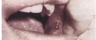
Fluorosis is a disease of tooth enamel associated with prolonged exposure to excessive amounts of fluoride. It can be endemic and occupational in nature - develop in connection with living in a region where drinking water contains a lot of fluoride or be associated with working conditions and the amount of fluoride in the air.
Endemic fluorosis is common in regions where the amount of fluoride in a liter of drinking water exceeds 1.5 mg. The occupational form of the disease is much less common, usually among employees of the aluminum industry.
Causes and mechanisms of development
Fluorine is a microelement that enters the body with water and food and is involved in many physiological processes. It is found in high concentrations in human bone tissue and teeth. The incoming fluoride is not completely absorbed; the main share is fluoride dissolved in water. If a lack of a microelement increases the risk of dental caries, then an excess becomes the cause of the disease.
Fluorosis is most often observed in children during the eruption of permanent teeth. This applies to children, including those who previously lived in regions with high concentrations of fluoride in water. It is enough to live in such a region for up to 3-4 years. Excess fluoride also affects the buds of permanent teeth. In this case, milk teeth are not affected by the disease, since their rudiments are formed in utero, and excess fluoride is retained by the placenta and is not passed on to the baby.
An element concentration of 1.5 mg per liter of water will not lead to the development of fluorosis in adults. Drinking water containing 6 mg of fluoride per liter leads to illness.
Ask a Question
Causes of the disease
The main cause of pathological changes is an excess of fluoride in the body. The optimal concentration of a chemical element is considered to be 1 mg/l; if mineralization indicators are exceeded, there is a high probability of developing fluorosis and structural defects. Fluorine easily undergoes substitution reactions and such transformations are not always benign for the body. The hard tissues of teeth are especially sensitive, and the crystal structure and strength of the protective coating are disrupted. A deficiency of a microelement is no less dangerous than an excess and entails growth retardation, defects and deformations of teeth, and pathologies of the skeletal structure. In addition, among the main reasons for the development of fluorosis are:
- eating plant and animal products with an excess of fluorides;
- irrational use of cleaning pastes and rinses enriched with microelements;
- increased fluorine content in the air (factories producing glass, metals, ceramics, optical and laser devices);
- living for a long time near industrial enterprises.
Children under fifteen years of age are at increased risk, which is explained by insufficiently strong enamel, unstable hormonal levels and lack of adequate oral hygiene.
Classification of fluorosis
By origin, endemic and occupational fluorosis are distinguished. Despite the similar clinical picture, recommendations for undergoing a course of treatment for these two types of disease will differ. An occupational disease causes disease not only of the teeth, but also of the bones - osteoporosis, osteosclerosis. The diseases are accompanied by impaired joint mobility.
As the disease develops, vegetative-vascular disorders and liver pathologies occur, and the risk of cancer—osteosarcoma—increases. It is worth noting that stains on teeth with such fluorosis may be completely absent.
Based on clinical manifestations, researchers distinguish five forms of the disease:
- spotted;
- dashed;
- chalky speckled;
- erosive;
- destructive.
The first two forms are considered a mild form of the disease. Chalky-mottled fluorosis is classified as moderate fluorosis. The last two are severe forms.
One patient may experience several forms of clinical manifestations at once - different symptoms develop on different teeth and groups of teeth. Doctors note that the resulting form of the disease persists for life and does not change even with changes in the concentration of the trace element in drinking water.
Symptoms and manifestations
Endemic dental fluorosis manifests itself as white spots or characteristic stripes on the enamel. As the disease progresses, they acquire a yellow or even brown tint. The incisors of the upper jaw are most often affected, but at high concentrations of fluoride, damage to all teeth occurs. Increased abrasion of the enamel is observed, chips and erosions form. Symptoms depend on the form of the disease.
Line
This form of the disease is characterized by the appearance of white stripes on the outer surface of the incisors. Sometimes they are pronounced, but more often they are noticeable only when the enamel surface dries. In some cases, the stripes merge into large spots, but the doctor will detect separate stripes in the structure of the spot.
Spotted
With this form, multiple white spots appear that merge with each other. The surface of the stain is shiny and smooth, without roughness. There are no clear boundaries of the stain - it smoothly transitions into healthy areas of the enamel.
Chalky mottled
With this form of enamel fluorosis, a matte tint appears on the entire surface of the hard tissues of the tooth. Clearly defined spots and dots appear on the surface. In some cases, the enamel becomes yellow. In addition, areas of destruction may be observed on the teeth: speckled depressions with a diameter of up to 1.5 mm and a depth of up to 0.2 mm. The bottom of such depressions is pigmented.
This form is characterized by increased abrasion of the enamel, which exposes dentin - the deeper tissues of the tooth, which have a dark brown color. Sensitivity of teeth when dentin is exposed is pronounced.
Erosive
This form is distinguished by the presence of larger areas of enamel destruction - erosions. In the area of such a depression, there is no enamel at all, and increased abrasion is observed on the chewing surfaces of the teeth.
Destructive
In the destructive form of the disease, abrasion and erosion occurs not only of the enamel itself, but also of other hard tooth tissue - dentin. Teeth become brittle, large chips occur, and the shape of the crowns is disrupted. The body strives to prevent the opening of the tooth cavity by forming replacement dentin. This is a severe form of the disease that can develop in regions where drinking water contains at least 10 mg of fluoride per liter of water.
Diagnosis of fluorosis
Typically, diagnosing fluorosis is not difficult for a dentist. The spotted form of the disease may resemble a so-called chalk spot - the initial stage of caries or an area of enamel demineralization. However, in the case of fluorosis, such stains are multiple in nature and affect permanent teeth after they erupt. And initial caries develops singly or on a small number of teeth. A standard examination by a doctor is usually sufficient to make an accurate diagnosis.
One of the diagnostic features is the recommendation to submit drinking water for analysis, this will determine the concentration of fluoride in it. If elevated levels are detected, the doctor will prescribe changing the source of drinking water or purifying it before drinking. If you do not follow this recommendation, a severe form of the disease may develop, accompanied by tooth decay.
Features of treatment
If you have enamel fluorosis, your doctor will recommend using fluoride-free toothpastes and mouth rinses. This recommendation applies to all cases and forms of the disease.
Removal of affected areas of enamel and filling are not used, since the likelihood of the filling falling out and further tooth destruction is very high. The doctor may prescribe phosphorus and calcium supplements to further strengthen hard tissues.
Mild forms of the disease are subject to cosmetic correction. Chemical, photo-whitening or laser teeth whitening can be used. After this, remineralizing therapy is necessarily performed: the doctor will apply preparations based on phosphorus and calcium compounds to the enamel using electrophoresis, ultraphonophoresis or application. Sometimes it is advisable to use mouthguards with a special composition inside - this procedure can be carried out both at home and in a dental office. The course of remineralizing therapy for this disease consists of 10-20 procedures, depending on the degree of tissue damage.
Chalky-mottled, erosive and destructive forms do not allow effective whitening. In this case, dental restoration can help achieve a cosmetic effect - installing lumineers or veneers. In severe cases, treatment of fluorosis is carried out by restoring teeth with artificial crowns made of metal-ceramics or ceramics.
Orthopedic treatment can not only eliminate a cosmetic defect, but also prevent further tooth decay, as well as cope with increased sensitivity.
Clinical researches
Clinical studies have proven that regular use of professional toothpaste ASEPTA REMINERALIZATION improved the condition of the enamel by 64% and reduced tooth sensitivity by 66% after just four weeks.
In addition, it has been proven that in order to eliminate increased sensitivity of teeth in people suffering from type 2 diabetes mellitus, long-term use of the domestic toothpaste “Asepta plus remineralization” is advisable, which is important to consider in the complex treatment of the dental pathology in question to ensure psychological balance, quality of life, restoration of chewing function and satisfaction with treatment of such patients.
Sources:
- Features of personal response in the prevention and treatment of hypersensitivity of teeth against the background of type 2 diabetes mellitus A.K. YORDANISHVILI, Doctor of Medical Sciences, Professor, North-Western State Medical University named after. I.I. Mechnikov, Military Medical Academy named after. CM. Kirova, International Academy of Sciences of Ecology, Human Safety and Nature, N.A. UDALTSOVA, Candidate of Medical Sciences, Associate Professor, St. Petersburg State University of the Government of the Russian Federation; dental clinic No. 29, Frunzensky district of St. Petersburg, O.V. PRYSYAZHNYUK, head. surgical department No. 2, dental clinic No. 29, Frunzensky district of St. Petersburg
- Report on the determination/confirmation of the preventive properties of personal oral hygiene products “ASEPTA PLUS” Remineralization doctor-researcher A.A. Leontyev, head Department of Preventive Dentistry, Doctor of Medical Sciences, Professor S.B. Ulitovsky First St. Petersburg State Medical University named after. acad. I.P. Pavlova, Department of Preventive Dentistry
- Report on determining/confirming the preventive properties of toothpaste “ASEPTA PLUS” GENTLE WHITENING” Author: doctor-researcher A.A. Leontyev, head Department of Preventive Dentistry, Doctor of Medical Sciences, Professor S.B. Ulitovsky First St. Petersburg State Medical University named after. acad. I.P. Pavlova, Department of Preventive Dentistry
Prognosis and prevention
The prognosis is favorable if the patient consults a doctor in a timely manner and follows all instructions. Even severe forms of the disease leave room for restoring the aesthetics of a smile using modern methods. But the sooner you contact a specialist, the less effort treatment of dental fluorosis will require.
The main direction of prevention is to reduce the amount of fluoride entering the body. When living in a region with a high concentration of trace elements in drinking water, it is important to find another source - consume purified water or purchase water brought from another region. You should also limit the consumption of fluoride-containing products: sea fish, spinach, butter, seaweed, etc.
It is important to stop using toothpastes, gels and rinses with fluoride. Please note that most children's toothpastes do not contain fluoride or aggressive abrasive particles.
The dentists at the STOMA clinic know how to treat fluorosis of all forms. We have ample opportunities for professional whitening and high-quality teeth restoration. You can make an appointment for an examination and consultation with a doctor by phone or through a special form on the website.
What causes dental fluorosis?
Dental fluorosis is caused by consuming too much fluoride during the formation of permanent teeth in children. This occurs before the age of 8 years. To avoid it, supervise tooth brushing so that children do not use too much toothpaste, mouthwash, and learn to spit out excess toothpaste rather than swallow it.
How do you know if your child has dental fluorosis?
Since there are many possible reasons for changes in the appearance of teeth, it is better to seek professional help. Your dentist can check your teeth for fluorosis or other problems. Regular visits to the dentist ensure complete control over the occurrence of this disorder.
How common is fluorosis?
Fluorosis first attracted the attention of scientists and doctors in the early twentieth century. Researchers were surprised by the high prevalence of what was called "Colorado Brown Stain" on the teeth of Colorado Springs natives. The stains were caused by high levels of fluoride in the local water supply. People with these spots also had an unusually high resistance to tooth decay. Fluoride is an important mineral for the normal development of children. Our mouths contain bacteria that combine with sugars in foods to produce acid that harms tooth enamel and damages the teeth themselves. Fluoride protects teeth, but increases the likelihood of fluorosis, which is why this disease is quite common these days.
How much fluoride should a child take to protect their teeth without risking fluorosis?
Children who use fluoridated dental products (toothpastes, rinses, etc.) will receive the fluoride they need for healthy teeth. There is no need to monitor your intake of water or food containing fluoride, since the body receives low doses of fluoride from these sources.
