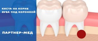
Tooth root caries is a common disease when the neck of the tooth is exposed. It is based on damage to the enamel by microorganisms that feed on food debris and produce acid that can destroy the surface of the teeth. Root caries is often secondary: the infectious process gradually penetrates into deeply located tissues. Inflammation spreads to the nervous tissue of the tooth - the pulp.
With gum recession and other disorders, root caries occurs without visible changes to the enamel. The disease is dangerously asymptomatic for several months or even years. The pathological process occurs inside the tooth and is invisible to the patient.
Make an appointment with a therapist by phone+7(985)532-21-01
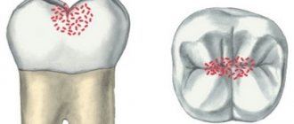
Types of caries localizations
Caries (lat. caries rotting) is a slow and hidden pathological process occurring in the hard tissues of the tooth. It is characterized first by focal demineralization of the enamel, then by the destruction of hard tooth tissues with the appearance of a cavity in the dentin. If left untreated, the tooth is lost or complications arise - pulpitis and periodontitis.
Cervical (circular)
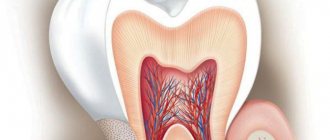
The structure of a tooth is divided into a root, a neck and a crown - the latter is located above the gum. The neck and root are located in the gum and are protected by soft mucosal tissue.
When the carious process begins to destroy the tooth in the area of contact of the crown with the mucous membrane, we can talk about the cervical form of caries.
This category is the most difficult to diagnose and treat. It most often affects the incisors. When biting food with incisors, the food is not only “cut off”, but also gets clogged into the gum pockets, moving towards the gums.
Crown caries
Caries has the ability to affect not only healthy, but also already treated teeth - under a filling or crown. If there is a crown, it is difficult to detect, because the tooth does not have a nerve and pain is not felt. There are no external signs for a long time.
The causes of this type of caries are:
- defect of the dental crown with the appearance of access to the tooth tissue underneath it
- poorly treated tooth before prosthetics;
- gum disease;
- poor oral hygiene.
The problem is that the process spreads into the deep layers and spreads to the bone structures, as a result of which the tooth is often completely removed. That is why preventive examinations and an orthopantomogram - a panoramic image of the entire jaw - are so important.
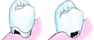
Radical caries
Basal and cervical caries are similar. Both incisors and chewing teeth are affected. Destruction begins with the enamel, gradually moving to the nerve in the root canal. When the process touches bone tissue, it threatens with osteomyelitis.
The main reason for the occurrence is a cariogenic situation in the oral cavity, when plaque accumulates especially quickly. The enamel in the root part of the tooth is much thinner and more easily destroyed. It is most often diagnosed between the ages of 30 and 60 years.
Root caries
Root or subgingival caries occurs hidden. It can be with or without a cavity. Along the flow - active, suspended, secondary. It most often affects molars.
Treatment
Treatment tactics are determined by the stage of the carious lesion.
Initial. The structure of hard tissues remains almost unchanged, there are signs of enamel demineralization, and white spots may appear. To stop the destruction of hard tissues, remineralization is performed. Remotherapy will help strengthen the enamel and stop its destruction. Before remotherapy begins, professional hygiene and cleaning of periodontal pockets are performed. Additionally, the doctor may prescribe rinsing with antiseptic solutions.
Dark spot. The dentist removes hard and soft deposits from the surface of the enamel and cleans the periodontal pocket. The affected tissue is removed and a filling is performed. If gum damage has already begun, a focus of inflammation has appeared, additional treatment is carried out: the doctor stops the infection.
Formed cavity. It will be possible to save the tooth provided that the inflammation has been stopped. The doctor removes the affected tissue, strengthens the crown with an inlay, and places an artificial crown.
Root destruction. If the root is destroyed by more than 50%, with severe inflammation or damage to periodontal tissue, the tooth must be removed. After this, implantation and prosthetics or restoration with a dental inlay are performed.
What is the danger of this disease?
Caries is dangerous because there are no symptoms for a number of years. It is detected more often in an advanced stage, when tooth extraction becomes the solution.
Visual diagnosis is difficult because all destructive processes occur inside the tooth. Pronounced plaque or tartar hides any stains on the enamel.
The root is hidden under the gum and external irritants do not affect it until a certain time. On the other hand, the root walls are thin, so they are destroyed quickly and with complications.
Diagnosis and symptoms of tooth root caries
Diagnosis of root caries is complicated by the fact that the disease can be asymptomatic for quite a long period of time. A visual examination of the oral cavity only in 11 - 13% of cases helps to identify the presence of the disease. Therefore, additional examination methods are required for diagnosis:
1) probing;
2) X-ray examination (orthopantomogram, radiography, etc.);
3) electrical odontometry (assessment of nerve response);
4) thermometry (checking the reaction to hot and cold).
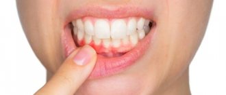
It is impossible to identify the presence of dental root caries on your own, but basal or cervical caries, which in most cases accompany root damage, is quite possible. This can be done with a toothpick and a mirror. With clean hands, take a toothpick and pass it between the gum and tooth, pressing lightly. If pain occurs or roughness is found on the enamel, there is a possibility of developing caries of the cementum of the tooth root. This means it's time for you to visit the dentist.
Symptoms of root caries, which, as we noted above, may not always appear:
- pain when exposed to high/low temperatures;
- pain when eating sour and sweet foods;
- increased sensitivity of the periodontium;
- discomfort when chewing;
- aesthetic defect at the border with the gum.
Causes of the disease
There are 3 main provoking factors that must act in a complex manner; none of them acts independently:
- Cariesogenic microflora - this includes Mutans streptococci, actinomycetes and certain types of lactobacilli. They should dominate the oral cavity. Bacteria produce organic acids from food carbohydrates, which cause demineralization of cement.
- Consumption of simple carbohydrates is the most cariogenic. Their breakdown produces glucan, which contributes to the appearance of plaque.
- Reduced caries resistance is a deterioration in the resistance of tooth tissue and the body as a whole. This is facilitated by a decrease in calcium content in hard root tissues, bad habits, and lack of saliva.
In addition, the anatomical features of the mouth can also have an effect - a small vestibule, a short frenulum, and bite pathology.
Gum disease, pocket formation
Gum pocket or otherwise periodontitis is a common occurrence in many patients. Many people do not treat it because it does not bother anyone for a long time.
The first manifestation of pathology is bleeding when brushing teeth, although outwardly they may look healthy. Dental pockets or depressions are normal, but their anatomical size should not exceed 3 mm.
Then the pocket is able to self-clean from food debris and epithelial particles. Reservoirs of bacterial accumulations form in deep gaps. This is accompanied by bad breath. Conventional treatment with antibiotics and brushing teeth with toothpaste have no effect.
Causes of periodontitis:
- insufficient hygiene;
- poor quality fillings;
- abnormal position of the dentition, malocclusion;
- hereditary predisposition;
- deficiency of vitamins and minerals;
- immunosuppression.
Dental root caries: causes
Root caries occurs as a complication of gum disease, in which pathological pockets form between the tooth and gum. They accumulate food debris and pathogenic microbes. They are difficult to remove with a toothbrush and require professional cleaning at the dentist's office.
Without proper oral care, cariogenic bacteria multiply rapidly. They secrete acids that corrode the cement of the tooth root. Gradually, microorganisms penetrate deep into hard tissues down to the pulp, causing pain.
Often this pathological process is caused by osteomyelitis, periodontitis, periodontitis or periostitis, that is, inflammatory diseases of the gums and hard tissues of the oral cavity. Indirect causes of the development of tooth root caries are past infectious diseases and reduced immunity. In addition, the likelihood of carious lesions increases with tobacco smoking, alcohol abuse, poor diet and lack of vitamins and minerals in the body.
If a person rarely brushes his teeth or does it incorrectly, the carious process spreads even faster. As a result, you can lose a tooth without even feeling symptoms of the disease. As a rule, the first signs appear after the pathology has affected most of the tooth root.
Lifestyle
If you have learned about the risk group for root caries (older age), this does not mean that you can relax. Caries has many causes and can occur at any age if basic precautions are not followed.
Those at risk include smokers, diabetics, pregnant women and even children. The provoking factors in this case are:
- smoking and lack of oral hygiene;
- infrequent brushing of teeth and lack of fluoride;
- poor nutrition with preference for desserts;
- frequent stress;
- alternating hot and cold;
- structural features of the oral cavity;
- alcohol abuse - alcohol breaks down into sugars and acids;
- abuse of coffee and strong tea, which creates an acidic environment in the oral cavity;
- lack of water intake, and therefore lack of saliva;
- gum injuries.
How to treat cervical caries
Treatment of the disease is carried out by a doctor. In the case of cervical caries, there are a number of reasons for immediately contacting a dentist:
- Rapid development of complications. In the cervical area, the thickness of hard tissue is minimal, so pulpitis and periodontitis occur more quickly than with other localizations of caries.
- High probability of crown fracture. Since the lesion tends to cover a large area in the gingival region, due to the softening of hard tissues over a large area, the tooth can break with a slight load.
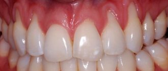
- Visible aesthetic defect. Since cervical caries often affects the front teeth, their destruction is noticeable to others, which negatively affects self-esteem, personal life, and career.
Depending on the stage of the pathological process and the damage to certain teeth, treatment methods may vary.
Treatment of cervical caries of chewing teeth
The treatment method for cervical caries depends on the advanced stage of the disease. If in the early stages conservative treatment is sufficient, then in later stages it is impossible to do without drilling the carious cavity (local anesthesia is performed before drilling).
- Treatment of “chalk spot” and suspended caries. The traditional method is long-term use of products containing fluoride. The dentist applies medications to the affected tooth. At home, the patient uses fluoride-containing toothpastes and rinses to care for his teeth. A modern alternative method is the use of a complex system that includes etching the stain with acid and “sealing” the affected area with a polymer resin (ICON). Treatment is carried out in one visit. Before conservative treatment (by any method), professional tooth cleaning is required.
- Treatment of enamel caries with the formation of cavity and medium caries. Includes preparation (removal of affected hard tissue), treatment of the cavity with an antiseptic solution, thorough drying and filling. After drying, the filling is ground and polished. The procedures are carried out during one appointment.
- Treatment of deep caries. It consists of the same stages as for median caries, but after drying the cavity is closed with a therapeutic demineralizing bandage and filling.
In the treatment of cervical caries, they always try to preserve the living pulp (depulpation is performed only when pulpitis develops). Preserving the pulp is important because it is responsible for the production of dentin, the natural restoration of hard tooth tissue. A tooth with preserved pulp lasts longer than a pulpless one.
Treatment of cervical caries of anterior teeth
The principles of treatment of cervical caries of the frontal group of teeth, depending on the spread of the process, do not differ from those with damage to molars and premolars. However, there are some features associated with the fact that the front teeth are visible when talking and smiling. When restoring them, it is important to obtain a good aesthetic effect. To achieve this you can use:
- Fillings. At the Academy Dent clinic, modern light-curing composite materials are used to fill carious cavities. They imitate tooth enamel well and can be used to cover a defect unnoticed. The seal is made of high-quality material, installed in compliance with all rules, and lasts 10-15 years.
- Veneers. The installation of plates on the front part of the frontal teeth is indicated for extensive or combined defects (if, in addition to cervical caries, there is a chip or a violation of the position of the tooth). Porcelain and, especially, zirconium veneers compare favorably with composite onlays and fillings in their durability, impeccable aesthetic results, and do not accumulate plaque. The disadvantage of installing any veneers is the preparation of the entire surface of the tooth (including the removal of the entire layer of healthy enamel).
Treatment with folk remedies
It is impossible to cure cervical caries at home. Attempts at self-medication lead to a loss of time for conservative therapy, further tooth destruction and often to its loss. The use of folk remedies is practically not justified.
Symptoms of the disease
The process usually occurs without symptoms, but pain may occur when brushing a toothbrush, eating sour, salty, sweet, cold or hot foods.
After eliminating the irritant, everything goes away. The patient can see a doctor if a stain is found on the front surface of the incisors, but often it is hidden under plaque or tartar.
The ongoing carious process gradually reaches the dentin junction, penetrating first into its surface layers and then further. The cavity deepens with bacteria and food debris. There is a smell from the mouth. Irritants cause pain more and more frequently.
With cement caries, teeth become mobile and lose their support, and bleeding gums occur. These are already symptoms of periodontitis. Now, even when chewing food or brushing teeth, severe discomfort occurs.
Digestion begins to suffer. Teeth become hypersensitive to hot or cold.
Next, the process follows the Leus classification:
- Active lesion - the edges of the cavity are undermined. The cavity is filled with softened tissues and tends to grow.
- Suspended caries - no increase in the affected area is observed. The cavity is clean, the bottom is shiny and smooth, the edges are even and dense.
- Secondary caries - occurs under a filling.
Stages of cervical caries
Cervical caries develops gradually, the process of disease development includes the following stages:
- Superficial caries. Characterized by damage only to tooth enamel. The disease begins with the stage of a “chalk” stain - the appearance of a white mark on the surface of the tooth. In the affected area, the enamel softens and becomes matte. Pain is not typical for the initial stage of caries, however, as the enamel further deteriorates and a cavity forms, short-term discomfort or pain may be experienced associated with brushing teeth, eating cold or hot, sour or sweet foods.
- Average caries. At this stage, dentin is involved in the pathological process. With caries in the middle stage, the pain is still associated with external influences, but lasts longer and becomes more intense.
- Deep caries. It is distinguished by deep penetration into the dentinal layer and severe pain. The reaction to external stimuli intensifies many times, the pain becomes unbearable.
Features of the flow
A distinctive feature of cervical caries is the extensive area of the carious cavity. Unlike other types of caries localization, the lesion quickly spreads not only in depth, but also in breadth. Cervical caries can be complicated by root caries when the cement covering the root of the tooth is involved in the pathological process.
The entire process from the beginning of demineralization of hard tooth tissues to the formation of a carious cavity can take from several weeks to several months (on average 3-6 months). Sometimes the progression of the disease stops - the growth of the stain stops, and the sensitivity of the teeth decreases. Suspended cervical caries is not a reason to refuse a visit to the dentist. It should also be treated, since the disease can become active at any time.
Diagnosis of the disease
The diagnosis is made in stages. Listening to complaints and visual examination make it possible to diagnose caries only in 13% of cases. Classic probing with a sharp probe and inspection with a mirror are informative. This allows you to examine the dentition.
Thermal diagnostics, electroodontometry and x-rays are also performed. If the patient has massive dental plaque, changes in the enamel will not be visible. In this case, professional teeth cleaning is first carried out to remove plaque and stone. All this is possible in a dental clinic.
At the dental clinic
The following types of studies can be performed at the clinic:
- Probing with a thin probe with a curved end - it is inserted under the gum, while the doctor has the opportunity to examine the structure of the tooth, its integrity, the presence of irregularities and chips. With rapidly progressing caries, the edges of the cavity are uneven and sharp. In the remission stage, the surface of the pathological focus is shiny, smooth, hard with smooth edges.
- Electroodontometry - can determine nerve damage and the depth of inflammation. The pulp reacts differently to the current strength.
- Thermal diagnostics - treatment of different areas of the tooth with a stream of water and heated wax. Unpleasant sensations that disappear after removing the irritant indicate caries.
- X-ray - a targeted photograph of one tooth or computed tomography. The presence and localization of obvious or hidden inflammation is determined with millimeter precision.
- Visiography is a special device - a visiograph scans the received data and transfers it to a computer, where the picture is studied from different angles and in detail, revealing the hidden process of inflammation.
Features of the progression of dental caries
In the area of cement, caries can occur in different ways, and on this basis it is classified into 4 types:
- progressive;
- rapidly progressive;
- remission;
- relapse.
Progressive root caries is characterized by a relatively slow rate of tooth root destruction. With rapidly progressing lesions, the lesion spreads in depth and breadth at high speed. The remission stage is when root cement caries does not progress. Relapse suggests that root caries develops along the edges of a previously placed filling.
Treatment or removal - what determines the outcome
The doctor makes a decision on treatment or tooth extraction after determining the depth of spread and the area of localization of the inflammatory process. It is mandatory to first carry out professional cleaning using an ultrasonic scaler, an AirFlow water-abrasive device and hand tools.
Initial caries is treated with conservative retherapy. For hard tissue defects, preparation and filling are performed. In severe cases, the tooth is removed.
Treatment of the initial stage of root caries
Conservative retherapy includes:
- fluoridation of teeth;
- covering teeth with protective agents
- training in proper oral hygiene
Different types of caries: WHO classification
Caries is a slowly occurring pathological process in hard dental tissues, which develops under the influence of unfavorable external and internal factors. According to statistics, this disease occurs in more than 90% of people in the world.
In this article
- Different types of caries: WHO classification
- How does dental caries develop in the root area?
- Features of the progression of dental caries
- Dental cement caries - main clinical manifestations
- Diagnosis of cement caries: basic and additional methods
- Treatment of cement caries: main stages
- Prevention of root caries
According to the classification of the World Health Organization, there are several types of caries: enamel, dentin, cement, as well as suspended, other, unspecified caries, and odontoplasia. In this article we will talk about dental cement caries, better known as tooth root caries.
Cleaning in dentistry from stone and plaque
1243900247100
Professional teeth cleaning involves removing the accumulation of all pathogenic microflora around the root.
It involves not only removing plaque, but also stones. This elimination of negative factors protects teeth for a long time.
Antiseptic treatment of affected areas
Treatment of root caries involves drilling out all affected areas with a drill and treating the carious cavity with antiseptics and special preparations. A 2% chlorhexidine solution or a gel based on it is used as an antiseptic for the treatment of carious cavities.
Treatment of cement caries: main stages
With root caries, the most difficult task is to gain access to the affected area, especially if the carious lesion is located under the gum. Traditionally, treatment of root caries takes place in several stages:
- Retraction. First, the dentist artificially exposes the neck of the tooth and part of the root, lowering the gums with special devices.
- Coagulation of the gums. It is carried out if indicated using laser exposure to gum tissue. The essence of the manipulation is to remove overgrown tissue.
- Direct treatment of caries. The choice of tactics depends on the degree and depth of the lesion. If caries is in the initial stage of the stain, then remineralizing therapy will be sufficient - restoring the mineral composition of tissues with the help of fluoride preparations. If the doctor reveals more extensive and deep caries with the formation of a cavity, then surgical treatment with preparation and filling will be required.
- Installation of an inlay or crown if the volume of destroyed tissue is more than 50%.
It is necessary to treat cement caries only in a trusted clinic with a reliable specialist, since violation of treatment technology and the use of low-quality materials can cause secondary caries, provoke the development of infection and inflammation of the gums, and lead to tooth loss.
What to do to protect your gums
If the gum is not treated, inflammation will remain and all treatment will become meaningless. The gums are protected by diathermocoagulation and retraction.
Diathermocoagulation
Diathermocoagulation – excision of excess areas of periodontitis with a coagulation knife. The knife immediately cauterizes the bleeding edges. The procedure is performed when there are severe changes in the gums. In such cases, filling is postponed until the soft tissue is completely regenerated.
Retraction
If the condition of the gums is advanced, treatment of caries becomes impossible - the mucous membrane bleeds and interferes with tooth treatment. This gum is excised or a special device is applied to move the gum back. But more often, retraction is performed - lowering the gingival contour or moving apart the overhanging edges of the gums using special hemostatic threads. A temporary filling is placed until complete healing.
Diagnosis of cement caries: basic and additional methods
Diagnostic methods that help identify dental caries are divided into basic and additional.
The main ones include:
- patient interview;
- external visual inspection;
- probing - determining the depth of the cavity, as well as the density and tenderness of tissues, using a dental probe;
- percussion (tapping on the tooth).
Additional diagnostic methods are:
- staining with a special dye to see the affected areas;
- thermal test - the tooth is irrigated with cold water and the pain reaction is assessed;
- radiography - helps to detect hidden carious cavities and caries of contact surfaces;
- electroodontometry - prescribed to determine the condition of the pulp;
- luminescent method - illumination of teeth with ultraviolet rays, under which tissues affected by caries change their shade.
Root caries itself is most often diagnosed through sequential manipulations:
- remove deposits from under the gums using ultrasonic and manual instruments;
- isolate the tooth root from saliva using a special plate;
- using a probe, assess the condition of the root surface;
- with the help of x-rays or radiovisiography, even small defects under the gum, in the gingival zone, and carious lesions at any stage are detected;
- if necessary, confirm the diagnosis of “cemental caries” or differentiate it from pulpitis, thermometry or electrical odontometry is prescribed.
What preventative measures are there?
To keep your teeth healthy for many years, you need to brush your teeth twice a day, use floss and a tongue scraper. After sweets, you should rinse your mouth and chew gum for 5-10 minutes.
Prevention also includes the removal of tartar. A sufficient amount of saliva is also important, so it is necessary to maintain a water regime.
Like any other pathology, caries is better prevented than treated. Therefore, it is important to visit the dentist twice a year for preventive examinations. People at risk should approach this especially responsibly.
Other
Is caries contagious?
Let us recall that dental caries is caused by cariogenic bacteria of the oral cavity - most often certain types of streptococci and actinomycetes. In adults, this microflora is present in everyone without exception. Therefore, you are unlikely to be infected with caries, for example, by kissing. But with children of early childhood the situation is completely different, because... children are born with a sterile oral cavity. In young children, caries is an infectious disease that is caused by infection of the oral cavity of a newborn child by those who care for him.
As a rule, cariogenic microorganisms enter the child’s mouth when the mother (or other persons caring for the child) licks the pacifier, the spoon with which the child is fed, and other objects intended for food. If you decide to try your child’s food with his spoon or kiss your child on the lips, it means you have just passed on your cariogenic bacteria to the child. And the earlier the infection occurred, the higher the intensity of the carious process will be.
Further, children experience rapid growth of cariogenic microflora, which leads to demineralization of tooth enamel. If you pay attention, in young children the carious process affects the teeth circularly - around their necks. Such carious lesions are called bottle caries. Most often, this happens when a child is not only infected with a cariogenic infection, but also when feeding rules are violated at the same time - drinking sweets at night, feeding on demand, etc.
Sources:
1. Higher prof. the author’s education in therapeutic dentistry, 2. Based on personal experience as a dentist, 3. National Library of Medicine (USA), 4. “Therapeutic dentistry: Textbook” (Borovsky E.), 5. “Pediatric therapeutic dentistry. National leadership" (Leontyev V.K.).
How children are treated
Many patients think that baby teeth need not be treated. But this is a common misconception that can lead to serious problems.
If the caries of a baby tooth is not cured, there is a high probability that, as a result of the advanced disease, problems will arise with the change of the time series to the root one.
When treating a child, it is very important to fulfill several basic requirements at once:
- An individual approach to the child is important. Our clinic employs doctors who specialize in treating children. They know well how to calm a small patient and what to do so that he does not worry. This is important because it is in this case that it is possible to rid the child of fear of dentists in the future.
- Only specially selected anesthesia is used. It is important to use medications that are needed specifically for a child at a young age.
- Doctors always try to detect the disease at an early stage and do everything to cure it. This way, you can ensure that you do not have to use more traumatic methods for treatment.
Our dentists know well how to treat caries in baby teeth without pain and potential threats to the baby’s health.
How long does it take to treat caries?
The duration of the treatment procedure may vary depending on the condition of the patient and how deep the tissue damage has gone.
Depending on the initial conditions, treatment can be carried out in one or two stages. In the first case, the process rarely takes more than half an hour. In the second case, this time is extended over two visits - you need to kill the nerve and only then carry out other manipulations.
Thanks to their extensive experience and level of professionalism, our dentists cope with the task with minimal time.