If you have ever had a magnetic resonance imaging scan, you know that before the examination you are asked to remove all metal objects. With rings and earrings everything is simple. Is it possible to do an MRI with dental implants? After all, they cannot be removed during the procedure.
Why are things made of metals removed before an MRI?
The principle of operation of a magnetic resonance imaging scanner is to create its own magnetic field in which the patient is located. Hydrogen atoms in the patient's body resonate with a magnetic field and the result is recorded as a series of images. These are virtual sections of organs and tissues that help make an accurate diagnosis.
Doctors ask that metal objects be left outside the room with the tomograph so that any metal or alloy that can be magnetized does not accidentally enter the magnetic field. All substances are divided into:
- Diamagnets
. Their magnetic susceptibility is negative. Silicon, gold, silver are diamagnetic. - Paramagnets
. They are capable of magnetization, but their magnetic susceptibility is very weak and difficult to detect, so only scientists understand the difference between dia- and paramagnets. Platinum and aluminum are paramagnetic. - Ferromagnets
. The magnetic susceptibility of such metals is high; they are capable of magnetization and change location under the influence of a magnetic field. Iron, cobalt, nickel are ferromagnetic.
Based on these properties, doctors ask to remove metal objects during magnetic tomography. Ferromagnets not only distort images and heat up in a magnetic field, but can also shift under its influence.
With which prostheses is it unacceptable to perform magnetic resonance diagnostics?
Although it is permissible to do an MRI with dental implants. During this examination, “internal” images of the body are obtained. A magnetic field is created in the tomograph, and the patient is placed in it.
The patient does not feel pain, and MRI does not damage tissues. The magnetic field created by the tomograph is 10 thousand times stronger than the Earth's magnetic field. It has the ability to attract objects that contain metal. If there are elements made of ferromagnetic materials in the magnetic resonance examination field, a potential threat arises. For this reason, people who have metal clamps on the aneurysm are not allowed to be diagnosed. The magnet can move the clamp - this will damage the vessel.
MRI diagnostics cannot be performed in the presence of the following types of implants:
- Cardio;
- Neuro;
- Ear;
- Containers that provide dosed dispensing of medications.
All electronic elements in the body, whose functioning can be disrupted under the influence of a magnetic field, the movement of which can create a health hazard - a limitation for MRI. But there are always exceptions, these include implants whose passport indicates their compatibility with the procedure.
Is it possible to do MRI with titanium implants?
Titanium is paramagnetic
.
Theoretically, it is magnetized. In practice, its magnetic susceptibility is 10 -5
. Such values are so small that in everyday life we say that titanium is not magnetized. It interacts with the magnetic field within limits that do not in any way affect the examination results. It is possible to do MRI with dental implants of all organs, including MRI of the brain. Fears that implants will move are unfounded, for the simple reason that titanium is not ferromagnetic. It does not heat up or change location under the influence of a magnet. Implantation is not a contraindication for tomography.
What about crowns and pins?
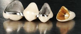
The question arises: what about the crowns that are placed on the implant? Some of them contain metals.
Modern metal dental crowns are created from alloys with weak or negative magnetic susceptibility. However, crowns that are many years old can distort the image and negate the diagnosis. Moreover, the more crowns, the stronger the distortion.
In principle, it is possible to do an MRI with metal crowns, provided that the patient warns the doctor about their presence. The doctor will take into account all factors: the examination organ, the fusion of the prosthesis, and will decide whether to carry out or refuse the procedure.
MRI with a pin in a tooth (stump inlay) has no contraindications if the pin is made of fiberglass. An MRI with a metal pin in the tooth is performed, because the amount of metal there is minimal and has virtually no effect on the results.
The doctor should be warned about metal crowns and pins. Ideally, he should be told what type of alloy was used. This can be found out in the dentistry where the prosthesis was installed. New tomographs take such information into account and correct the data.
Should you tell your doctor that you have dental implants?
A patient with installed dental implants must notify the doctor about this. Then the specialist makes the necessary adjustments, taking into account the location and composition of the prostheses. These settings eliminate the appearance of blurred distortions (also called field artifacts). And then the results of the examination will be clear and precise.
But if a person decides to keep silent about “non-native” teeth, then the picture will turn out blurry, because implants, although slightly, do affect the quality of the pictures. And such a careless patient will have to pay for the procedure and undergo diagnostics again. Why does the blame in this case fall on the patient? Because before the MRI, he will definitely be asked about the presence of implants and what kind of metal they are made of.
“Mom 2 years ago had an MRI of the head and blood vessels, using a high-field tomograph. And she’s had implants installed in her upper jaw for about 5 years now. And nothing, everything went fine. There are no special sensations during the procedure. Just be sure to tell the girls at the reception that there are implants before starting the procedure. They warn the doctor themselves, and then the device is reconfigured.”
Vitalina, from correspondence on the woman.ru forum.
conclusions
- There is no reason not to get an MRI if you have implants. Dental titanium is practically not magnetized. Titanium implants do not affect the examination results.
- Metal pins are also not a contraindication to tomography. The amount of metal there is minimal, and in fiberglass products it is completely absent.
- Metal dental crowns can distort results. This depends on the metal from which they are made, the number of prostheses and the organ being examined. Modern materials for prosthetics have low magnetic susceptibility. They are safer. In general, it is better to choose metal-free designs when installing crowns. This will prevent problems with MRI in the future.
Expert of the article Alexey Pavlovich Nesterenko Implant surgeon
Experience more than 10 years
How to avoid inaccuracies during MRI?
As mentioned above, implants can cause distortions in MRI images.
But this can be completely avoided if you provide the doctor with accurate information about the implants and their location. The doctor can change certain settings in the device, and distortion will be minimized. Therefore, if you feel discomfort in the oral cavity, you can safely contact the doctors of our clinic. Even if you need an MRI and you have dental implants, this will not be a problem. Come to us for a consultation, and we will select the optimal examination method just for you!
How can MRI affect iron crowns?
Prostheses made of iron, steel, cobalt alloys and other ferromagnetic materials are still most often installed. Yes, there is titanium, porcelain and zirconium dioxide - but they are expensive, so not everyone can afford them.
Iron orthopedic structures are durable and cheap. But they become a problem if you need to do an MRI for 4 reasons:
- They distort the picture. The results will not be accurate. This is especially critical when diagnosing oncological pathologies: MRI is the best method for identifying tumors at the initial stage, when recovery success is high.
- They get very hot. This leads to a burn of the mucous membrane.
- They move from their place. After displacement, the crowns will have to be adjusted, re-attached, or new ones made.
- Possible loss. The magnetic field attracts metal crowns - it literally rips them out of your mouth.
Iron crowns get very hot and distort MRI results
Patients with metal prostheses are recommended to remove the structures before MRI or undergo another study - CT, PET. The above applies to closed-type tomographs or when examining the head, cervical or thoracic region. There is no risk when diagnosing other parts of the body.
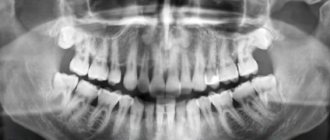
What does a 3D image of teeth show?
Tomography in dentistry is used to study the structure of teeth, their location, vessels and nerves of the jaw area. The examination area also includes the paranasal sinuses and nasal cavity.
The tomogram determines:
- Structure, injuries of the jaws, condition of the skull bones.
- Defects in root position.
- Inflammatory phenomena, complications of caries.
- Pathological gum pockets, general periodontal condition.
- Anomalies in the development and growth of the dental system.
- Number and nature of channels.
- Pathologies of the paranasal sinuses, deviated nasal septum.
- Tumor process at an early stage, metastases.
- Quality of installation of seals.
- Success and progress of treatment: therapeutic, endodontic, surgical, orthopedic (prosthetics), orthodontic.
Computed tomography examination facilitates diagnosis before implantation, surgery in maxillofacial or ENT surgery.
How long does a crown stay on a tooth?
The service life of entire crowns primarily depends on the material. So:
- metal ones last an average of 5 years;
- metal-ceramic – 8-10 years;
- ceramic and zirconium dioxide - 10-15 years and above.
The duration of wearing dentures is also affected by: how well the supporting tooth was treated and ground, the skill of the dental technician and the quality of oral care. The dentist is visited every six months to remove plaque from products and adjust them. And after 7-10 years, most crowns become unusable and are replaced with new ones.
But loose and broken dentures will not last long: from several days to a couple of weeks. Therefore, people contact the clinic immediately: the faster the doctor adjusts or replaces the bridge or crown, the greater the chance of saving the abutment tooth.
This is how a crown is sawed
How often can you do it
Cone beam tomographs have been developed for dentistry and maxillofacial surgery. They have a clearly directed narrow beam of rays, which allows you to speed up the procedure and reduce the radiation dose to 40-70 microsieverts.
According to sanitary rules, a person can receive a total dosage of 1000 μSv in a year without health consequences. Therefore, tomography is allowed more than 10 times a year.
3DCT is performed for pregnant and nursing mothers according to strict indications. The decision on how often a dental CT scan can be done during pregnancy and breastfeeding is made by a consultation of a gynecologist, pediatrician, and dental specialists.
MRI option
The cost of MRI varies depending on the clinic and the type of machine. For those who have dentures on pins, it is important to choose modern devices, because they have the function of adjusting individual parameters. As a result, the diagnosis is adjusted to the characteristics of each patient, and the doctor receives accurate and not distorted results.
Such modern MRI machines are not available in all clinics in Kyiv and Ukraine. When receiving referrals for diagnostics, it is worth discussing this point with your doctor. Prices for diagnostics will also vary accordingly.
Private: Kozharin Oleksandr Mikhailovich
Dental surgeon, implantologist
Did you like the article?
Rate: (no rating)
Loading…
How is it carried out?
The procedure is painless and takes 5 - 10 minutes. Before starting, the patient will be asked to remove metal accessories and hearing aids, which cause false darkening in the image. The safety of internal organs susceptible to radiation (thyroid gland, reproductive system, heart) is ensured by protective pads and vests.
In dentistry, the injection of a contrast agent is rarely used. If such manipulation is indicated, you should come on an empty stomach. Tests are carried out in advance to determine individual sensitivity to the drug.
To perform it, you need to sit or stand, pressing your face tightly against the frame of the device. The X-ray room employee will tell you the correct position. The head is fixed with a special stop to prevent “smearing” of the images. You cannot move while a 3D photograph of your teeth is being taken.