From this article you will learn:
- symptoms of lymphadenitis in children and adults,
- reasons for the development of inflammation,
- acute and chronic lymphadenitis - treatment, antibiotics.
The article was written by a doctor with experience in maxillofacial surgery.
Lymphadenitis is an inflammation of one or more lymph nodes that occurs against the background of acute or chronic infection. Most often, lymphadenitis occurs in children (due to the imperfection of their immune system), and at an outpatient appointment with a pediatric dentist, lymphadenitis is diagnosed in approximately 5-7% of all children. Moreover, lymphadenitis in children under 5 years of age occurs primarily due to acute respiratory viral infections, infectious processes in the tonsils, inner and middle ear, and in children after 6-7 years of age - due to foci of purulent inflammation at the roots of the teeth.
In approximately 40% of cases, inflammation of the lymph nodes is not diagnosed in time. This is due to the fact that pediatricians and pediatric dentists, to whom parents most often bring their children for examinations, do not have much experience in working with this pathology. As for adults, lymphadenitis occurs in them much less frequently than in children, and its development is usually caused by a combination of factors - the presence of acute or chronic purulent inflammation + a weakened immune system.
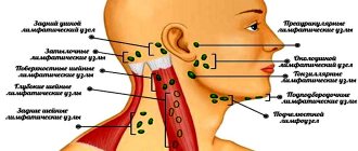
Lymphadenitis: photo
There are many groups of lymph nodes on the face and neck, the main ones being the buccal, parotid, submandibular (submandibular), retromandibular, submental, retropharyngeal, as well as superficial and deep cervical lymph nodes. In healthy children and adults, the lymph nodes are never enlarged, but if they are enlarged, this almost always happens as a result of invasion of pathogenic microorganisms. But we must remember that in some cases, enlarged lymph nodes may indicate tumor growth or blood diseases.
Structure and function of lymph nodes –
In the body, lymph nodes play the role of a biological filter. Lymph nodes retain and destroy pathogenic bacteria and toxins that enter them through lymphatic vessels - from teeth and bones affected by inflammation, tonsils, soft tissues of the face, and other organs and tissues. However, with the constant chronic sedimentation of microorganisms in them, they lose the ability to neutralize them, and in some cases they themselves turn into sources of purulent infection.
Each lymph node has a connective tissue capsule on the outside, from which lymphatic vessels depart, providing inflow or outflow of lymph. Thin connective tissue septa (trabeculae) extend from the capsule into the lymph node, between which the parenchyma is located. Closer to the capsule, the parenchyma consists of lymphoid follicles, and closer to the center of the lymph node - of strands of lymphocytes. The lymph passing through the lymph node is cleared of infectious agents and other antigens, and the so-called “immune memory” is also formed here.
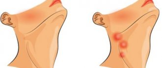
Symptoms of pathology
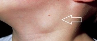
The fact that a person has “caught” a lymph node usually means its increase in size. Occasionally it hurts, and the skin over it becomes red. In this case, doctors talk about lymphadenitis.
An important point is the mobility of the lymph node. Normally, it easily moves a short distance. If the lymph node is motionless and enlarged, a doctor’s consultation is urgently needed.
Not all lymph nodes can be examined independently. When magnified, they can be felt on the neck, under the armpits, and in the groin area. It is important where the inflamed area is located. If a lymph node in the neck has caught a cold, how to treat it will differ from the situation when the groin or axillary area is affected. By localization, one can judge a preliminary diagnosis, since lymphadenopathy is often only a symptom of the underlying disease.
Complications of lymphadenitis –
As for complications, lymphadenitis can be complicated by adenophlegmon, and the latter can lead to the development of phlebitis, thrombophlebitis, and sepsis. The causes of complications are usually:
- a sharp weakening of the immune system due to viral and infectious diseases,
- if in the source of inflammation, in addition to the usual microflora (staphylococcus, streptococcus), there are also anaerobes (for example, clostridia and fusobacteria),
- errors in diagnosis,
- untimely start of treatment,
- improper treatment, including attempts at home self-medication.
Prevention of lymphadenitis is timely sanitation of the oral cavity, as well as foci of acute and chronic infection in the body. We hope that our article: How to treat neck lymphadenitis was useful to you!
Sources:
1. Higher medical education of the author of the article, 2. Based on personal experience in maxillofacial surgery and dental surgery, 3. National Library of Medicine (USA), 4. The National Center for Biotechnology Information (USA), 5. “Outpatient surgical dentistry" (Bezrukov V.), 6. "Pediatric surgical dentistry and maxillofacial surgery" (Topolnitsky O.).
What diseases does it occur in?
First, you need to look for inflammation near the affected lymph node. Usually bacteria or viruses enter it from a nearby organ.
Diseases in which the cervical lymph node becomes inflamed:
- acute respiratory infections and acute respiratory viral infections;
- tonsillitis (acute or chronic);
- dental diseases (focus of infection in the mouth).
Enlarged inguinal lymph nodes are often associated with sexually transmitted diseases, including herpes infection. Axillary units are inflamed with hidradenitis - a pathology of the sweat glands.
Sometimes inflammation of the lymph node occurs against the background of a general weakened state of the body. This is observed in malignant neoplasms, blood pathologies, HIV infection and viral hepatitis B.
In a chronic specific inflammatory disease, an enlarged lymph node is a normal variant. This is a reaction to the underlying pathological process and does not require treatment.
Enlargement of more than one lymph node often indicates infection. This is how tuberculosis, syphilis, and tularemia manifest themselves. Inflammation of a large number of lymph nodes indicates a serious infectious process - HIV, toxoplasmosis, brucellosis, CMV, mononucleosis and others. This requires urgent consultation with an infectious disease specialist.
Causes of enlarged cervical lymph nodes
This group of lymph nodes drains the area of the head, neck, upper chest, and proximal parts of the upper extremities. Accordingly, enlarged cervical nodes most often indicate the presence of thyroid diseases, inflammatory processes in the oropharynx, bacterial and viral infections. The symptom is pathognomonic for rubella, develops with measles, and can be detected with more rare infectious pathologies - psittacosis, Ebola and Marburg fever, mycoplasma infections.
Thyroid cancer
Thyroid neoplasia accounts for about 1.5% of all malignant neoplasms and in most cases is asymptomatic. The most typical variant is papillary cancer. Follicular tumors are quite common. With these space-occupying formations, there is always an enlargement of the cervical lymph nodes, which indicates increased proliferation of malignant thyrocytes and metastasis of tumor cells. Typically, the lymph node remains soft and mobile for a long time, since the process does not affect the capsule and surrounding tissues.
Patients, as a rule, consult a doctor about an accidentally discovered nodule in the thyroid gland when it reaches 1 cm or more. As the tumor grows, other manifestations occur: cough, hoarseness, associated with compression of adjacent anatomical structures. Large tumors can compress the airways, causing shortness of breath and suffocation. When cancer spreads beyond the organ capsule, an expansion of the subcutaneous venous network and deformation of the contours of the neck occurs. In elderly patients, cachexia increases.
Cervical lymphadenopathy is one of the signs of thyroid lymphoma - an aggressive neoplasia characterized by intensive growth and involvement of neighboring organs in the pathological process. The disease often develops against the background of autoimmune thyroiditis. The tumor grows rapidly, occupying an entire lobe of the organ. Patients often themselves discover a node of woody density, which is combined with enlargement and hardening of the cervical lymph nodes on the affected side. Compression of surrounding tissues provokes dysphagia, paresis of the vocal cords, and displaces the esophagus and trachea.
Organic diseases of the thyroid gland
In other lesions of the endocrine gland, enlargement of the lymph nodes is caused by an increase in blood flow by 10-15 times, increased production and differentiation of normal lymphocytes in response to stimulation by foreign antigens. The size of the lymph nodes is more than 1 cm, they are elastic, not fused with the surrounding tissues, and are sometimes sensitive to palpation. The symptom is characteristic of acute inflammatory processes, but also occurs in benign neoplasms and chronic autoimmune thyroiditis. Cervical lymphadenopathy is caused by:
- Acute thyroiditis
. The disease begins suddenly with a sharp pain in the thyroid gland, which radiates to the lower jaw and ear. Lymph nodes enlarge on both sides, become very painful, and redness of the skin is noted. Purulent thyroiditis occurs with an increase in temperature to febrile levels and severe symptoms of intoxication. There may be complaints of a feeling of pressure and fullness in the neck, and increased symptoms when coughing. - Nodules and cysts.
According to statistics, various benign formations of the thyroid gland are detected in 10% of the population, but more often they are asymptomatic. Enlargement of lymph nodes in the cervical region occurs with inflammation or suppuration of cysts, hormonally active neoplasia. Lymph nodes are elastic, practically painless, the skin over them is not changed. A detailed clinical picture of thyroid damage is observed with overproduction of hormones - thyrotoxicosis.
Rubella and measles
Cervical lymphadenopathy involving the posterior cervical and occipital nodes is an important symptom of rubella. Lymph nodes are moderately enlarged in size, painless, and not fused to the surrounding skin. For children, the appearance of “bullet” lymph nodes is typical - multiple small formations in the neck. Simultaneously with lymphadenopathy, a rash occurs - pinpoint or papular rashes are localized on the extensor surfaces of the limbs, in the torso and head. The rash disappears after a few days, leaving no peeling or pigmentation.
Lymphadenitis with enlargement of the cervical lymph nodes develops in the catarrhal stage of measles. Lymphadenopathy is combined with rhinitis, conjunctivitis, hyperemia of the pharynx and puffiness of the face. In adults, the manifestations of the catarrhal period are less pronounced. A pathognomonic sign of measles is Belsky-Filatov-Koplik spots on the mucous membrane of the cheeks. After 4-5 days, the second wave of fever begins, which coincides with the appearance of a maculopapular rash. On the 1st day, the rash is located on the face and neck, by the end of the first day it spreads to the torso, and on the third day the rash spreads to the limbs.
Other infectious diseases
Cervical lymphadenopathy is detected in various infections occurring in the oral cavity and ENT organs. Symptoms may occur with systemic bacterial and protozoal lesions - anginal-bubonic form of tularemia, sleeping sickness, diphtheria. Enlargement of lymph nodes is associated with the primary penetration and proliferation of pathogenic microorganisms, intensive proliferation and accumulation of specific clones of lymphocytes in the follicular and paracortical zones. With damage to the cervical lymphoid formations, the following occurs:
- Congenital listeriosis
. Infectious pathology develops during transplacental or intrapartum infection of the child and manifests itself in the first days after birth. Damage to the cervical nodes is combined with febrile body temperature, roseola or hemorrhagic rash, granulomas on the oral mucosa. The late form is accompanied by muscle tremors, convulsions, enlargement of the liver and spleen. - Syphilis
. After entering the body, Treponema pallidum multiplies in regional lymph nodes, causing them to enlarge. Cervical lymphadenopathy is often observed when the pathogen penetrates through the mucous membrane of the oral cavity or lips, where the primary affect is localized - chancroid. A month later, the chancre disappears on its own, then a polymorphic rash appears, which indicates the generalization of the infection and the development of secondary syphilis. - Brucellosis
. In the prodromal period, patients complain of myalgia, arthralgia, and headaches. Then a fever occurs, lasting from several days to 3 weeks and alternating with heavy sweats. At high temperatures, there is facial hyperemia, enlargement of the cervical and axillary lymph nodes, which can be painful on palpation. In the acute form, small fibrous formations appear along the tendons. - Inguinal lymphogranulomatosis
. Enlargement of the lymph nodes of the neck and submandibular region is observed in the secondary period when the primary affect (ulcer) is localized in the mucous membrane of the mouth and pharynx. As the disease progresses, lymphoid formations turn into large-lumpy tumors and lose mobility. Subsequently, the nodes suppurate, fever and intoxication develop, and fistulas form. - Tonsillitis, pharyngitis
. The reaction of the cervical lymph nodes is detected in tonsillitis, which is due to increased antigenic stimulation of lymphoid formations. The symptom is accompanied by sore throat, redness of the throat and tonsils, and fever. You may notice yellowish dots or widespread plaque on the surface of the tonsils. Enlarged lymph nodes are also found in herpetic pharyngitis, which is characterized by a vesicular rash on the pharyngeal mucosa.
Severe cases of tonsillitis can be complicated by a retropharyngeal abscess, a purulent inflammation of the pharyngeal tissue. In addition to lymphadenopathy of the upper cervical and occipital lymph nodes, the patient is bothered by sharp pain in the throat, difficulty swallowing, and if the abscess is large, breathing disorders are possible. The general condition is disturbed, body temperature rises to 39-40° C. Enlarged lymph nodes, combined with damage to the jugular vein and septicemia, are pathognomonic for Lemierre's syndrome. In children, cervical lymphadenopathy often indicates adenoiditis.
Head and neck tumors
Lymph from the face and neck goes directly to the cervical lymph nodes, therefore, for various malignant tumors of this area, they are a typical site of metastasis. Lymph nodes are usually woody in density, tightly connected to the skin and surrounding tissue, and are not painful. Lymphadenopathy in certain types of tumors serves as the initial sign of the disease, when the primary tumor does not yet cause clinical symptoms. Enlarged lymph nodes are observed with such malignant neoplasias as:
- Neoplasms of the jaws.
The tumor may be characterized by exophytic growth with the formation of a protruding node with ulcerations. It may occur as a long-term non-healing ulcer with purulent discharge. Bone damage (osteosarcoma) is indicated by shooting pains, loosening and loss of teeth. The lymph nodes are enlarged on both sides, have a rocky density, and are fused to the skin and subcutaneous tissue. - Tongue cancer
. Although with neoplasia of this localization there is often an enlargement of the submandibular lymph nodes, sometimes metastasis also occurs in the cervical group. Neoplasia of the tongue externally looks like a diffuse thickening of the organ with the formation of ulcers or local tissue growth. Pain syndrome appears early, eating disorders are pronounced, and weight loss progresses. - Neoplasms of ENT organs.
Cervical lymphadenopathy is a symptom of epithelial tumors of the nasopharynx, which also manifest as nosebleeds and difficulty breathing. The symptom is determined by esthesioneuroblastoma and is combined with anosmia, nasal congestion, and mucous discharge. Sometimes unilateral enlargement and hardening of the lymph nodes indicates ear neoplasia (basal cell carcinoma, epithelioma, sarcoma). - Eye tumors.
Enlarged nodes are observed at an advanced stage of conjunctival neoplasms, when malignant cells grow into the surrounding tissues and spread through the lymphogenous and hematogenous routes. Enlarged lymph nodes of the submandibular region and neck occur in ocular melanoma, an aggressive neoplasm of pigment cells that rapidly progresses with the development of distant metastases. - Timoma
. An increase in cervical lymphoid structures is detected in benign and malignant tumors of the thymus. Invasive growth of the tumor causes compression syndrome with intense chest pain, dry cough, and difficulty breathing. When peripheral nerves are compressed, Horner's syndrome, hoarseness, and dysphagia occur. About 30% of cases of the disease are accompanied by myasthenia gravis.
Systemic lesions of lymphoid tissue
Enlarged lymph nodes in the neck can be the first sign of lymphogranulomatosis - malignant hyperplasia of lymphoid tissue with the formation of specific granulomas. The disease is characterized by the appearance of dense, painless nodes, which are arranged in the form of a chain. In the local form of the lesion, one group of lymph nodes enlarges; in the generalized form, total lymphadenopathy develops with damage to the internal organs. Cervical lymph nodes are affected in chronic lymphocytic leukemia, an autoimmune lymphoproliferative syndrome.
Diagnostics
The first thing to do if a lymph node is inflamed is to visit a doctor. He will inspect not only the disturbing group, but also the neighboring ones. It’s easy to miss this on your own, but detecting such changes will help make a diagnosis. If several lymph nodes are affected, the mucous membranes and skin are also examined for rashes.
The phased examination includes:
- General and biochemical blood test, general urine test.
- Testing for hepatitis B and C, HIV, syphilis.
- Fluorography, if necessary, an x-ray of the lungs.
- Ultrasound of the abdominal organs.
- Ultrasound of lymph nodes.
- Biopsy of lymph node tissue.
Based on the research results, a diagnosis is made and treatment tactics are formulated.
Content:
- Causes of pathology
- Signs of inflammation of the submandibular lymph nodes 1.1. First stage 2.2. Second stage 2.3. Third stage
- How to cure an enlarged lymph node in the submandibular area
- Prevention of the inflammatory process
Inflammation of the submandibular lymph node is one of the most common types of lymphadenitis.
Its development is caused by inflammatory processes occurring in the oral cavity and less often in other parts of the body. Often the problem occurs with advanced caries, pulpitis, gingivitis, and inflammatory lesions of the tonsils. It is also caused by throat diseases. Let's take a closer look at why the submandibular lymph nodes are enlarged and what should be done to normalize the situation.
Treatment
If a patient has a cold lymph node, treatment is selected taking into account the underlying disease. The treatment regimen depends on the type of pathogen.
Treatment methods:
- Antibiotic therapy - for bacterial infection.
- Antiretroviral drugs - for HIV and AIDS.
- Antiherpetic drugs - for herpetic infection.
- Antimycotics - for fungal infections.
- Bed rest and plenty of warm drinks - for mild ARVI.
Self-medication is dangerous not only due to the incorrect selection of drugs, but also the development of the underlying disease. It is better to detect it early and treat it than to waste time and money on inappropriate medications.
Read also: Dry paroxysmal cough
Dear patients! Remember that only a qualified doctor can make an accurate diagnosis, determine the causes and nature of the disease, and prescribe effective treatment. You can make an appointment with our specialists or call a doctor at home by calling 8-(4822)-33-00-33
Be healthy and happy!
The structure and structure of the lymphatic system in the human body
The structure of the multi-level and complex mechanism includes lymphatic vessels - these are cylindrical cavities through which lymph flows, lymph nodes (small accumulations of lymphatic tissue located in different places throughout the body), lymphatic organs - the thymus, tonsils and spleen.
Content:
- Structure and structure of the human lymphatic system
- What are cervical lymph nodes
- Inflammation of the cervical nodes: characteristics of the concept
- Causes of inflammatory processes in the lymph nodes
- Symptoms of inflammation of the cervical lymph nodes
- Treatment of inflammation of the lymph nodes of the neck
In addition, two ducts participate in the system - the left and the thoracic, flowing into the right and left subclavian veins, respectively. All these elements are united by a liquid that circulates throughout all cavities - lymph.
The capillaries of the lymphatic system are tubes closed on one side that form a network in human tissues and organs. Capillaries have very thin walls, through which proteins, liquid and large particles freely enter the cavities. Due to the structural features of the walls of blood vessels, these elements cannot penetrate into the vessels, so they enter the blood through the capillaries of the lymphatic system.
In turn, lymphatic vessels are a collection and fusion of small capillaries. In fact, they resemble veins in their structure, but have thinner walls. In addition, they have a large number of valves that regulate the outflow of lymph.
Each vessel passes through its corresponding lymph node. All nodes are combined into several groups located along the vessels. The mechanism of lymph movement looks like this: through a large number of small capillaries, lymph enters the node, and from it it leaves through several efferent vessels.
The nodes themselves are tissue formations, shaped like ellipses or beans, less often ribbon-shaped, up to 2 centimeters long. In these “beans”, lymph is filtered, during which various foreign inclusions are separated and destroyed. Lymphocytes, cells that form part of the body’s immune system, mature in them. The vessels leaving the nodes unite into trunks that form the thoracic and right lymphatic ducts.
Through the right duct, lymph is collected from the right arm, right side of the head and chest into the right subclavian vein. The thoracic duct moves fluid from the left upper half of the body to the left subclavian vein. In this way, lymph moves from the interstitial spaces into the blood.