The inlay is a one-piece structure, the lower part of which is similar to a stump (therefore a stump). It takes into account the characteristics of the destroyed tooth, so it can be single, double or triple. In complex cases, it may consist of individual elements. The upper part is made of any shape, in accordance with the impression taken. The inlay can be installed without a crown, completely repeating the shape of the tooth. Such microprosthetics are often used in orthopedic dentistry, because even a severely damaged tooth can be completely restored, obtaining an aesthetic dental unit.
Advantages and disadvantages of tabs
- There is no danger of destruction or cracking - the strength and reliability of the inlays is high.
- Resistance to food dyes, color stability.
- The characteristics of the bite are taken into account.
- The shape of the tooth is completely restored and remains unchanged in the future.
- There are no unpleasant sensations at all.
- The result of tooth restoration is highly aesthetic.
- The possibility of the spread of harmful organisms is eliminated.
Negative aspects may arise if you go to clinics with insufficient equipment or inflated prices. In these cases, patients note the lengthy process of manufacturing and installing the inlay and the high cost.
Stump inlays: what do you need to know about them?
At the very beginning of our article, we said that stump inlays should not be confused with restorative microprostheses - they are used to restore pulpless and severely damaged teeth, in which they will serve as the basis for installing a crown or dental bridge.
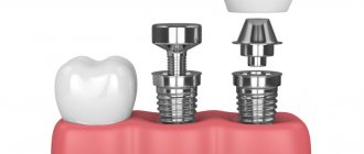
Stump inlays are structures consisting of two parts: a pin installed in the root canal and a support on which the crown will be fixed. Stump inlays can be monolithic or collapsible, and most often this type of structure is used when the tooth has less than 50% of healthy tissue left.
Many people are interested in the question: why is it best to use core inlays rather than fillings on a pin to restore severely damaged teeth? The fact is that the pin may not withstand the load and begin to wobble in the canal, and this will either lead to the filling breaking or falling out, or, worst of all, to damage to the tooth root, in which the tooth is most often removed. Stump inlays allow you to avoid such risks because they distribute the load more correctly and themselves have much greater strength compared to pins.
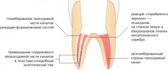
Stump inlays are also made in the laboratory and the construction material is selected to match the type of dental crown that is planned to be placed in the future. Metal core inlays are used for metal-ceramic crowns, and zirconium inlays are used for ceramic crowns. If you make a metal inlay for a ceramic crown, it can shine through the thin and transparent material of the crown, which will have a bad effect on the aesthetics of the restoration.
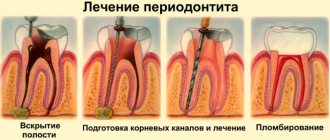
Tooth restoration using a core inlay will also be divided into several stages: diagnostics, preparation, during which a cavity is formed in the tooth for the inlay, manufacturing of the inlay, and its installation. After installing the core tab, the crown is fixed on it and this completes the process of tooth restoration.
If the tooth is destroyed, but there is a living nerve in it, before installing the stump tab, depulpation is carried out, as well as treatment and filling of the root canals. It is important that these procedures are carried out without errors, since poor canal processing leads to various kinds of complications, the elimination of which requires complex and lengthy treatment.
Indications and contraindications for use
It’s worth putting a tab if there is:
- Extensive caries damage (more than 60%) of the tooth tissue;
- Large cavity size;
- Traumatic tooth damage;
- Hyperplasia, dysplasia;
- Wedge-shaped defect;
- The need for protection against tooth wear;
- Installation of bridges simultaneously with root structures.
The use of inlays is not recommended if the patient has:
- Small tooth cavity;
- Carious processes are active;
- There is no quality oral hygiene.
Reconstruction of the functionality of the dentition using an inlay allows you to cope with high loads. This is a great solution, but it can be painful some time after installation. What to do if a tooth hurts under the tab?
The cause of pain may be high sensitivity. If the situation worsens over time, you need to see a dentist and get an x-ray.
If a tooth hurts under the stump tab, then the following reasons are possible.
- Pulpitis is damage to the pulp of a tooth with a preserved nerve. When installing the inlay, the pulp chamber was accidentally damaged. After the anesthesia wears off, the pain becomes severe and intensifies with chewing and contact with cold and warm foods. The patient feels pain under the tab.
- Pressing the inlay into the tooth tissue. A bursting, aching pain occurs, which does not allow establishing a specific location.
- The tab is too large. The patient feels pain only when chewing. The pain is dull, there is no reaction to hot and cold food. The sensations are more like discomfort. In the absence of correction, inflammation of the periodontium or gums develops.
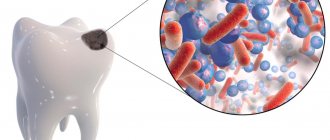
- Infection. Before installing the inlay, the doctor cleans the carious cavity to prevent further spread of the destructive process. If at this moment there is already an infection in the tooth, but has not yet been identified externally, after installing the tab the process will spread deeper, to the pulp and to the roots. If left untreated, the pain under the ceramic inlay will become very severe, the nature of the “sensations” will be pulsating and sharp.
Clinical case
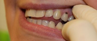
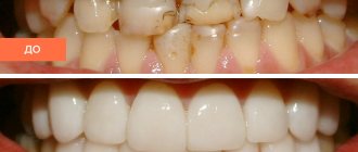
A 30-year-old patient came to the clinic with complaints of darkening of fillings. The patient was interested in an aesthetic restoration with a long service life, so it was decided to restore the teeth with ceramic inlays. The initial situation is shown in Fig. 1. After collecting complaints, anamnesis, and conducting an examination, a diagnosis was made: K02.1 Dentin caries of a tooth 3.6, class II according to Black. K02.1 Dentin caries of tooth 3.7, Black class I.
Rice. 1.
At the clinical stage, the tooth was cleaned of plaque. After anesthesia, the color was determined using the standard VITAPAN Classical scale, the color of future indirect restorations is A2.
The preparation and formation of cavities of class I and II according to Black were carried out according to generally accepted principles in compliance with all stages with the obligatory use of a rubber dam to isolate the working field.
A small carious cavity was found on the medial contact surface of tooth 3.6, without destruction of the marginal ridge and without communication with the main carious cavity on the chewing surface. After preparing the carious cavity on the medial contact surface of the tooth, filling was performed using a fluid composite material.
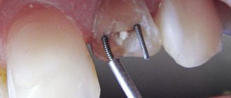
Before taking the main impression, immediate dentin bonding was performed according to a traditional adhesive protocol. After polymerization of the adhesive, the undercuts in the area of the cavity walls were closed with a layer of flowable composite. The next step was to apply glycerin to the inner surface of the prepared cavity and illuminate it with a photopolymer lamp to remove the oxygen-inhibited layer in order to prevent possible interaction with the vinyl polysiloxane impression material during impression taking. The final view of the prepared cavity before taking an impression is shown in Fig. 2.
Rice. 2.
Impressions were taken with A-silicone impression material, temporary restorations were made using Clip material.
A follow-up visit took place 1 week later to fix the IPS e.max ceramic inlay. Previously, before admitting the patient, we assessed the marginal fit of the ceramic inlays on dismountable models (Fig. 3-6).
Rice. 3.
Rice. 4.
Rice. 5.
Rice. 6.
The second clinical stage began with cleaning the tooth from plaque. After anesthesia and the application of a rubber dam, the temporary restorations were removed, the cavity was cleaned of temporary cement residues, followed by fitting and preparation of ceramic inlays for fixation (Fig. 7-10). The indirect restoration was cemented using Calibra dual-curing adhesive composite cement (DENTSPLY).
Rice. 7.
Rice. 8.
Rice. 9.
Rice. 10.
After positioning the restoration, excess fixing material was removed in the area of the contact surfaces. Next, excess material was removed from other surfaces of the teeth. After 3 - 4 minutes, the surface of the fixing material was covered with a layer of glycerin and the final illumination of the restorations was carried out from each surface for 20 seconds.
At the final stage, an occlusion check was carried out. The view of the restoration immediately after fixation is shown in Fig. 11.
Ceramic restorations look natural, no different from your own tooth tissue.
Rice. eleven.
The appearance of the restorations 5 years after fixation is shown in Fig. 12.
Rice. 12.
What complications are possible?
An incorrectly installed inlay can result in the development of a cyst. Treatment will be lengthy and expensive - resection of the root apex will be required.
If you have pain under a microprosthesis or the inlay under the crown hurts, contact dentistry at amazing prices. An endodontist will examine the oral cavity and prescribe additional examination.
- X-ray - to assess the condition of the root and surrounding tissue (the image will show whether there is a cyst;
- EDI – Electroodontodiagnosis to assess the condition of the pulp – the doctor has the opportunity to determine:
- The degree of damage to nerve tissue;
- Is it necessary to open root canals, treat or remove the pulp?
Taking into account the results obtained, effective treatment will be carried out.
Causes of tooth pain under a crown or denture
- If you have not had root canal treatment to remove the pulp before getting a crown, pressure on the damaged nerve may cause pain.
- Patients with malocclusion and bruxism may experience pain at night from pressing on ridges or areas of the tooth that are higher than normal.
- If the denture has shifted, exposing part of the tooth, or, worse, is pressing on the dental nerve, then the slightest pressure or change in temperature can send strong pain signals.
- Teeth under a crown are also at risk for all the problems associated with regular teeth, meaning they can become infected, break, and become vulnerable if the enamel wears down. And this can cause pain.
Emergency help
If you can’t see a doctor, and the tooth under the tab hurts, the following measures will help.
- Chewing and temperature stress on the teeth should be completely avoided. Food should be consistent with body temperature and should not require chewing or biting. Broths and pureed soups are the best solution.
- For unbearable pain, take an analgesic or anti-inflammatory drug. The main thing is that the product does not cause allergies. It could be Nimesil, Nise or Analgin.
- If the situation worsens, accompanied by headache and chills, you should contact the nearest medical facility. The staff will call an emergency dentist, and the problem will be solved.
Author:
Mayorov Andrey Mikhailovich
Specialization:
orthopedic dentistry, dental prosthetics, implant installation
In what cases is a pin placed?
Dentists prescribe pin implantation when the crown is destroyed by more than 50%. In this way, the thinned walls are strengthened. The pin is also installed when building up stumps for artificial crowns.
In what situations is it not recommended to install a pin:
- Is it possible to do an MRI if there are metal crowns?
- the periodontium does not hold the tooth well;
- small root size;
- root caries damage
- the channel is perforated;
- cysts were diagnosed at the roots;
- for blood diseases;
- The walls of the tooth root are very thin.
There are situations when the installation of pins is contraindicated
Let's sum it up
Pain after insertion of a pin occurs for various reasons, but moderate pain is normal. The maximum time through which they must pass is up to five days. If the pain lingers for a longer period of time, it is necessary to consult a doctor who will find out what caused the pain.
However, if erroneous and unprofessional actions of the dentist were noticed, which include installing the rod too deeply, non-compliance with the rules for filling the root canals, damage to them with dental instruments, an incorrectly fixed pin and pieces of dental instruments accidentally left in the canal, then the pain is usually very intense and you need to get rid of it only after consulting a doctor.
Removing a dental post
The pin is removed in the following situations:
- if the apical part of the root becomes inflamed;
- when a filling needs to be replaced;
- during the preparatory stage before implantation;
- with unprofessional and poor-quality installation of the pin.
Removal of the pin is carried out in several stages. First, old fillings, crowns and other structures installed earlier are removed. Then, using ultrasound, the cement holding the pin is destroyed.
After this, you can directly remove the pin using forceps.
Important! If the pin is very firmly fixed in the root canal of the tooth, then ultrasound treatment is performed - due to vibrations, the pin moves, and this makes it easy to remove it.
After the procedure is completed, the doctor must check the patency of the root canals.
The doctor will definitely check if everything is in order after the procedure.
The pins are removed painlessly; this procedure does not pose any danger to the patient. Even anesthesia is not used in this case - only vibration and light pressure are felt.
How to build teeth onto pins, what is this process?
Prosthetics are performed after the pin is installed. Nowadays in dentistry, modern materials are used to make prosthetics; crowns are installed, as well as bridge prostheses if several teeth have been damaged at once.
First, the doctor must shape the filling material into the tooth around the post. It is shaped, filed and polished. What the result will be depends primarily on the experience of the dentist who works with this problem.
- Frequent urination or why do I pee so often?
If the tooth is seriously damaged, a crown is installed. It is a special prosthesis that cannot be removed and is performed individually for each patient. Usually the cost of crowns is quite high.
If the tooth is seriously damaged, a crown is installed
The entire prosthesis imitates a damaged tooth. It is fixed on the pin using a special strengthening solution. After the crown is installed, some time must pass. Then the doctor checks whether her body rejects it or not. Implantation is considered successful if the prosthesis is installed tightly and there are no signs of inflammation.
Other causes of pain
One of the common causes of pain is the development of inflammation. This process begins approximately 1-2 days after the pin is installed and may indicate that the soft tissue was damaged during the procedure and became infected. In the oral cavity, infection occurs quite quickly, so you should not postpone a visit to the doctor for treatment - this is fraught with complications.
There is also such a thing as congenital tooth sensitivity. Usually the patient is aware of this peculiarity. Pain occurs, for example, due to chewing hard food or when eating food that has a contrasting temperature (very cold or hot).
Interesting ! The pain occurs due to congenital sensitivity of the teeth.