Quite often in their practice, ENT doctors are faced with such a problem as paranasal sinus cyst. Despite the fact that the disease occurs frequently, in almost every tenth person, it is mainly discovered by chance, for example, during an X-ray examination for a completely different reason. This is explained by the fact that a cyst in the nose may not bother the patient for a long time and may not manifest any symptoms. Therefore, it is important to understand that timely treatment of cysts is necessary to avoid possible complications and to improve the patient’s quality of life.
What is a maxillary sinus cyst?
A maxillary cyst is a voluminous benign formation that originates from the mucous membrane of the maxillary sinus and has the appearance of a bubble containing fluid inside. At the same time, the contents of its cavity vary, depending on the mechanism of appearance and the nature of the ongoing pathological process.
Depending on this, there are several types of maxillary sinus cysts:
- mucous membranes (mucocele);
- serous (hydrocele);
- purulent (pyocele).
According to their structure, cysts can be true (with an internal epithelial lining in a thin wall) or false. A pseudocyst in the nose does not have clearly defined walls and looks like cavities in the tissue; they are usually formed in the thickness of the overgrown mucous membrane and can consist of several chambers of different sizes.
Cysts do not have a tendency to malignant degeneration, however, they cannot be classified as harmless formations. They can fester and burst, compress adjacent vessels and nerves, and gradually destroy the bone walls of the sinus.
Cystic formations negatively affect ventilation and drainage of the entire system of paranasal sinuses and impair nasal breathing. Therefore, surgery to remove a cyst often becomes a prevention of chronic recurrent inflammation of the ENT organs.
Why does a cyst appear in the nose?
The main reasons for the appearance of maxillary cysts include:
- Recurrent or chronic maxillary sinusitis (sinusitis). Inflammatory changes in the mucous membrane lead to blockage or scarring of the excretory glandular ducts. The produced secretion has no outflow, accumulates and stretches the gland. This is how true mucous and serous cysts are formed.
- Chronic non-infectious inflammation (usually of an allergic nature), accompanied by hyperplasia of the mucous membrane of the nose and paranasal sinuses.
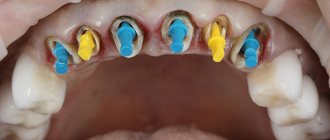
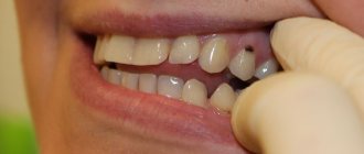
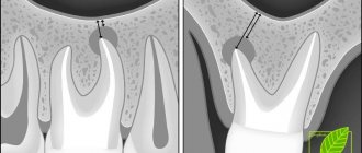
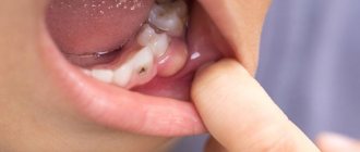
- Dental pathology, the resulting cysts are called odontogenic. The most common reason for their appearance is inflammation around the root of a carious tooth or near the tooth germ of the upper jaw. The purulent process leads to atrophy and destruction of bone tissue, spreading to the walls of the maxillary sinus. More rare causes include abnormally deep tooth roots and excessively traumatic tooth extraction.
- Predisposing factors are injuries to the facial part of the skull, congenital anomalies with asymmetry of the hard palate and nasal bones, and immunodeficiency states. Occasionally, a cyst in the sinus is formed against the background of a congenital defect in mucus production, when the secretion of the glands has an excessively viscous consistency.
Prevention
There are no specific preventive measures to prevent the appearance of cysts in the sinus. To reduce the risk of cyst formation, you should:
- treat inflammatory diseases of the nasal cavity in a timely manner;
- if you are allergic, avoid contact with the allergen to avoid allergic rhinitis;
- visit the dentist for preventive maintenance, and if dental problems are diagnosed, correct them in a timely manner;
- correct the anatomical features of the structure of the nasal cavity (for example, a deviated nasal septum) and abnormalities in the development of the upper jaw.
What are the symptoms of a cyst in the nose?
A cyst of the upper jaw does not lead to strictly specific symptoms, and sometimes even occurs without obvious manifestations. Complaints from patients with this pathology include:
- Nasal congestion, clinical signs of chronic rhinitis.
- Unpleasant sensations, a feeling of fullness and pressure in the projection of the maxillary sinus (on the side of the cyst). Discomfort often increases when bending forward.
- A feeling of pressure from the inside on the eyeball, and with large neoplasms, transient double vision may occur.
- Recurrent headaches with a predominant localization in the forehead and bridge of the nose. Their appearance is mainly associated with impaired ventilation of the paranasal sinuses.
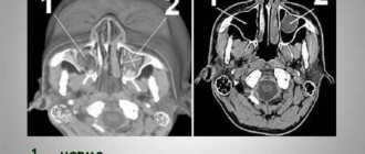
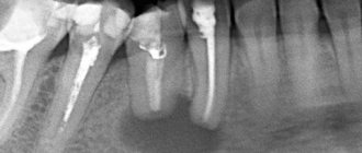
Reliable diagnosis is based on visualization of the maxillary sinus and its contents. Traditional examination methods include radiography of the facial part of the skull. Currently, CT scans of the paranasal sinuses are widely used.
Diagnostics
If the symptoms described above appear, you should contact an otolaryngologist for examination and diagnosis. Diagnosis of a cyst includes interviewing the patient for complaints, collecting an anamnesis of the patient’s life and health, as well as methods of physical examination and other types of research. Informative methods for identifying sinus cysts are:
- Rhinoscopy - during the study, the doctor evaluates the condition of the mucous membranes, their color, swelling, the presence of mucous or purulent discharge, and the size of the inferior turbinates.
- Endoscopic examination - allows you to examine distant parts of the nasal cavity, including the sinus anastomosis.
- X-ray examination of the paranasal sinuses - it allows you to assess the condition of the sinuses and detect a cyst in them.
- Computed tomography, magnetic resonance imaging - is carried out if the x-ray is insufficiently informative, and the doctor has doubts regarding the diagnosis. These studies help to accurately determine the presence of a formation and its size.
- Puncture - in some cases, a diagnostic puncture is performed to assess the nature of the contents of the cyst.
When is surgery needed to remove a maxillary sinus cyst?
Formed cysts are often not prone to regression, and conservative therapy is also usually not able to lead to their disappearance even after the elimination of inflammation. Therefore, the only effective treatment method is surgical removal of the cyst in the sinus.
Indications for surgery include:
- Signs of suppuration of the contents of the cyst.
- Progressive growth of the tumor.
- The diameter of the cystic cavity is more than 6 cm.
- The appearance of double vision (diplopia) and blurred vision, which is a sign of excessive pressure of the formation on the bottom of the orbit.
- Often recurrent or continuous rhinosinusitis, chronic nasal congestion.
- Significant discomfort experienced by the patient even with a small tumor size.
Surgery to remove a cyst is usually performed as planned. The decision on surgical treatment is made by the ENT doctor after examining the patient and assessing the clinical situation.
Preventing inflammation
In addition to preserving the wound, preventive therapy is carried out aimed at preventing the development of the inflammatory process. Most often, antibacterial drugs and anti-inflammatory drugs are prescribed. In addition, drops are prescribed for instillation into the nasal passages, which have a vasoconstrictor effect. Treatment can be carried out both on an outpatient basis and while staying at home.
If the examination reveals the presence of a foreign body in the maxillary sinus, then treatment is carried out only in a hospital setting. Therapy consists of surgical intervention to open the cavity and remove the tooth root from the maxillary sinus.
How is surgery to remove a cyst in the nose performed?
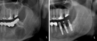
For a long time, surgery to remove a sinus cyst was carried out only by opening the wall of the maxillary sinus through the oral cavity or through the cheek. This manipulation is called maxillary sinusotomy; it is quite traumatic and requires a long recovery period.
Currently, classical surgery is rarely performed, with preference given to minimally invasive interventions. But for extensive purulent cases, just such a radical intervention is used. The dangerous formation is completely removed, the sinus is washed with an antiseptic and drained.
In other cases, surgery on the maxillary cyst is performed endoscopically, with minimal disruption of the integrity of the sinus walls.
If necessary, additional plastic surgery of the anastomosis is performed and adjacent polypous growths are removed.
Endoscopic surgery to remove a maxillary sinus cyst is performed on an outpatient basis and usually lasts 20–40 minutes. After its completion, the patient remains in the clinic for 1–2 hours under the supervision of a doctor, then returns to his normal life.
Using an endoscope allows you to remove the maxillary cyst in a gentle way. This operation does not disrupt the drainage and ventilation of the sinuses, reduces the risk of chronic rhinosinusitis and does not require long-term recovery for the patient.
Treatment
The choice of treatment for maxillary sinus perforation depends on the changes that occur as a result of the rupture. Non-surgical treatment seems possible in case of violation of the integrity of the tissues during tooth extraction, when the pathology was detected immediately, and according to the results of the radiographic examination it is clear that there is no infection of the sinus cavity and there are no foreign bodies in it. With such a clinical picture, the doctor, as a rule, tries to preserve the blood clot formed in the hole as much as possible. In addition, preventative measures are taken to prevent wound infection.
For this purpose, a small gauze swab soaked in an iodine solution is inserted into the hole. As a rule, the tampon is self-fixed in the wound cavity. However, sometimes it may be necessary to place a stitch in the gum. Treatment with an iodide solution should be carried out for 6-7 days until the defect disappears and full-fledged granulations are formed. It is not recommended to remove the tampon from the socket during treatment, as this can damage the clot and lead to infection of the wound.
When a tooth root is found in the maxillary sinus, everyone should know what to do. In some cases, the doctor decides to close the defect with a special plastic plate, which is secured to adjacent teeth with clasps. Thus, it is possible to achieve separation of the sinus and oral cavity.
Removal of a maxillary sinus cyst at the ENT clinic of Dr. Korenchenko
See also Treatment of ENT diseases Cyst in the maxillary sinus Treatment of a cyst in the maxillary sinus Surgery to remove a cyst in the maxillary sinus
Endoscopic ENT surgeries are not performed in all clinics. After all, they require modern equipment, the doctor having the appropriate skills and certificates. Dr. Korenchenko’s ENT clinic is a modern, specialized and well-equipped medical center. Our specialists are highly qualified and have rich clinical experience, all the necessary certificates and skills. When treating patients, we use only modern, clinically proven and highly effective techniques.
Endoscopy at Dr. Korenchenko’s Clinic is an important and widely used therapeutic and diagnostic procedure. It is included in the basic examination of all patients who apply and are observed, which allows doctors to receive reliable and accurate information about the current condition of the ENT organs. Our specialists also perform removal of maxillary cysts and most other operations endoscopically, with high results and without long-term rehabilitation of patients.
Treatment at Dr. Korenchenko’s ENT clinic is a modern and competent approach, using effective technologies and effective therapeutic regimens.
Clinical researches
Repeated clinical studies have proven that the two-component mouth rinse ASEPTA ACTIVE more effectively combats the causes of inflammation and bleeding compared to single-component rinses - it reduces inflammation by 41% and reduces bleeding gums by 43%.
Sources:
- The role of anti-inflammatory rinse in the treatment of periodontal diseases (L.Yu. Orekhova, A.A. Leontyev, S.B. Ulitovsky) L.Yu. OREKHOVA, Doctor of Medical Sciences, Prof., Head of Department; A.A. LEONTIEV, dentist; S.B. ULITOVSKY, Doctor of Medical Sciences, Prof. Department of Therapeutic Dentistry of St. Petersburg State Medical University named after. acad. I. P. Pavlova
- Report on the determination/confirmation of the preventive properties of personal oral hygiene products “ASEPTA PLUS” Remineralization doctor-researcher A.A. Leontyev, head Department of Preventive Dentistry, Doctor of Medical Sciences, Professor S.B. Ulitovsky First St. Petersburg State Medical University named after. acad. I.P. Pavlova, Department of Preventive Dentistry
- Clinical studies of antisensitive toothpaste “Asepta Sensitive” (A.A. Leontyev, O.V. Kalinina, S.B. Ulitovsky) A.A. LEONTIEV, dentist O.V. KALININA, dentist S.B. ULITOVSKY, Doctor of Medical Sciences, Prof. Department of Therapeutic Dentistry, St. Petersburg State Medical University named after. acad. I.P. Pavlova