A pin is a structure made of durable materials that is installed in the root of the tooth and strengthens it. With its help, the tooth is maintained in normal condition and can take part in chewing food. Sometimes the pin needs to be replaced.
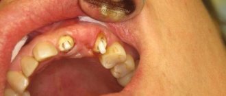
The pin is considered a foreign body at the root, although fragments of a dental instrument are also foreign bodies. A foreign body is detected in the root canal using x-rays. The photo clearly shows a piece of a tool or a pin. To remove a foreign body, you first need to remove the seal and depressurize the canal. After removing the foreign body, the canals are sealed again.
The pin has the appearance of a spoke. It is used to strengthen root canals and damaged teeth. The use of a pin helps restore a tooth when at least a fifth of it is preserved. A dental post extends the “life” of the tooth by slowing down further decay of the crown after caries has been treated. The pin is also used as a basis for tooth restoration.
Restoring the lower front tooth using a CEREC inlay
The work was quite serious. The tooth under the filling and the root of the lower canine were affected by caries.
First I removed the filling:
Then I removed the anchor pin:
In the video below you can see how the anchor pin was removed, and then I got ahead of myself a little, showing you a fitting of the finished CEREC module on the lower canine: The next step was to go quite deep inside the root, because the root of the lower canine was in caries.
IMPORTANT!
Each stage described above was accompanied by
3D scanning
with a Dentsply Sirona scanner, this is one of the best scanners in the world. The CEREC technological complex is equipped with just such a scanner. Scanning each stage, the CEREC hardware system stores all the data in memory, and when the time comes to plan and manufacture the missing tooth module, CEREC does this with filigree precision down to hundredths of a millimeter.
See for yourself by watching this short video:
Watch the video as I trace the border of this tooth.
The destroyed lower canine has a very complex geometry. However, the tooth will be completed using the “tooth only and missing module only” system. The canine will be installed and we will have a tooth that consists of only two components, no metal with one seam. The result will be a perfect lower canine. The restoration conditions are very difficult, deep, but in principle in this system it does not matter how deep they are, the main thing is that the tooth tissue is healthy. Our root has no caries, it has the classic dark color of the root, a non-carious color. Complex volumetric geometry in this tooth.
I create a tooth
, which will play the role of a reliable and strong fang for our patient. It has a super secure connection because there is only one seam, not two. In the video you can also watch the upper jaw, how we will close the teeth. You can also view contact points.
Yes, a very difficult tooth for a classic restoration. Difficult, but not for us. In CEREC, with a sufficiently professional level of the doctor, everything looks like “everything is the same”: this tooth for the Cerec system is simple like all the others. Well, in reality, of course, it looks and all the work looks fantastic:
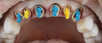
As a result of the preparatory work I carried out, only the root of the lower canine 3.3 remained from intact tissue, and I made the missing module for it using CEREC technology: only the tooth and only the missing module without any anchor pins. It is this approach, without intermediaries in the form of pins, that ensures the best quality of treatment and further use of the tooth.
I created a CEREC inlay in A3.5 color with a color transition from the gum to the incisal edge. This transition ensured the absolute naturalness of the final design and the aesthetics of the tooth.
Sergey Samsakov
Indications
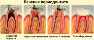
Often, after some time, caries develops under the crown around the pin devices. As a result, the oral cavity requires repeated sanitation. And the main task of canal re-treatment is to remove the foreign body from the root canal. Indications for removal are pain under the crown, tooth decay and deep caries. Most often this concerns pins that were installed a long time ago.
One of the serious indications for re-treatment of canals is a foreign body, or rather a fiberglass rod or its fragments. Before starting this procedure, be sure to take an x-ray.
Do you want to restore a tooth with a CEREC ceramic inlay?
8
or order a free call
Request a call
This is how the dental inlay for the lower canine turned out:
The inlay was fixed with double-curing cement Relix U200 (RelyX U200) - this is one of the most expensive cements, German cement. The patient was super satisfied. The restored tooth looks like its own:
All work FROM and TO was carried out in 1 appointment
, on a fresh tooth surface. Great job.
PS
As you can see in the video, the patient is facing the next stage of dental restoration. But we'll talk about this some other time.
Author:
Sergey Samsakov
orthopedic dentist
born 02/02/1989
Education:
2011 — Graduated from the Moscow State Medical and Dental University named after. A.I.Evdokimova
2012 — Internship in the specialty “Orthopedic Dentistry”, Moscow State Medical University named after. A.I.Evdokimova
2014 - Residency in Orthopedic Dentistry, Moscow State Medical University named after. A.I.Evdokimova
Types of foreign bodies that may end up in the root canal
Most often, foreign objects end up inside root canals during complex dental treatment. Depending on the situation, these could be:
- Broken fragments of endodontic instruments (in particular, files for pulp extraction);
- Anchor fiberglass pins, which are specially installed to strengthen the tooth and its further prosthetics;
- Elements of stump inlays (in particular, their pin parts, which can break off during use).
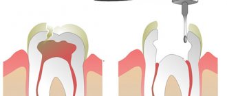
Fragments of instruments end up in the canal during its treatment - as a rule, removal of necrotic pulp from it during:
- Carrying out manipulations with narrow or tortuous channels in which the instrument breaks, unable to withstand the stress of bending;
- Repeated use of hand tools whose tips may break off due to metal fatigue or defects.
During endodontic treatment, instruments do not break very often, but such a possibility cannot be excluded. Today it is one of the pressing problems of dentistry. Treatment under a microscope helps reduce its risk to almost zero, in which the doctor is able to notice the broken fragment immediately and remove it in a timely manner.
More examples of treatment
Caries under filling Depulped tooth Absence of the crown part of the tooth
Restoring a tooth with a CEREC inlay after removing a metal inlay
Tooth 3.5 initially had a filling; a metal inlay was installed in the tooth, quite deep, and a filling on top. This is a “puff sandwich”.
Module crown + tooth root CEREC Removal of metal inlay
more details
Pulpless tooth Destruction of the front tooth in the upper jaw
Restoring a front tooth damaged by super glue with a CEREC inlay
Superglue is toxic to the body. Is it possible to glue a piece of tooth with this glue? Of course not. But you can easily ruin a tooth!
Module crown + tooth root CEREC Professional hygiene
more details
Complications after removing pins from the root canal of a tooth
As a rule, experienced dentists calculate the risks associated with the procedure and in every possible way reduce them to zero. For example, removing a post made of a material such as zirconium carries a high risk of breakage due to its brittleness. For this reason, the specialist needs to act as carefully as possible, and upon completion of the procedure, monitor its quality. Other complications may include:
- perforation of the tooth root due to its mechanical damage;
- fracture of the tooth root due to the appearance of cracks in it and the retreatment procedure;
- damage to the pin itself and preservation of its fragments in the canal.
An important role is played not only by the occurrence of complications, but also by the availability of the resources of the endodontist in order to properly cope with them. Most often, perforation or root fracture requires removal of the tooth root and placement of an implant. At the same time, in some cases it is possible to use the mineral trioxide of the unit, the properties of which allow it to close the perforation and preserve the functionality of the unit, extending its service life.
Do you want to restore a tooth with a CEREC ceramic inlay?
Always at your service - Sergey Samsakov, orthopedic dentist with more than 10 years of successful experience, Moscow. Expert in 3D digital smile modeling, veneers and CEREC technology.
Patients come to me from all over Russia:
Indications for removing the stump tab, pin, dental instruments from the canal
The procedure is carried out in two ways:
- Simple - indicated when identifying broken fragments of pins. In the process, the endodontist destroys the filling by applying ultrasound to it. Thus, he provides himself with access to the required area. After this, he carefully loosens and removes the foreign body;
- Complex – indicated for identifying fragments of endodontic instruments. The process involves a dental microscope. The specialist prepares the root canal, loosens the discovered fragments and carefully removes them using a special device. If the foreign body is not visible in the lumen of the root canal, the procedure will require microsurgical intervention.
CELT dentists individually select treatment tactics and use modern equipment (in particular, a microscope and endodontic ultrasound attachments, which allow the procedure to be carried out as carefully as possible, without affecting healthy tissue). When choosing treatment, the nature of the patient’s complaints is taken into account, as well as the duration of the pathological processes, the identification of inflammatory processes and the location of the pathological focus.
Ways to get rid of pain
Under no circumstances should you delay a visit to the doctor if you experience prolonged or progressive pain after installing the pin. The doctor will conduct a full examination, determine the cause and prescribe appropriate therapy. Usually, fluoridation of tooth enamel or building up a layer of enamel using filling material is prescribed.
Fluoridation of tooth enamel
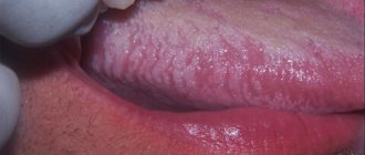
If the examination shows that everything is in order with the installation of the pin, and the pain occurs on a tooth that is devoid of a nerve, most likely the cause is congenital sensitivity. The patient can cope with it independently, using a special toothpaste with a high content of calcium and fluoride. It is also recommended to use dental floss to remove food debris that causes discomfort and irritation.
When installing a dental pin, it is recommended to rinse your mouth with soda. For about three days, mechanical influences on the tooth from the outside are prohibited; you cannot touch it while chewing. You can take analgesics and apply cold compresses to relieve pain after the procedure.
It is recommended to rinse your mouth with baking soda
Is it possible to glue the bridge in place yourself?
What to do and how to secure a dental bridge if it falls out at home? Let us immediately point out that putting a fallen bridge back on your own is not the best idea. Since this can injure the supporting and neighboring teeth, harm the gums (if you suddenly decide to glue the bridge with pharmaceutical cement or household glue). The consequences can be unpredictable - up to the removal of “damaged” teeth or implants. Therefore, it is better to entrust your teeth to a professional dentist.
What to do
If the crown falls out along with the pin, it is necessary to save the orthopedic product. It is not recommended to throw it away. To prevent deformation or damage, the structure is placed in a special container or box.
Since the stump of a dental unit becomes very fragile after treatment, it must be treated with extreme caution until the dentist installs the product again, since at this moment the risk of damage increases.
It is not recommended to put excessive chewing load on the tooth, or bite food with it. The hole in which the pin was placed must be covered with a cotton swab while eating.
All hygiene procedures before visiting a specialist are carried out carefully, the oral cavity is treated with an antiseptic solution.
Causes of crown loss
There are many factors that can cause a crown to fall out. The most common ones include:
- resorption of the cement composition that was used for fixation;
- breakage or abrasion;
- excessive pressure;
- the use of temporary material instead of permanent;
- formation of a carious process under the product;
- violation of the rules in the process of creating a structure or incorrect grinding of a dental unit.
A crown can fall off for several reasons. But only a specialist can determine what exactly led to this condition. In this case, you should not blame only the technician or the orthopedic surgeon. The patient himself can also become the culprit of the incident if all recommendations are not followed or due to negligence.
When the client contacts him again, the dentist must take a more responsible approach to the process of preparing the tooth for prosthetics and correctly fixing the structure using better quality material.
The dental technician, in turn, should not neglect all the rules and stages of creating a product. The patient is required to strictly follow all recommendations for oral care, perform high-quality hygiene procedures and exclude solid foods from the diet.
Preventing Looseness
The service life depends on precise manufacturing, the quality of the materials used and the required hygiene. If the product is formed poorly, it will break under chewing pressure. A hole may form in it, even if the structure is metal-ceramic. Here the cause does not depend on the patient; the lost tooth is replaced by a dentist. Why is this happening? Perhaps the treatment was carried out in a low-level clinic, or the technician made mistakes at the stage of manufacturing the prosthesis.
If the product is made correctly, is well fixed, and can withstand the stress of chewing food, then often the loss occurs due to the fault of the client. The dentist will be able to restore the tooth immediately after the patient contacts him. To prevent complications, the following is recommended:
- Once every six months you need to have an examination with a dentist.
- Professional teeth cleaning is performed periodically.
- Do not use toothpicks.
- It is forbidden to eat hard or tough foods.
- Do not put excessive pressure on the prosthesis.
- After eating, you should use dental floss and mouthwash.
- Hygiene products must be correctly selected in accordance with the recommendations of the treating dentist.
The basis for preventing crown loss is proper hygiene care. If problems arise, you should not indulge in self-medication; you should contact your dentist.