Headaches often occur due to dental errors or chronic dental diseases. A number of factors can cause the problem:
- remnants of roots or fragments in the socket after complex tooth extraction;
- pulpitis;
- periodontitis;
- galvanism due to fillings and dentures made of different metals;
- poorly installed or too high dentures;
- periodontal diseases;
- osteomyelitis of the jaw bones and other diseases.
More often, headache in pathologies of the dental system is a symptom of odontogenic neuralgia of the trigeminal nerve (more than 60% of cases) or dental plexalgia (diagnosed in 12% of patients), so we will consider these diseases in more detail.
Treatment of odontogenic trigeminal neuralgia
Therapy usually involves a combination of medications and physical therapy. At the first stage, patients are prescribed non-narcotic analgesics. If an inflammatory process is present, then it is recommended to take anti-infective drugs in parallel.
Physiotherapy at this stage may include:
- modulated currents,
- ultrasound,
- Ural Federal District,
- moderate heating.
After the pain subsides, they move on to the second stage of treatment. At this stage, electrophoresis of novocaine or calcium chloride is recommended. If the process is inflammatory, then phonophoresis with hydrocortisone is prescribed. In addition, it is important to begin treatment for the dental disease that caused the problem.
At the last recovery stage, biostimulants, treatment with mud or paraffin, and local use of ozokerite are useful.
Reasons for the development of dacryocystitis
The causes of the disease in adults and children are different. For adults, the list is wider, and it is also supplemented by a number of factors that can provoke pathology. Dacryocystitis can develop due to:
- the presence of polyps in the nose, sinusitis, which, when swollen, compress the nasolacrimal duct, causing stagnation of fluid there and disrupting its outflow;
- rhinitis of allergic, vasomotor and hypertrophic types, which are associated with swelling of the mucous membrane;
- sinusitis - ethmoiditis or sinusitis;
- injury to the nasal septum;
- penetration of a foreign body;
- viral or infectious pathologies affecting the mucous membrane, such as conjunctivitis;
- injury to the eyelids in the area of the nasolacrimal duct;
- tumor neoplasms.
Factors in the development of dacryocystitis are decreased immunity, allergic reactions, complications of which include rhinitis and conjunctivitis, decreased body defenses, severe chronic pathologies, systemic diseases, such as diabetes, metabolic disorders, and work in hazardous conditions.
The causes of dacryocystitis in children are different. Manifestations of pathology can be noticed as early as 8–10 days after birth. Doctors note that dacryocystitis is caused by:
- congenital pathologies of the nasolacrimal duct (fusion, narrowing);
- incorrect location of the holes, which impedes the normal flow of fluid;
- the child retains a gelatinous plug, which should dissolve in utero (for this reason, for example, dacryocystitis is diagnosed in prematurely born babies);
- delayed opening of part of the bone near the nasolacrimal system;
- infection during childbirth - syphilis, herpes, chlamydia, staphylococcus or streptococcus.
Signs and features of treatment of dental plexalgia
In this disease, the source of headaches are the structures of the dental plexus. The pain can be localized in the upper and lower jaw. Moreover, 50% of patients report discomfort only in the upper part of the jaw due to the fact that many people lack the lower nerve plexus.
If the upper jaw is affected, the pain can radiate to the palate, cheekbones, eyes, and back of the head. If the lower plexus is affected, pain may be felt in the upper neck, buccal and masticatory areas. In some cases, the pain affects the entire half of the head.
Signs of dental plexalgia:
- excruciating pain that affects the area of 3-7 teeth;
- numbness of the skin of the face or tongue may occur;
- sensations may intensify for several hours and then return to normal intensity;
- the pain becomes stronger when pressing on the area of the dental plexus and decreases while eating.
If you suspect this disease, it is important to consult an experienced doctor, because the problem is often confused with trigeminal neuralgia. Due to an incorrect diagnosis, the patient may be prescribed anticonvulsants or blockades, which are ineffective for this pathology.
Trigeminal neuralgia is the most common lesion. Neuralgia is understood as a sensitivity disorder, which is expressed in paroxysmal pain in the zone of innervation of the corresponding nerve. The essence of trigeminal neuralgia is a dysfunction of afferent animal and autonomic fibers.The sensation of pain is formed in the cerebral cortex. In response to painful stimulation, changes occur in the central nervous system. At first, the painful sensation, until it becomes long-lasting, causes subtle changes in the body. Continuing and becoming long-term (chronic), the pain syndrome in trigeminal neuralgia creates a persistent focus of excitation in the cerebral cortex, the revival of which occurs with any additional irritation.
Etiology and pathogenesis. There is no single point of view on the nature of trigeminal neuralgia. There are several reasons for the development of trigeminal neuralgia:
- narrowing of bone openings and canals when nerve compression occurs;
- chronic local infections in the area of the terminal branches of the trigeminal nerve, when the nerve plexuses of the teeth, paranasal sinuses, etc. are affected;
- prolonged hypothermia of the face;
- anatomical, inflammatory changes in the meninges, gasserian node;
- constitutional features that create a high position of the pyramid of the temporal bone, which creates irritation of the trigeminal root on the crest;
— violation of occlusion and changes in the TMJ and the adjacent branch of the trigeminal nerve;
-vascular disorders due to atherosclerosis, which leads to malnutrition of the semilunar node and its compression
-dysfunction of sympathetic innervation;
-destructive changes in central thalamocortical structures
Clinical picture. Trigeminal neuralgia is a chronic disease accompanied by sharp paroxysmal pain lasting from several seconds to 1 minute. During an attack, the patient “freezes” with a grimace of fear, pain, and sometimes twitching of facial muscles is noted. Pain is usually limited to the innervation zone of one of the affected nerve branches. Their intensity is different. Over time, they become drilling, cutting, burning, beating like an electric current. Without treatment, attacks of pain become frequent and severe. At the onset of the disease or under the influence of treatment, remissions between painful attacks are long. An attack of pain occurs both spontaneously and as a result of any irritation: movement, changes in ambient temperature, touching allogeneic or trigger (trigger) zones. These zones are small areas of the mucous membrane or skin, usually in the zone of innervation corresponding to the affected branch of the nerve: if the first branch is affected - in the sub-brow region of the inner corner of the eye, the back of the nose, the second branch - in the area of the nasolabial fold, wing of the nose, upper lips, mucous membrane of the upper vault of the vestibule of the mouth, sometimes small molars, the third branch - in the area of the chin, lower lip, mucous membrane of the lower vault of the vestibule of the mouth. Provoking factors for an attack of neuralgia can be talking, chewing, swallowing, washing, or blowing wind. With a long course of the disease, hyperesthesia is observed. At the height of the attack, contraction may occur in the form of “twitching” of the facial and masticatory muscles.
With strong pressure on the affected branch of the nerve, the attack of pain stops, sometimes abruptly. Usually the exact localization of pain is determined, but sometimes it may not correspond to the topography of the nerve and becomes diffuse. Pain often radiates to intact teeth, which is why sometimes wrong decisions are made and healthy teeth are unreasonably removed. In some cases, attacks of pain are accompanied by vegetative symptoms: sweat appears on the affected side of the face, redness (rarely paleness) of the skin, dilation of the pupil, swelling, lacrimation are observed, and increased secretion of saliva and nasal secretions. Bilateral trigeminal neuralgia occurs more often in women. Pain, starting on one side, moves to the other side and rarely occurs on both sides. The second or third branch of the trigeminal nerve is most often affected. The main pathogenetic factors of bilateral neuralgia are age-related, allergic and vascular changes, as well as infections, hypothermia, and mental trauma. The symptoms of bilateral neuralgia are similar to unilateral ones.
Treatment. Treatment methods are divided into conservative and surgical. Conservative methods include: 1) physical - darsonvalization, Bernard currents (diadynamic therapy), fluctuarization, electrophoresis, etc.; 2) medicinal - vitamin therapy (B1, B12, nicotinic acid), the use of sedatives (seduxen, meprobamate, trioxazine, medinal bromide mixture). A solution of sodium bromide is administered intravenously according to the Nesvizhsky method (10 ml daily; up to 25 injections per course of treatment). The concentration of the solution is gradually increased from 0.5 to 10%.
For treatment, nonspecific agents (different blood group, insulin, snake and bee venoms) are widely used, and tissue therapy is carried out. Antiepileptic drugs are effective: carbamazepine (finlepsin), diphenin, tegretol, stazepin, baclofen, etc. The most effective drug for the treatment of trigeminal neuralgia is carbamazepine, which is able to block the passage of pain signals. Carbamazepine is prescribed daily at 100 mg (½ tablet) 3 times a day for 2 days, in the next 2 days - 200 mg 3 times. If there is no effect, the dose is increased to 1200 mg. The drug can be used in combination with pipolfen (1 ml of 2.5% solution). It is advisable to periodically change medications, as well as combine them with antidepressants. When treated with antiepileptic drugs, a gradual weakening of their effect is observed, and when they are prescribed in subtherapeutic doses, toxicity develops. In patients with vascular diseases, vasoactive drugs (Trental, Cavinton) should be included in the treatment complex. The homeopathic drug Traumeel in the form of tablets or injections, as well as an ointment applied to the area where the affected branches of the nerve branch, has a good effect. In the absence of a therapeutic effect, surgical treatment is indicated.
Local trimecaine and lidocaine blockades and intravenous infusion of anesthetic solutions provide a good therapeutic effect. Treatment consists of applying a 0.5% or 1% anesthetic solution to the nerve exit sites in a dose of up to 5 ml 2-3 times a week (15-20 injections per course of treatment). Blockades of autologous blood administered perineurally are effective. Local anesthetic ointments applied to trigger areas are also used.
For trigeminal neuralgia, the following operations are performed: 1) operations on the three branches of the trigeminal nerve (transection of the nerve trunk, alcoholization); 2) operations on the trigeminal ganglion and the sensitive root of the trigeminal nerve (transection, decompression, electrical destruction); 3) cutting the pathways of the trigeminal nerve and its sensory nuclei in the medulla oblongata and midbrain, at the level of the thalamus and pain pathways from the thalamus to the cerebral cortex.
Surgical methods such as alcoholization or nerve cutting were previously widely used. Currently, when conservative treatment is ineffective, they are used selectively, sometimes only in very elderly patients when treatment is ineffective. A 2-4% solution of novocaine, trimecaine or lidocaine in 80% ethyl alcohol (no more than 0.5 ml) is injected endoneurally into the affected branch of the trigeminal nerve. The ensuing degeneration of the nerve impairs its conductivity. Perineural injection of alcohol usually does not cause nerve degeneration, and the painful condition is aggravated by the addition of neuropathy. The effect of alcoholization decreases with each subsequent procedure and the period of remission becomes shorter. In addition, with frequent alcoholization, nearby vegetative nodes are excited and gangliolitis may develop. For the same indications, for trigeminal neuralgia, surgical intervention is performed - cutting the peripheral branches on the face and at the base of the brain.
One of the effective methods is percutaneous stereotactic destruction of the trigeminal nerve. It is based on coagulation to turn off the sensory root of the trigeminal nerve, but while maintaining tactile sensitivity. Usually 2 to 8 coagulations are performed. The sensitive root of the trigeminal nerve is destroyed by hydrometric destruction using bidistilled water at a temperature of 95 °C. High-frequency thermoneurolysis and thermogangliolysis are effective in the treatment of neuralgia. Rhizolysis with glycerol is also used. Percutaneous retrogasseral injections of glycerin are made, after which sensitivity in the area of innervation of the nerve may decrease for some time, but the pain syndrome is quickly relieved.
An effective surgical method is microvascular decompression of the trigeminal nerve root, which consists of trephination of the posterior cranial fossa and revision of the trigeminal nerve root, superior cerebellar artery and superior petrosal vein. If compression is detected, the vessels and nerves are isolated and isolated with biomaterial. Decompression operations are also performed to free the peripheral branches of the trigeminal nerve from the bone canals as they exit to the surface of the face. Resection of the peripheral branches of the trigeminal nerve with all its branches is not always effective; in 30-40% of cases, relapses are observed after surgery.
Trigeminal neuropathy
In dental practice, lesions of the trigeminal nerve - neuropathy - are often encountered. They have different etiologies and pathogenesis and can be infectious, infectious-allergic, traumatic, ischemic, immunological, etc.
In dental practice, the disease can be caused by various reasons associated with odontogenic inflammatory processes, trauma and surgical interventions in the maxillofacial area. The alveolar nerves are most often affected after tooth extraction, during injection for anesthesia and endodontic treatment.
Neuropathy of the small branches of the trigeminal nerve can be caused by wearing dentures, as well as the toxic and allergic effects of materials - plastics and metals used in dental prosthetics.
For the development of neuropathy, injuries are of great importance: damage to teeth, bones of the facial skeleton, adjacent perimaxillary soft tissues, as well as damage to the branch of the trigeminal nerve during surgery. Therapeutic measures in the form of alcohol-vocaine blockades and neuroexteresis can also be the cause of the development of trigeminal neuropathies.
Clinical picture. Most often small ones are affected (plexalgia), less often - the main branches of the trigeminal nerve. Odontogenic plexalgia of the trigeminal nerve is manifested by constant pain in the area of innervation of the affected branch, a feeling of numbness of the teeth, gums, skin of the upper and lower lips and chin, sometimes paresthesia in the form of crawling, tingling and other unpleasant sensations.
Damage to small branches of the upper or lower dental plexus is referred to as dental plexalgia. The dental plexuses of the trigeminal nerve system are affected and are more often localized in the branches of the upper dental plexus, less often in the lower dental plexus. Women are predominantly affected.
Dental plexalgia is characterized by acute, burning pain in the teeth and gums radiating to the opposite side. The pain often intensifies with hypothermia or stress.
If the dental plexuses in the upper jaw are affected, pain is observed and radiates along the second branch of the trigeminal nerve and vice versa - the branches of the lower dental plexus irritate the third branch. When examining the exit point of the nerves - at the infraorbital, greater palatine, incisive foramen, at the tubercle of the upper jaw - on the upper jaw and along the inner surface of the branch of the lower jaw, at the mental foramen, no pain is detected. The presence of dental plexalgia requires elimination - treatment of periapical destructive foci and monitoring of osteogenesis for 6 months, treatment of periodontal diseases. Removing intact teeth for dental plexalgia is usually a mistake.
Provoking moments of damage to the branches of the upper dental plexus can be traumatic factors - complex removal, including cutting out impacted and semi-impacted teeth, treatment of teeth with filling material getting behind the root apex, after conduction anesthesia, when the lower alveolar nerve is injured, removal of several teeth. The development of dental plexalgia may be associated with changes in the TMJ in the cervical spine. The occurrence of pain in the area of branches of the maxillary plexus can be influenced by inflammatory processes in the paranasal sinuses.
Bilateral dental plexalgia is often observed, mainly in women aged 40-45 years. The disease is characterized by the development of pain on both sides of the upper jaw or pain in the area of the upper and lower jaws. Pain often radiates to the temporal, frontal, zygomatic and infraorbital areas. When studying the sensitivity of the mucous membrane in the area of branching of the branches of the nerve plexus, hyperesthesia is noted during an attack of pain and the absence of this symptom during remission.
When examining a patient, symptoms of a disorder of all types of sensitivity are detected in the form of a persistent increase (hyperesthesia), persistent decrease (hypesthesia), loss (anesthesia) or distortion (paresthesia) of the sensitivity of the facial skin, oral mucosa, and teeth. The leading symptom of neuropathy of the small branches of the trigeminal nerve is pain, which can occur spontaneously, be constant, aching, intensify with pressure on the affected nerve, periodically become more or less pronounced, but persist for a long time. Characterized by the absence of paroxysms and allogeneic (trigger) zones.
Large branches - the buccal and lingual nerves - may also be affected. Depending on which nerves are affected, numbness of almost half of the tongue is noted with neuropathy of the lingual nerve, pain and numbness of the mucous membrane of the cheek when the process is localized in the buccal nerve, burning and pain in half of the palate with damage to the anterior palatine nerve. The severity of clinical manifestations may also depend on the degree and form of the lesion. In its mild form and chronic course, trigeminal neuropathy does not cause significant disturbances. In severe cases, the patient may develop shock from unbearable pain. If the process continues for a long time, then trophic changes are observed - swelling, redness of the mucous membrane, desquamation of the epithelium. In case of damage to the third branch of the trigeminal nerve due to the involvement of motor nerves in the process, spasm or even paresis of the masticatory muscles often occurs.
In case of neuropathies due to trauma, among the clinical signs, in addition to pain, there is a violation of the sensitivity of the soft tissues of the face and, as an important diagnostic criterion, a violation of the sensitivity of the teeth. Degeneration of the nerve elements of the dental pulp was noted already on the 5-10th day after injury. These phenomena intensify at a later date, but are reactive in nature. Neuropathy of the peripheral branches of the trigeminal nerve is observed after various operations on the jaws: after radical maxillary sinusotomy, removal of jaw tumors, and osteoplastic operations.
Treatment. The basis of treatment for trigeminal neuropathy is elimination of the cause and complex therapy, including local analgesics, tranquilizers, antihistamines, antidepressants, antiserotonin drugs, B-blockers, biostimulants, biocorrectors, as well as drugs that have a resolving effect. In addition to individual and targeted treatment, acupuncture and physical influences are carried out. Carbamazepine remains the main drug for treatment.
For all types of neuropathy, carbamazepine drugs (finlepsin, tegretol, stazepin, baclofen) are used. In case of insufficient effectiveness of these drugs, it is recommended to prescribe sodium hydroxybutyrate or phenibut. Effective tranquilizers are gidazepam, phenazepam in solution, as well as intravenous administration of 10 ml of a 1% solution of trimecaine, mepivacaine, bupivacaine and their combination with nicotinic acid.
For neuropathies associated with inflammatory diseases, conventional anti-inflammatory treatment is supplemented with the prescription of desensitizing drugs (suprastin, tavegil, diphenhydramine, diazolin, pipolfen). At the same time, vitamin therapy is carried out (B1, B12, nicotinic acid).
In case of immunity disorders, especially in the case of deficiency of immunoglobulins of classes M and G, complex treatment is supplemented with the use of immunomodulatory drugs, including milopeptite - B-activin.
In the complex treatment of patients with traumatic neuropathy, biostimulants and immunocorrectors should be prescribed. In case of traumatic pinching of a nerve, it should be freed from traumatic factors - foreign bodies, bone fragments, and if it is ruptured, an epineural suture should be applied.
Neuropathies of ischemic nature are treated with the use of cardiac glycosides and neurotropic drugs. In the case of a combination of trigeminal neuropathy with vascular pathology of the brain, it is advisable to use vasoactive and neurotropic drugs - aminophylline, nicotinic acid, Cavinton, cinnarizine, nootropil, enduracin, which are especially effective if patients have hypertension, cerebral atherosclerosis, or a combination thereof. It is recommended that such patients be prescribed antispasmodic and antihypertensive drugs.
Physical methods of treatment are also recommended: fluoridation, hydrocortisone phonophoresis, ultrasound, diadynamic currents, longitudinal galvanization of the nerve using lidase, vitamin B, anesthetics, thiamine. Acupuncture gives positive results.
Rational prosthetics are required, especially with a reduced bite.
"Surgical Dentistry" edited by Robustova T.G.
Fourth edition. Moscow "Medicine" 2010
Surgical treatment of dacryocystitis
If conservative treatment is ineffective, surgery is performed to free the canal from purulent contents. There are several treatment methods used today:
- bougienage - probing the lacrimal canal and rinsing with an antiseptic solution, which allows you to restore patency and disinfect the canal;
- balloon dacryocystoplasty is the same procedure, only a balloon is inserted along with the probe, which expands in volume as it passes, thereby restoring the lumen of the canal;
- dacrycystorinomia - the formation of a new lacrimal sac if it is not possible to restore patency by other means (sometimes surgical treatment requires turbinate plastic surgery).
The choice of surgical treatment method is determined by the severity of the pathology and the changes that have already occurred during the inflammatory process. Unfortunately, the mucous membrane suppurates very quickly, and there is a risk of adhesions and infection of the passage, so you should not delay your visit to the doctor.
Drugs for therapy
The basis of conservative therapy is various eye drops that have a pronounced antimicrobial effect. Among them are recommended:
- Tobrex - available in the form of ointment or drops. It works well against various types of pathogenic microorganisms, therefore it is widely used not only for the treatment of dacryocystitis, but also for the treatment of other ophthalmological pathologies associated with infection. You need to use 1-2 drops for several days. Eye drops are given every three hours, but a specialist will write out a detailed treatment regimen. Tobrex is usually well tolerated by patients, but sometimes it provokes inflammatory reactions in the form of itching, burning, and swelling of the soft tissues around the eyes. Not for use in children or pregnant women.
- Albucid are antibacterial drops, but they are highly irritating and therefore not recommended for children.
- Floxal is a broad-spectrum antibiotic that is available in the form of ointment or drops. Application is quite economical - just one drop or a small amount of ointment is enough to effectively treat the affected surface. The course of treatment lasts 10 days.
- Vitabact is a very effective medicine against dacryocystitis, which is effective against most known pathogens that provoke pathology. It is noteworthy that the active substance does not enter the blood, but works exclusively locally, and therefore does not cause side effects.
- Collargol is a silver-based drug that has a high antiseptic effect and perfectly kills pathogenic microflora. It is prescribed for instillation and washing of the inflamed area.
In severe cases of the pathology, the doctor will prescribe antibiotics for internal use for an average of 7 days. Mostly this is a group of cephalosporins and penicillins.
Treatment
The prescription of any treatment is carried out in accordance with world practice - treatment tactics are determined at an interdisciplinary council, at which specialists of various profiles are represented. In particular, when a diagnosis of “trigeminal neuralgia” is made, the interdisciplinary consultation necessarily involves a neurosurgeon and a radiosurgeon - a radiation therapist specializing in high-precision treatment with high (ablative) doses of ionizing radiation. The choice of treatment tactics, which primarily includes surgery or CyberKnife radiosurgery (treatment without surgery), is carried out in accordance with international protocols.
Surgery
If the area of the affected trigeminal nerve is located in a place where the surgical operation is not particularly difficult for the surgeon and does not pose a risk of damage to other structures of the person’s face, surgical removal is performed. However, not many such cases are known. Most often, treatment is required by a section of the trigeminal nerve, located in such a way that during surgical access, significant complications are extremely likely to occur, including changes in facial expression.
CyberKnife: treatment without surgery
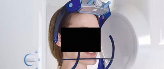
CyberKnife System
In the radiosurgical treatment of the trigeminal nerve on the CyberKnife, the same principles are applied as in the radiosurgical treatment of tumor diseases - high-precision destruction of biological tissues in a volume specified based on the results of preliminary diagnostics.
The advantage of this treatment for trigeminal neuralgia in Kiev is a specialized diagnostic department that serves the needs of the high-precision CyberKnife system. In addition, the cause of the disease can often be a tumor process in any part of the nerve or in surrounding tissues, which is difficult to identify in the early stages without special training and significant experience.
The key to the success of radiosurgical intervention is the precise determination of the location, shape and size of the damaged area of the trigeminal nerve, as well as the location in the potential zone of radiosurgical intervention of a zone of healthy structures through which the passage of radiation beams during treatment on the CyberKnife is unacceptable.
Plan for radiosurgical treatment of neurocytoma using the CyberKnife system
After calculating and visualizing the spatial model of treatment, the patient will receive the required number of sessions (fractions) of radiosurgery on an outpatient basis, during which a CyberKnife emitter placed on a robotic “arm” - a manipulator - will move around a conscious person.
Acting according to a given program, CyberKnife will deliver multiple single beams of radiation, from the intersection points of which a zone of uniformly high dose of ionizing radiation will be formed. Thus, CyberKnife will destroy a section of the nerve without irradiating healthy tissue, which makes it possible to cure trigeminal neuralgia in the shortest possible time and without traditional surgery.
It is obvious that radiosurgical treatment of trigeminal neuralgia using the CyberKnife system has a number of significant advantages over surgical intervention:
- no hospitalization is carried out,
- no anesthesia is required - there is no damage to healthy tissue, under the layer of which the damaged part of the nerve is hidden,
- there is no need for plastic reconstructive surgery to hide traces of surgery, rehabilitation, etc.
The absence of these elements also makes it possible to reduce the cost of CyberKnife treatment compared to traditional surgery.
Causes
The most common cause of the development of trigeminal neuralgia is compression of the trigeminal nerve by the arteries or veins of the posterior cranial fossa, or by a vascular loop in the presence of a vascular anomaly. The branches of the trigeminal nerve may also be compressed in the bony canals of the skull base through which they pass. Narrowing of bone canals can be congenital or acquired during chronic inflammatory processes in adjacent areas (sinusitis, caries).
Trigeminal neuralgia can occur as a result of mechanical compression of the trigeminal nerve by a tumor, with the development of a tumor process in one of the areas of the brain or the nerve itself. There are frequent cases of this pathology occurring against the background of multiple sclerosis, as a result of pinching of the trigeminal nerve by a sclerotic plaque.
Other causes of trigeminal neuralgia include:
- various types of head injuries involving the trigeminal nerve;
- jaw development abnormalities;
- infectious diseases;
- inflammatory processes in the nerve itself.