The inflammatory process of the periodontal soft tissues that occurs during complicated or improper teething is called pericoronitis. Inflammation during teething can occur in both adults and children, regardless of the location of the problem tooth, but most often pericoronitis of the wisdom tooth develops (it is also often called the “eight” or third molar).
Causes of inflammation and swollen gums during teething
The main prerequisites for the development of pericoronitis are:
- Mechanical injuries to the gingival tissue over the tooth germs due to hard foods, too hard toothbrushes, as well as due to other damaging factors.
- Long-term or difficult teething.
- Favorable conditions in the mouth for the accumulation of bacterial plaque and food particles under the gingival margin (insufficient hygiene, peculiarities of microflora in the oral cavity).
Swollen gums during teething are easily susceptible to traumatic influences, which leads to the development of inflammation. In this case, teething itself can be difficult for the following objective reasons:
- Dense bone tissue in the “eight” zone; bone compaction is most often formed because there are no milk teeth in this zone.
- There is not enough space for the “eight” in the dentition.
- Thickening of the gums and periosteum in the posterior part of the jaw is usually explained by the characteristics of embryogenesis.
Even one of these factors can cause the disease.
A lump on an area of the gum where there is no tooth yet: what to do in this case?
A couple of weeks before the tooth erupts, a cyst may appear on the gum, looking like a lump filled with a clear or bluish liquid. Such bumps are by no means common: dentists do not consider such formations to be a pathology requiring treatment. In addition, such formations on the gums in no way indicate an inflammatory process.
According to statistics, a small percentage of children are affected by the appearance of such a cyst. The child may not even suspect that he has a lump in his mouth, because if he touches it, there is no pain. However, a dental examination is still necessary, because the inflammatory process may still begin, in which case intervention will be required. The presence of inflammation is indicated by such signs as increased body temperature, pain when touching the lump, and swelling of the mucous membrane.
Most parents do not like the fact that their child has a lump in his mouth: in this case, you can ask the dentist to make an incision under anesthesia, as a result of which the fluid inside the cyst will come out. As a rule, when part of the cyst is removed, the crown of the tooth about to erupt is visible.
Features and risk factors
During difficult and prolonged eruption, the tooth germ practically does not change its position, and a significant part of its chewing surface is covered with a flap of gingival or mucoperiosteal tissue, the so-called “hood”. The flap covering the tooth germ forms a kind of pocket in which food debris and bacterial plaque accumulate. A moist nutrient medium creates optimal conditions for the intensive reproduction of pathogenic microorganisms, which, in turn, actively secrete waste products that are toxic to our body. This provokes the development of an infectious-inflammatory process in the tissues surrounding the problematic tooth germ.
Most often, inflammation of the gums occurs when a wisdom tooth erupts in the lower jaw, which is due to the anatomical structure of the jaw and the lack of space for third molars. In modern people, the jaw arch is approximately 1-1.2 cm smaller than in our distant ancestors, but the size of the teeth has remained the same. This is why the “eights” are cut much later, when the body as a whole and the dental system in particular have already formed - and because of this, it is difficult for the new tooth to find the “right” place on the dental arch.
Since wisdom tooth pericoronitis is the most common type of disease, people over the age of 18 are primarily at risk (usually “eights” are cut at the age of 20-25, but for some this process can take up to 30 years) . As for pericoronitis in children, it usually occurs against the background of insufficient oral hygiene in a child or with infectious diseases of the oral cavity (caries, gingivitis, stomatitis), which complicate teething in children and require treatment. Accumulations of plaque and hard dental deposits significantly increase the risk of developing the disease, so timely and regular removal of tartar can serve as a good preventative method.
Reasons for the development of gingivitis in children
The abundance of causes of the disease can be divided into two large groups.
Common reasons include:
- colds, especially if they occur frequently;
- sore throat and chronic sinusitis;
- hormonal disorders;
- gastrointestinal diseases.
Among the local reasons it is worth highlighting:
- poor-quality fillings, which, when destroyed, injure soft tissues;
- caries (infection from the cavity quickly enters the gums);
- crowded teeth;
- violations during the installation of braces;
- Tight lips that interfere with quality teeth cleaning.
Separately, it is worth noting the most important reason, since all of the above are only a catalyst for the development of the disease. So, the main reason why gingivitis begins to develop is poor oral hygiene, which can manifest itself both in improper brushing of teeth and in incorrectly selected toothpaste and brush.
Symptoms of the disease
Gingivitis has quite a few characteristic signs by which one can guess the onset of the disease:
- bleeding gums;
- pain during cleaning;
- swelling of the gums;
- severe redness of the gums in the case of an acute phase of inflammation;
- when the disease becomes chronic, the gums may acquire a bluish tint.
Often with the naked eye you can notice a large amount of accumulated plaque and tartar, and there may also be many teeth affected by caries.
In addition, gingivitis can be caused by diseases that have nothing to do with the oral cavity:
- heart and vascular diseases;
- disorders affecting the gastrointestinal tract;
- respiratory diseases.
The above factors reduce immunity, as a result of which the gums lose their ability to resist toxins formed in plaque.
What is the best way to treat?
As was said a little above, gingivitis occurs due to accumulated soft plaque, so the first thing you need to do is get rid of it. Plaque can be removed using ultrasound - this is a simple and absolutely painless procedure. Using a special attachment, the dentist touches dental plaque, which is destroyed by ultrasound. By the way, ultrasound allows you to get rid of not only soft plaque, but also hardened formations on the teeth.
It is necessary to understand that plaque can be effectively removed, especially if inflammation has begun, only with ultrasound: rinsing with various means, using special toothpastes and gels with the addition of antibiotics can temporarily remove the symptoms of gingivitis, but these methods are not able to eliminate its cause. As soon as you stop using these medications, bleeding will return and the disease will continue to progress. Therapy aimed at reducing the inflammatory process is justified only after the deposits have been removed.
Anti-inflammatory therapy is usually prescribed as follows:
- Rinse with Chlorhexidine solution. The maximum duration of use of the drug is 10 days. Rinsing should be done twice a day for at least 30-40 seconds per procedure. The solution, which has a bitter taste, which not all children will like, has no contraindications based on the child’s age.
- Rinse with Miramistin (prescribed after the child reaches the age of 3 years). You need to rinse three times a day. The drug is inferior in its effectiveness to Chlorhexidine and is significantly higher in price.
- Rinse with infusions of chamomile or sage (it is not recommended to use oak bark).
In addition to rinses, many doctors prescribe ointments and gels. It is believed that the gel is more effective than ointment because, due to its consistency, it has the ability to remain on the gums for a long time, which promotes better absorption. In addition, the medicinal substances contained in the product penetrate much better into the tissues from the gel than from the ointment.
The most effective and proven gels on the positive side are the following:
- Cholisal is a drug that simultaneously has two effects: analgesic and anti-inflammatory. It is used for gingivitis and the beginning of teething (the drug is rubbed directly into the place where the tooth is emerging). There are no age restrictions. The gel can be used for a maximum of one and a half weeks, twice a day. After applying the product, it is recommended to eat within two, or preferably three, hours.
- Metrogil Denta. Used after 6 years. Apply directly with your finger or a cotton swab to the gums near all teeth; there is no need to rinse off the gel.
Many parents often decide to treat their child on their own, not wanting to see a dentist. But you need to understand that the use of anti-inflammatory drugs will have no effect if you do not get rid of the deposits, so you will still have to set aside time to visit the dentist. If you ignore the advice, then:
- symptoms of bleeding will disappear, but will reappear after completion of the course of therapy;
- gingivitis can become chronic, causing periodontitis.
In addition to anti-inflammatory therapy, oral cavity sanitation and silvering are carried out - a method that does not require drilling, but has a lot of disadvantages.
How to avoid the occurrence of gingivitis?
The following remedies will help prevent gum bleeding:
- High-quality oral hygiene. Parents should instill in their children oral care skills from an early age. Not all adults know that hygiene measures must begin before the first tooth begins to erupt.
- Using the right paste. If the child does not have gingivitis, then you can use any paste designed for children. If your child does not brush their teeth well enough, you can purchase a paste with amino fluoride.
- Stick to your diet - avoid snacking, limit the consumption of fast carbohydrates (carbonated drinks, sweets). Of course, you don’t need to completely deprive your child of sweets, but you can give them only immediately after the main meal, after which you should brush your teeth.
Pericoronitis, symptoms and types
With pericoronitis, the symptoms largely depend on the form of the disease - acute or chronic. Acute pericoronitis is characterized by:
- Aching intense pain in the area of the cutting tooth, the pain intensifies under the influence of food and mechanical irritants (hygienic procedures, chewing, touching, etc.).
- Difficulty chewing food, complicated swallowing. A few days after the onset of inflammation, it becomes difficult to open the mouth and severe pain appears.
- Swelling and hyperemia of the mucous membrane.
- Enlargement of regional lymph nodes, sometimes the lymph nodes become painful when touched.
- Externally noticeable swelling of the cheeks a few days after the onset of the inflammatory process.
- General deterioration in health - weakness, lethargy, prolonged low-grade fever (increased temperature to 37-37.5 degrees).
- Bad breath, which is caused by the decomposition of food particles and the active activity of microorganisms in the “hood” space.
Acute pericoronitis can occur in a serous (catarrhal) or purulent form: in the first case, the signs of inflammation are more pronounced, the pain is more intense, and there is no purulent discharge. In the second case, purulent exudate flows freely from the inflammatory focus, due to which tissue tension is somewhat reduced and the symptoms are less pronounced. A person may feel like their condition is gradually improving, when in fact the infection is simply spreading to other tissues in the mouth.
If treatment for acute pericoronaritis was not carried out or was carried out at home, the disease gradually becomes chronic: the severity of symptoms decreases, while exacerbations of pain and discharge of pus into the oral cavity are periodically observed. In this case, there is a high risk of developing severe complications.
Clinical manifestations of the disease
Each of the clinical forms has its own typical manifestations and features of the course of the disease.
The following symptoms of gingivitis in children are distinguished:
- Catarrhal - this form appears in babies at the time of teething (by 1 year) or their change. They experience swelling and soreness of the mucous membrane. They intensify when eating spicy and hot foods. There is also bleeding, itching of the mucous membranes, and bad breath. Upon examination, a loose, thickened mucous membrane is revealed, its color changes to purple with a bluish tint. Low-grade fever and general malaise are noted.
- Hypertrophic – refers to chronic forms. It is divided into edematous and fibrous. The edematous form is manifested by swelling, bleeding, itching, and pain. Fibrous tissue is characterized by a pronounced proliferation of gingival tissue. Upon examination, gum hypertrophy is revealed, it hangs over the crown, partially covering it. The mucous membrane is loose, edematous, and false gum pockets are formed. There is plaque in large quantities. This option is more common in teenagers.
- Ulcerative – is a complication of catarrhal gingivitis. It is characterized by ulcers on the mucous membrane, covered with a grayish-greenish coating. There is an increased viscosity of saliva. The patient's mouth has a putrid, unpleasant odor. There is a deterioration in the general condition, there are signs of general intoxication, decreased appetite, and disturbed sleep.
- Atrophic – its development is possible due to defects in orthodontic treatment, anomalies in the location of the frenulum. There are no signs of inflammation. Dystrophy of the gingival tissue progresses, and the necks of the teeth are exposed. There is an increase in reaction to temperature changes.
With all these forms, the baby is capricious and refuses to eat.
Complications of the disease
In the case when treatment of the gums during complicated tooth eruption was not carried out, serious complications of the inflammatory process develop:
- Periostitis, or flux, is inflammation of the periosteum.
- Osteomyelitis - in this disease, inflammation affects both the bone tissue itself and the bone marrow, gradually leading to bone resorption. This can cause abnormal jaw fractures.
- An abscess when the infection spreads to nearby tissues. Purulent inflammation leads to the melting of muscles and fatty tissues with the formation of a large accumulation of pus.
- Cellulitis is the next stage of a purulent process after an abscess, which does not have clear boundaries. Such purulent inflammation is a serious threat to the patient and is treated in a maxillofacial hospital.
These complications are usually difficult to treat, require a serious systemic approach and can lead to irreversible consequences. Therefore, if a person suspects he has pericoronitis, treatment should begin without delay in order to prevent the condition from worsening. You need to be especially careful if gum inflammation occurs during teething in children, since the children's immune system is still immature, the body's resistance to infections is lower and the process quickly becomes more severe.
Types of boils
The choice of treatment method, as well as drug therapy, depend on what kind of abscess the child has. Only a specialist can accurately determine the type of inflammatory process and its cause, therefore any home treatment methods without consultation with a pediatric dentist are strictly prohibited. Improper treatment can lead to the spread of the purulent process to the area of the tooth root and to tooth loss (in severe cases, the lesion can also affect adjacent teeth).
Table. Types of abscesses in children and their symptoms.
| Type of education | Description |
| Granuloma | The formation is a cavity that looks like a cystic growth. The inside of the granuloma is filled with pus and necrotic cells. Such an abscess is located in the periodontal tissues and affects the tooth root, therefore, in the absence of timely treatment, the pathology will quickly lead to rotting of the tooth root system. |
| Abscess | A cavity formed in the tissues surrounding the tooth, filled with exudate or pus. The difference between an abscess and a granuloma is that it does not affect the root system. |
| Cyst | A cyst is a cavity in which pus accumulates, so the second name for this pathology is “purulent sac.” It is the result of chronic inflammatory processes and in most cases requires surgical treatment. |
Important! Some parents try to open the abscess themselves to squeeze out the pus and bacterial exudate. Do this under no circumstances - in 97% of cases such actions lead to serious complications and worsen the child’s condition due to secondary infection of soft tissues.
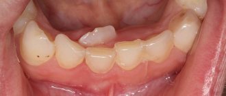
Swelling near the upper canine
Pericoronitis, treatment in children and adults
It should be noted right away that for pericoronitis, treatment at home can only reduce the symptoms of inflammation, but not solve the problem. Therefore, all home methods (rinsing with antiseptics, applying anti-inflammatory ointments, etc.) can only be used as a way to relieve inflammation during the eruption of wisdom teeth and reduce pain - this is a temporary measure before visiting the dentist.
In a dental clinic, in case of complicated teething, treatment will depend on whether the problem tooth should be preserved or whether it should be removed. If preserved, the doctor performs surgical excision of the “hood” under local anesthesia. This eliminates conditions for the accumulation of food and the growth of bacteria, and also facilitates teething. After excision of the “hood,” the patient is prescribed antiseptic therapy (rinses, baths) and, if necessary, antibiotic therapy.
In cases where tooth extraction is indicated due to their incorrect position on the jaw, lack of antagonists, or insufficient space for full eruption, the doctor immediately removes the problematic tooth and prescribes antiseptic therapy to the patient.
In some cases, at the initial stages of the inflammatory process, conservative therapy can be preliminarily applied - local treatment with antiseptic rinses and the introduction of an iodoform tampon into the “hood”. However, local therapy does not always give a positive result; moreover, it is appropriate only at the initial stage of the disease; in other cases, surgical intervention will be required.
When it comes to teething in children, treatment is always aimed at preserving the teeth (except for those cases when dystopia is clearly expressed and the doctor sees that preserving is useless).
Treatment
Depending on whether a permanent or baby tooth caused a lump on the child’s gum , the doctor carries out appropriate treatment. If a permanent tooth is damaged, seek help immediately while there is a chance to save the child’s tooth. The doctor will have to remove the nerve and fill the canals. In this case, adult dental treatment is a necessity, because if high-quality treatment and appropriate treatment are not carried out, the child may lose a tooth.
Milk teeth are removed immediately - their root system has not yet had time to form, and it is impossible to properly fill the canals. In addition, if a diseased tooth is left, the molar may grow back with defects (stains, damage.
At the Family Dentistry Center, you can remove a child’s tooth without pain or fear. All procedures in the clinic are performed under medical sedation. While the baby is sleeping peacefully and does not feel pain or discomfort, the doctor can carry out all the necessary procedures. Being able to treat your teeth while you sleep is a great way to cope with dental phobia.
Diagnostic features
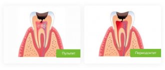
History of the disease Pericoronitis sometimes looks like a completely different disease of the oral cavity - for example, its symptoms can be confused with periodontitis (inflammation of the peri-apical tissues of the oral cavity) or pulpitis (inflammation of the dental pulp). The pain in these diseases is very similar, and periodontitis is often accompanied by hyperemia and swelling of the gums. One disease can be distinguished from another by the following signs: with pulpitis there is no pain when opening the mouth and no noticeable swelling of the gums, and periodontitis develops only at the tops of the roots of fully erupted teeth. You also need to pay attention to the presence of a characteristic “hood” - a photo will help you assess how it looks with pericoronaritis.
Why does an abscess occur?
There can be many reasons for the formation of a lump (fistula, swelling), but the most common of them is undetected or ignored caries. Not all children maintain oral hygiene and eat properly, and not all parents carefully control this, which causes caries to appear on one or several teeth at once.
If caries is not treated, it will develop into pulpitis, extending beyond the tooth area and affecting the upper part of the root. As a rule, a lump appears just near the tooth whose root is inflamed. Typically, a lump can be noticed near a tooth with caries or with a filling that was placed long ago or poorly; Another common reason for its appearance is an injury received from a strong blow or an accidental fall, which can trigger the onset of the inflammatory process.
Stages of development of an abscess:
- As a result of caries, the infection penetrates into the pulp, affecting the root tip.
- Pus begins to form near the top of the root.
- Purulent formations fall under the mucous membrane of the gums.
- A cyst appears, looking like a small lump from the outside.
The most important symptom of an abscess is a soft and painful swelling, even with slight pressure.
Sometimes, due to an excess of pus, the lump may burst under pressure, and then a fistula appears - a small hole in the gum, which is connected with the source of inflammation located in the upper region of the root. A feature of the fistula is the constant release of purulent formations.
If inflammation decreases for some reason, the fistula may close on its own. However, when the child’s immunity decreases and the inflammatory process begins again, accompanied by pus formation, the appearance of a fistula will not be long in coming.
Prevention
In some cases, especially when it comes to inflammation of the gums during teething in children, preventive measures will help prevent the disease: careful regular oral hygiene, timely removal of tartar and soft deposits, visits to the dentist for diagnostic examinations at least once every six months.
If pericoronitis is caused by the anatomical features of the structure of the dental system and the lack of space for the lower “eights” on the dental arch, preventive measures will only smooth out the course of the inflammatory process (due to timely cleaning of the oral cavity from bacterial plaque and food particles that create favorable conditions for inflammation) and identify the problem in time (subject to regular dental examinations).
Why is gingivitis dangerous in children?
Gingivitis is an inflammation of the gums that occurs as a result of the adverse effects of local and general factors. In children, it occurs quite often: between the ages of two and four years, its prevalence is 2%, and by the age of thirteen, gingival inflammation occurs to one degree or another in 80% of children. Often the disease does not progress beyond gingivitis, since it can be easily cured, avoiding more serious problems. However, if the process is started, the inflammation from the gums will spread to other periodontal tissues (the child will develop periodontitis) and will become a chronic condition, the symptoms of which will need to be relieved with each relapse.
Is it possible to cope with an abscess at home?
Treatment of abscesses and inflammations at home is quite possible, but only if the process has not turned into a purulent form. In all other cases, the child requires emergency dental care. Traditional medicine recipes can be used to slightly reduce the severity of the inflammatory process before consulting a doctor, as well as during the recovery period for faster healing of damaged tissues.
Linden lotions
Mix 1 spoon of linden flowers and crushed oak bark and pour 300 ml of boiling water. Leave covered for at least 1 hour. Use the infusion to rinse the mouth 4 times a day for 10 days. Young children can make lotions: to do this, moisten a cotton swab with the infusion and apply it to the abscess for 10-15 minutes. The procedure should also be repeated 3-4 times a day.
Linden flowering branch
Oil mixture with eucalyptus
The product copes well even with severe abscesses, but it will take a day to prepare. Mix 2 tablespoons of a mixture of olive and pumpkin oil with a spoon of crushed eucalyptus leaves. Place the product in the refrigerator and leave for 24 hours. The next day, add juice from half an onion, mix everything and put it in the refrigerator for another 1 hour. Apply the resulting mixture to abscesses and ulcers 5-6 times a day for 15 days.
Treating an abscess on the gum at home
Abscesses on the gums are a fairly serious pathology that can be easily treated if you do not delay visiting a dentist. If treated incorrectly, there is a high probability of complications in the form of tooth root decay, which is an indication for extraction of the affected tooth. To prevent this and maintain the integrity of the dentition, there is no need to self-medicate - the most reasonable choice would be to go to the treating dentist and follow all his prescriptions and recommendations.