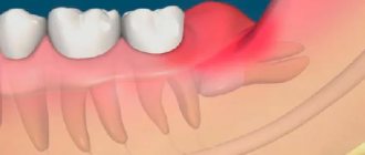
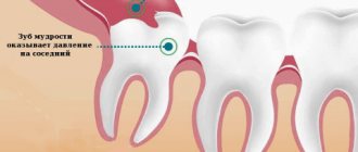
Periocoronaritis is a dental disease characterized by inflammatory processes in the soft gum tissues that surround the erupting units (in the vast majority of cases, wisdom teeth). It manifests itself as intense pain, the inability to open the mouth normally, swelling of the gums, as well as an unpleasant odor from the oral cavity. Inflammation develops from the accumulation of plaque in the opening of the gum through which the unit erupts, or due to the difficulty of the process itself. The latter causes pressure on the adjacent unit along with damage to the bone and gum tissue.
The CELT Surgical Department of Dentistry invites you to undergo treatment for pericoronitis in Moscow. Our multidisciplinary clinic has been operating in the paid medical services market for more than two decades and enjoys a good reputation among patients. We have all the necessary certificates and licenses and provide treatment with the conclusion of an official contract and provision of a guarantee. You can find out the price of excision of the hood for periocoronitis or removal of a wisdom tooth by going to the “Services and Prices” section.
Consultation with a dentist-therapist - 1,000 rubles.
Treatment of pericoronitis with medication - 1,000 rubles.
Surgical treatment of pericoronitis (excision) - 2,500 rubles.
At CELT you can get advice from a dental specialist.
- The cost of a dental consultation is 1,000
Make an appointment
Causes of pericoronitis
As the tooth erupts, it rests on the mucous membrane. He does not always immediately overcome this barrier. A tooth can remain under the gum from 4 weeks to 6 months or more, gradually lifting and injuring the gum. This leads to the fact that the gums cease to protect the tissues from pathogenic bacteria and small food particles; they easily penetrate under it and provoke an inflammatory reaction.
The inability to perform hygiene under the hood is the main mechanism of pericoronitis. But other factors can increase the risk of inflammation or worsen the situation:
- anatomical features of the structure of bone tissue and gums - thickened periosteum or gums, lack of space in the jaw;
- violations of the rules of oral hygiene - in this case, it is easier for bacteria to begin to actively multiply;
- trauma - damage to periodontal tissues by solid food particles, excessive brushing with bristles during cleaning;
- chronic diseases of the oral cavity - erosions and ulcers, stomatitis, gingivitis, caries, pulpitis, periodontitis, including near the erupting tooth;
- dystopic or impacted tooth, eruption at an angle, impossibility of complete eruption.
More often, pericoronitis is observed in the area of erupting eights or wisdom teeth. This is due to the fact that they appear in adulthood, when the dentition is formed and the jaw has stopped growing. At the end of the dentition there is simply no room left for a large tooth, and therefore the figure eight grows at an angle or is partially hidden by the gum. If wisdom teeth begin to erupt before the age of 20, the process is usually easy. At older ages, the process is more often complicated by pericoronitis.
Ask a Question
Prevention
In some cases, especially when it comes to inflammation of the gums during teething in children, preventive measures will help prevent the disease: careful regular oral hygiene, timely removal of tartar and soft deposits, visits to the dentist for diagnostic examinations at least once every six months.
If pericoronitis is caused by the anatomical features of the structure of the dental system and the lack of space for the lower “eights” on the dental arch, preventive measures will only smooth out the course of the inflammatory process (due to timely cleaning of the oral cavity from bacterial plaque and food particles that create favorable conditions for inflammation) and identify the problem in time (subject to regular dental examinations).
Symptoms of pericoronitis
Pericoronitis of the tooth begins with pain in the gum area, which intensifies with pressure. You can usually notice discomfort when chewing or brushing your teeth. The gum area becomes swollen and red, and subsequently an unpleasant odor appears from the mouth.
In the absence of timely medical assistance, other manifestations also occur:
- spread of pain radiating to the ear, temple;
- difficulty swallowing, sore throat;
- speech difficulties;
- limitation of mouth opening due to swelling of the peripharyngeal area;
- enlarged lymph nodes;
- low-grade fever;
- swelling of the cheek due to inflammation.
A person experiences difficulty chewing food and may suffer from a general deterioration in health and headaches.
Symptoms may subside after some time, which means that the disease has entered a chronic stage. Purulent complications may also occur, and if a passage opens in the gum to remove purulent contents, the acute pain goes away. However, the inflammatory process persists.
Types and forms of the disease
There are acute and chronic pericoronitis. Acute, in turn, is classified into the following types:
- Catarrhal. The disease begins with swelling and redness of the gums, pain, and itching. This is the simplest form of the disease.
- Ulcerative. It is characterized by the formation of ulcerations covered with a white coating on the mucous membrane of the gums, necrotization of tissue along the edges of the ulcer.
- Purulent. In the area of inflammation, serous and subsequently purulent contents are released. It is characterized by throbbing pain, general intoxication of the body, and bad breath.
Chronic pericoronitis is a consequence of an untreated acute period. With it, the symptoms become less pronounced, but serous or purulent contents continue to form. It can lead to the formation of a pathological passage through which the contents are discharged into the oral cavity. Chronic pericoronitis is characterized by periodic exacerbations; they can be provoked by tooth movement and other unfavorable factors, a general decrease in the body's defenses.
In the chronic form, lymphadenitis is often observed. The mucous membrane, even in the absence of unpleasant symptoms, is swollen and has a reddish tint. Despite the absence of serious difficulties when speaking, chewing, or opening the mouth, it is important to get help from a dentist, since the risk of complications is quite high, and the disease may not go away on its own.
Classification
Pericoronitis is divided into acute, which occurs most often, and a rarer form - chronic pericoronitis. Despite the fact that the latter type is not reflected in ICD-10, it occurs with improper treatment of pericoronitis.
According to the flow, the following types of pericoronitis are distinguished:
- Catarrhal – characterized by slight swelling and pain when touched;
- Purulent - constant pain and the presence of purulent discharge;
- Ulcerative – leads to the formation of ulcerative defects on the gums;
Retromolar is distinguished as a separate form - it is distinguished by its location, which does not allow it to be easily seen and treated, which makes it difficult to combat this disease and increases the likelihood of complications.
Diagnosis of pericoronitis
Manifestations of acute pericoronitis may resemble pulpitis and periodontitis. But even an untrained person can distinguish these diseases from each other. Thus, with acute pulpitis, throbbing pain does not interfere with opening the mouth, and the gums often remain calm, without swelling and redness.
With periodontitis, swelling and redness of the gums are common symptoms. But periodontitis develops under a fully erupted tooth and is most often a consequence of advanced caries. With pericoronaritis, the tooth has not fully erupted.
A dentist can make an accurate diagnosis. Radiography is mandatory - a targeted image will help to accurately assess the condition of the tissues, the position of the erupting tooth in the jaw, the extent of the spread of the inflammatory process, and also exclude diseases with similar symptoms.
Development of acute pericoronitis
Acute Pericoronitis is considered as a type of acute periodontitis with primary symptoms of damage to the gum tissue. Damaged tissues located above the crown of a growing tooth involve in the inflammatory process all areas of the gums located near the resulting lesion. This course of the process is most often observed during the growth and eruption of the third, less often, fourth large molars of the lower jaw.
As a reason, experts consider both the lack of space for the formation of new teeth due to the development of a short jaw, and the abnormal development of the dental crown itself, which has assumed an incorrect location in the dentition.
What happens if pericoronitis is not treated?
It is important to understand that pericoronitis will not go away on its own. Even if after 4-5 days the symptoms subside, this does not mean that the inflammatory process is over. From time to time the disease will go into the acute stage and cause a lot of inconvenience. In addition, pericoronitis can cause serious complications.
- Periostitis, or flux, as well as osteomyelitis. The spread of the inflammatory process to the periosteum and bone tissue can be explained by a decrease in local and general protective forces and other factors. Such complications will require serious medical intervention.
- Mobility of adjacent teeth.
- Cellulitis, abscess, lymphadenitis.
- Formation of fistula tract, cysts. The appearance of ulcerative stomatitis.
- Damage to nearby tissues, otitis, pharyngitis and other inflammatory complications in the area of ENT organs.
- Sepsis.
Therefore, treatment of pericoronitis is mandatory, regardless of the area in which the inflammation has spread. Lack of timely medical care can lead to inflammatory consequences and even loss of healthy adjacent teeth.
Surgical methods of treatment
Treatment of pericoronitis is almost always surgical. And if we talk about molars and premolars, dentists choose the tactic of excision of the gingival hood. But treatment in the area of eights is almost always performed by removing wisdom teeth, and further we will explain why.
Excision of the gingival hood opens access to the growing tooth; this measure allows not only to remove excess inflamed tissue and thoroughly rinse the area, but also to help the tooth erupt completely and take its correct place in the dentition. This method has several conditions or indications:
- integrity of the crown and root system of the erupting tooth;
- correct location of the tooth in the bone tissue of the jaw;
- availability of sufficient space for complete tooth eruption in the row.
That is, this approach is used in all cases where the gums are the only obstacle to normal tooth eruption. If a unit of the dentition is not very healthy, for example, caries is observed, the integrity of the crown is compromised, or there are diseases of the root system, then the decision is made individually. If it is advisable to preserve the tooth, the doctor will also perform an intervention and take measures to eliminate the pathology.
Impacted and dystopic teeth, as well as situations in which complete tooth eruption will inevitably lead to malocclusion or displacement of adjacent teeth are indications for a different type of surgical treatment. The doctor may suggest tooth extraction, and in cases of disease above the “eight” level, this is almost always the only option to solve the problem.
The fact is that wisdom teeth do not bear a functional load, very often erupt at an angle, interfere with neighboring teeth and increase the risk of complications, including bite defects. Therefore, it is considered appropriate to remove the “eight” and further measures to eliminate inflammation in the soft tissues.
The only exceptions are cases when the “eight” could potentially be used as a support for a prosthesis or be important in planned orthodontic treatment.
In general, the following cases and features are indications for removal of both the hood and the tooth:
- excision of the hood has already been performed previously, but the inflammation has not gone away and/or has intensified, there is no effect from the treatment;
- the tooth does not have enough space in the dentition;
- impacted, dystopic tooth, the preservation of which jeopardizes the health of neighboring teeth and the oral cavity;
- the gingival hood grows over the erupting tooth again (this rarely happens);
- the tooth is affected by caries/destroyed, its treatment and preservation are impractical.
Since it requires the removal of a tooth that has not yet fully erupted, the procedure is considered a complex extraction. Most often, it requires preliminary dissection of the gums and extraction of the tooth from deep-lying tissues. Therefore, in many cases, sutures will be required on the gums, which will speed up tissue healing.
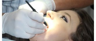
Surgical treatment of pericoronitis is performed under local anesthesia, so the procedure will not cause discomfort. An obligatory step is washing the mucous membrane with antiseptic solutions.
Removal of tissue hanging over the tooth can be done with conventional instruments or a laser.
Features and risk factors
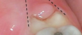
During difficult and prolonged eruption, the tooth germ practically does not change its position, and a significant part of its chewing surface is covered with a flap of gingival or mucoperiosteal tissue, the so-called “hood”. The flap covering the tooth germ forms a kind of pocket in which food debris and bacterial plaque accumulate. A moist nutrient medium creates optimal conditions for the intensive reproduction of pathogenic microorganisms, which, in turn, actively secrete waste products that are toxic to our body. This provokes the development of an infectious-inflammatory process in the tissues surrounding the problematic tooth germ.
Most often, inflammation of the gums occurs when a wisdom tooth erupts in the lower jaw, which is due to the anatomical structure of the jaw and the lack of space for third molars. In modern people, the jaw arch is approximately 1-1.2 cm smaller than in our distant ancestors, but the size of the teeth has remained the same. This is why the “eights” are cut much later, when the body as a whole and the dental system in particular have already formed - and because of this, it is difficult for the new tooth to find the “right” place on the dental arch.
Since wisdom tooth pericoronitis is the most common type of disease, people over the age of 18 are primarily at risk (usually “eights” are cut at the age of 20-25, but for some this process can take up to 30 years) . As for pericoronitis in children, it usually occurs against the background of insufficient oral hygiene in a child or with infectious diseases of the oral cavity (caries, gingivitis, stomatitis), which complicate teething in children and require treatment. Accumulations of plaque and hard dental deposits significantly increase the risk of developing the disease, so timely and regular removal of tartar can serve as a good preventative method.
Laser treatment
Laser excision of the hood provides better results. The main advantages include:
- lower risk of inflammatory complications, including secondary wound infection;
- coagulating effect of the laser - no risk of bleeding;
- short recovery period;
- reduction of pain after intervention;
- no need to additionally treat intervention areas with turundas and antiseptics.
There are few contraindications for laser intervention, one of them being cancer.