Have you been prescribed a resection of the apex of a tooth root, and would you like to study the essence of the operation in more detail? Do you want to know about all the possible consequences and recovery after all manipulations? Read our material to the end, where we have collected answers to the most popular questions regarding resection of the apex of the tooth root.
Resection of the upper part of the tooth root or apicoectomy is a surgical operation that allows you to remove the inflammatory process in the root of the tooth and bone without affecting the crown itself. It should be carried out if there is a cyst, granuloma on the very root part of the tooth or near the diseased tooth. In the process, not only the infected area is removed, but also part of the root - this is done in order to prevent repeated inflammatory processes.
What is an apexectomy
Stages of tooth root resection
This is a surgical procedure that allows you to get rid of the source of infection located inside the gums without removing the tooth unit. Otherwise called resection of the apex of the tooth root. Often, infectious foci form in the area of the tooth root, and apexectomy allows you to cut out and remove the affected part of the root without affecting healthy tissue. This treatment method is considered the best for granuloma, periodontitis and similar serious pathologies, in which the affected teeth were removed in the past.
Contraindications for tooth root resection surgery
Like any operation, tooth root resection has its contraindications:
- Chronic inflammatory gum diseases (periodontal disease, periodontitis);
- Increased tooth mobility;
- Severe destruction of the root part of the cyst and granuloma and of the tooth itself, which cannot be a reliable basis for a crown;
- Close contact of healthy tooth roots with pathological ones;
- Spread of cysts and granulomas over more than 60% of the root part;
- Diseases of the body, such as oncology, tuberculosis, bleeding disorders, diabetes mellitus, central nervous system diseases, disorders of the heart and blood vessels.
It is important! Sometimes it will be more effective to remove a pathological tooth than to restore it by resection of the root apex. The dentist will weigh the pros and cons of the operation and, based on the diagnosis, recommend the optimal solution to the problem.
What causes complications?
Negative consequences often arise either due to medical error or the fault of the patient during rehabilitation. Causes of resection complications:
- Late filling. This procedure is carried out 1-2 days before the apexectomy, otherwise there is a risk of inflammation occurring after the operation as a negative reaction.
- Poor quality filling. The canals inside the tooth must be sealed, otherwise an inflammatory process will occur if the infection continues to spread.
- Tooth decay. If the treatment is handled incorrectly during surgery, there is a risk of damage to the tooth, which will lead to its loss.
- Unprofessionalism. Sometimes serious damage during surgery is caused by the doctor due to improper use of the instrument or careless handling of it.
- Incomplete resection. If the inflamed tissue and source of infection are not completely removed, then a recurrence of the cyst is possible in the future.
- Tissue infection occurs during surgery due to incomplete sterility or after improper oral care.
- Root destruction. The operation involves removing only the tip of the root, a minimal part of it, but if the dentist makes a mistake, the root can completely collapse, causing the tooth to become loose.
- Failure of the patient to comply with postoperative appointments. Proper care, lack of physical activity, diet and other medical prescriptions speed up the rehabilitation process.
What are the consequences after tooth root apex resection surgery?
Apexectomy can result in various complications, for example, soft tissue swelling (this is a temporary reaction to injury) or even recurrence of the cyst. All negative consequences are usually divided into two types: conditionally normal and pathological.
Fever
This is the body's natural reaction to injury during surgery. An increase in temperature occurs on the first day after apexectomy. Typically varies between 37.5°C and 38°C. In the evening the temperature rises and drops in the morning. It normalizes on its own in 1-2 days. It is not recommended to knock it down if the mark on the thermometer is below 38°C. But if you feel very unwell (general weakness, dizziness, body aches), you are allowed to take an antipyretic drug. Sometimes fever and chills signal the development of serious complications. In this case, the high indicator lasts 2-3 days after the operation, increases every day and is not knocked down by pills. If there are complications, it is accompanied by additional symptoms:
- pain;
- severe swelling of the soft tissues of the face;
- putrid odor from the mouth;
- the presence of purulent plaque on the wound;
- pressure instability.
With such a set of symptoms, you should immediately contact the dentist.
The tooth is loose
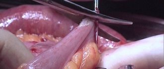
result of tooth root resection
Resection involves removing the tip of the root, which often leads to a decrease in the stability of the tooth. If the root is destroyed during surgery, the tooth will become very loose and may fall out. Also, tooth instability is caused by the patient himself if he loads the operated area with solid food.
The dentist returns the stability of the tooth if you contact him immediately after the problem occurs.
To avoid such a complication, it is necessary to follow a gentle diet in the postoperative period. The tissue healing process lasts 4-6 months, during which time the cavity formed during the operation will completely heal, and then the stability of the tooth will become the same. Until this moment, you should not chew nuts, dry foods, or hard candies on the operated side.
White gums
White plaque can be a sign of a natural healing process or complications: gum inflammation, purulent infection:
- A day after surgery, a white fibrin coating forms on the gum.
- After two to four days, the plaque becomes most noticeable.
- By day 7, swelling subsides, pain disappears, but the gums remain white.
- By day 14, the plaque disappears, new mucous tissue forms underneath it, and the gums acquire a normal color.
The following symptoms indicate the development of a purulent infection:
- severe increasing pain;
- bleeding or profuse ichor (the wound is bleeding);
- rotten smell;
- swelling and redness of the gums for more than a week;
- change in color of plaque to yellowish, gray, brown;
- poor general condition.
This consequence will have to be treated with anti-inflammatory and antibiotics, and can be prevented by rinsing with antiseptic solutions (Miramistin, Chlorhexidine).
Polypous purulent sinusitis
Apexectomy of the upper dental units is accompanied by the risk of breaking through the wall of the maxillary sinus. If this happens, a fistula forms between this cavity and the vestibule of the mouth, which subsequently provokes purulent polypous sinusitis. Its symptoms:
- pain in the lower part of the eye sockets;
- nasal congestion;
- speaking through the nose (nasality);
- it hurts to chew, press on the disturbing area and move your mouth;
- constant headaches;
- weakening or loss of sense of smell;
- purulent fluid from the nose;
- sensation of the presence of a foreign object in the nasopharynx.
This complication is treated with conservative and surgical methods. Conservative treatment consists of taking antibiotics and intranasal hormonal medications (such medications include Mometasone furoate). Surgical treatment involves closing the fistula.
Bleeding from the nose
Nosebleeds during surgery or immediately after indicate perforation (break through) of the bone of the nasal sinus. It can open during resection of the upper teeth, since the roots in the jaw are located close to these sinuses, which is why the sinuses are easily injured at the slightest careless movement of the doctor. If a sinus perforation occurs, the doctor must stop the bleeding, make sure that nothing gets into the hole (perhaps a piece of the root has fallen off and pressed in there), disinfect and close the resulting wound. Then antiseptics for rinsing, antibiotics and anti-inflammatory drugs are prescribed. If fragments and foreign objects get into the sinus, a more serious operation will have to be performed.
Painful sensations
The resection itself is completely painless thanks to anesthesia, but when it wears off (2-3 hours after the procedure), the patient may experience soreness, which is a natural reaction to injury. In the first few hours after surgery, the gums ache very much. The drugs Diclofenac, Dicloberl, Ketanov and the like, which will be recommended by the dentist, will help relieve pain. With each subsequent day, the severity of pain decreases. After 3-5 days it goes away completely. If the pain throbs, the sensation does not go away after 3-5 days, you need to contact the dentist again.
Damage to the maxillary, nasal sinuses, and blood vessels of the jaw
problems associated with resection of the roots of teeth
Damage to the nasal maxillary sinuses during resection of the roots of the upper tooth is manifested by severe bleeding with air bubbles in the blood, a nasal voice of the patient and bleeding from the nose. This postoperative complication is eliminated on the spot. Damage to large blood vessels is manifested by prolonged bleeding from the wound. Stops with sterile swabs with vasoconstrictor medications. If it opens at home, you need to apply a gauze swab to the wound, press it and change it periodically. If the bleeding does not stop within 2-3 hours, you should seek help. To prevent bleeding, you should stop taking blood thinning medications before surgery and not take them after the procedure.
Numbness of the face
This symptom is possible when the trigeminal nerve and its branches are damaged or irritated. Main symptoms:
- nagging pain;
- burning sensation in soft tissues;
- numbness of the soft tissues of the cheek (the cheek hangs and cannot be felt);
- weak sensation when touching the cheek.
Damage to the trigeminal facial nerve is extremely rarely caused by an instrument. More often, irritation occurs due to the pressure that the filling material exerts on the branches. If there is numbness in the face, a course of neurological treatment should be carried out in combination with addressing the cause of the nerve damage.
Suppuration, fistula, lump, abscess
A fistula is a channel connecting foci of infection to the surface of the gum, through which pus comes out. Its appearance indicates poor quality cleaning of the dental canal and wound during the operation. This can be avoided by sealing the perforated area with a special filling, which the dentist must do. Also, the complication is often provoked by the patient by improper oral care in the postoperative period. To prevent gum decay (formation of a purulent lump), it is necessary to regularly disinfect the oral cavity after surgery: rinse with antiseptics (for example, Chlorhexidine) or soda solution. Treatment of complications in the form of a purulent fistula is carried out in two ways:
- Medication with antibiotics (Levomekol, Lincomycin, Metranidazole).
- Surgically (the seal is opened and cleaned so that all the pus comes out).
The success of resection depends not only on the professionalism and experience of the dentist, but also on the patient’s compliance with all prescriptions during the rehabilitation period.
Filling the root canal using the single pin method with a sealer based on tricalcium silicate
Randall Cohen, DDS , Consultant, Alleman-Delipieri Center for Biomimetic Dentistry, General Dentist, Temple University (Philadelphia, PA, USA). INTRODUCTION
Lateral condensation and vertical compaction of gutta-percha have been widely used in endodontic practice in recent decades. The need to seal gutta-percha historically arose due to the fact that sealers themselves did not allow achieving an adequate result. The sealers were hydrophobic, unstable in volume, not biocompatible, subject to degradation and caused periodontal irritation when removed beyond the root apex. As a consequence, all modern techniques - lateral condensation, vertical compaction, hot gutta-percha on a carrier - have been developed to achieve a minimum volume of sealer. It was generally accepted that increasing the amount of sealer leads to shrinkage, microleakage and failure, and all root canal filling techniques were aimed at achieving a minimum thickness of the sealer layer.
This article provides an overview of the goals of endodontic treatment and the main obturation techniques. Its author proposes a simpler method of root canal filling using one gutta-percha point and a new material based on tricalcium silicate, which does not have the disadvantages of traditional sealers. Goals of endodontic treatment
The triad of biomechanical preparation, antibacterial treatment and complete obturation forms the basis of endodontic treatment.1
The tooth cavity and root canals must be thoroughly cleaned of organic residues and given the correct shape. This is achieved by instrumental and medicinal treatment of the root canal, after which the root canal is free from infection and ready for obturation.
A good sealer for filling destroys all bacteria remaining in the root canal system, depriving them of nutrition and creating conditions that make it impossible for them to reproduce. In addition to this, the root canal filling material must have antimicrobial properties to prevent further bacterial growth. It is also important to provide both coronal and apical sealing to prevent bacterial entry and reinfection.
For filling root canals, today it is generally accepted to use gutta-percha as a material with the necessary properties. Gutta-percha is insoluble, has minimal impact on tissue, is well tolerated by the body, is stable in volume and, if necessary, can be dissolved using a solvent.
Another component used for obturation is a sealer, which complements gutta-percha.
The sealer must:
- Easily inserted into the root canal.
- Seal the apex and lateral canaliculi.
- Do not shrink.
- Be impermeable to moisture.
- Possess bacteriostatic properties.
- Be radiopaque.
- Do not stain tooth tissue.
- Do not irritate periapical tissues.
- Subject to sterilization.
- If necessary, it can be easily removed from the root canal.1
Modern methods of root canal obturation
There are several methods of root canal obturation, each of which uses gutta-percha. The first technique is called lateral condensation (Fig. 1), under which root canals are usually treated using the traditional “step back” system.
Rice. 1. Lateral condensation The master pin, coated with sealer, is set to working length, and then the dentist, using a spreader, compacts additional pins until the entire space between the master pin and the root canal wall is filled.
Another method of filling root canals - vertical compaction (Fig. 2), first proposed by Schilder2 - involves installing a master pin coated with gutta-percha to the working length.
Rice. 2. Vertical compaction of hot gutta-percha Then, with a red-hot plager, the gutta-percha is thermoplasticized and compacted. This filling technique creates hydraulic pressure to fill the laterals and other branches of the root canal system. The coronal two-thirds of the master pin are removed when the heated plug is removed, and a “plug” is formed in the apical third, allowing controlled filling of the root canal with softened gutta-percha. This technique has many advantages, but also disadvantages compared to lateral condensation. One of the problems is the need to widen the orifice of the root canal sufficiently to insert a hot plug into the apical third (less than 4 mm from the root apex).
Other problems with vertical compaction are heterogeneity, relatively large amounts of sealer in the apical zone, poor adaptation to the root canal walls, and extension of gutta-percha beyond the apex.3
For the long-term survival of the tooth, it is critical to preserve as much coronal dentin as possible. This must be taken into account not only when creating the minimum possible endodontic access, but also when choosing a restoration technique. Unfortunately, both root canal filling methods require root canal preparation, which removes large amounts of coronal dentin and reduces the strength of the tooth. Method of filling a root canal using one pin
Recently, dentists have increasingly used the single pin method4 (Fig. 3), especially with a large taper corresponding to the taper of root canals after treatment with nickel titanium endodontic instruments (NiTi).
Rice. 3. Single pin method This technique does not require additional pins, lateral condensation, or vertical compaction of hot gutta-percha. In other words, the root canal is filled with a master pin corresponding to the last nickel-titanium instrument used. This combination of the single pin and sealer method allows you to avoid mistakes that are possible when using other root canal filling methods. This method is much easier for the dentist, requires less time, which makes treatment easier for the patient, and also does not result in pressure on the canal walls. Root canal treatment with nickel-titanium instruments
The use of rotary nickel-titanium root canal instruments allows for a simpler obturation method using a single pin (corresponding to the size of the last file used) tightly adjacent to the walls throughout the entire length of the root canal. By supplementing this technique with a bioactive, biocompatible, non-shrinking sealer, the requirements for successful preparation, disinfection, shaping and filling can be met without excessive dentin removal and a high long-term treatment success rate can be achieved. Sealer based on tricalcium silicate
Sealer based on tricalcium silicates BioRoot RCS (Septodont) has many advantages over materials of previous generations. Due to its alkaline pH (providing antibacterial properties), calcium ion release, sufficient radiopacity and flowability, BioRoot RCS is superior to all materials used today. BioRoot RCS is volume stable, biocompatible, hydrophilic, stimulates bone regeneration and provides sufficient adhesion to dentin in root canals. CLINICAL CASE
A 68-year-old patient consulted a dentist with complaints of pain in the left lower jaw. According to the patient, she took the antibiotic Augmentin, prescribed to her by her general practitioner. An x-ray showed a focus of bone tissue destruction and a diagnosis was made: periapical abscess of tooth 35.
The root canal was prepared to size 30.06. Irrigation was carried out with a 5% sodium hypochlorite solution. The root canal was washed with an anesthetic solution (Septocaine, Septodont) and solutions of EDTA and chlorhexidine. After repeated treatment with anesthetic, the canal was again washed with 5% sodium hypochlorite solution. A 30.06 master pin was then fitted and BioRoot RCS sealer was prepared according to the manufacturer's instructions. The root canal was dried with paper points and filled with BioRoot RCS using a master point. After this, the master pin was removed from the root canal, re-coated with sealer and set to working length. The master pin was cut at the level of the orifice with a heated plugger, and excess sealer was removed with a No. 2 spherical diamond bur (Fig. 4–7).
Rice. 4. Diagnostic radiograph
Rice. 5. The root canal is processed, the master pin is adjusted to the working length
Rice. 6. The master pin coated with BioRoot RCS (Septodont) at the mouth is cut off with a hot plugger
Rice. 7. A follow-up radiograph after one year shows complete healing of the periapical lesion. DISCUSSION
For the success of endodontic treatment, thorough instrumental and medicinal cleaning of the walls of the root canals, as well as high-quality filling, is necessary.
Sealers have disadvantages - shrinkage, degradation, irritation of surrounding tissues. As a result, traditional filling methods are aimed at filling the space of root canals with gutta-percha in such a way as to minimize the amount of sealer, which requires a lot of time, so success highly depends on the qualifications of the doctor, their use is tedious for both the dentist and the patient, and can even cause root cracks due to excess pressure.
The new method uses a single pin in combination with a tricalcium silicate sealer, overcoming the challenges associated with previous generations of materials. The new sealer based on tricalcium silicate has antimicrobial and anti-inflammatory properties, binds to dentin, remains stable in volume, that is, it most fully meets the requirements for an ideal material for filling root canals. Accordingly, there is no need to apply significant force to push the sealer, since it fills all the voids in the root canal, due to its hydrophilicity, and prevents the growth of bacterial colonies. Due to the lack of shrinkage and degradation, there is no apical and coronal microleakage. There is also no risk of tooth root fracture, since the master pin used for filling is inserted to the working length without pressure. In addition, there is no need to weaken the tooth by removing coronal dentin to allow the plugger to reach the apex. LITERATURE
- Cohen S, Hargreaves K. Pathways of the Pulp 9th ed. Mosby, St. Louis, MO, 2006.
- Schilder H. Filling root canals in three dimensions, Dent Clin of North Amer 1967;723-44.
- Tasdemir T, Er K, Yildirim T, Buruk K, Çelik D, Cora S, et al. Comparison of the Sealing Ability of Three Different Techniques in Canals Shaped with Two Different Rotary Systems: a Bacterial Leakage Study Oral Surg Oral Med Oral Pathol Oral R adiol Endod 2009 Sep;108(3):e129–34.
- Pereira C. A., et. al. Single-Cone Obturation Technique: a literature review. RBSO Oct/Dec 9(4):442–7 2012.
Go to the website of the exclusive distributor of Septodont in Russia Dental Times 37