Causes of development of cleft palate in the fetus
The list of factors that can provoke this pathology is quite wide. Among them are both related to the expectant mother and external:
- genetic predisposition;
- toxicosis;
- exposure to bacteria and viruses;
- chronic diseases of the mother (gynecological, respiratory organs);
- infectious diseases of the mother (herpes, sore throat, influenza, otitis and bronchitis);
- smoking and alcohol abuse during pregnancy;
- taking medications (antibiotics, salicylates, sulfonamides);
- work in hazardous production;
- stress;
- sudden changes in atmospheric pressure;
- radiation, including solar;
- environmental violations [2, 3].
Environmental influence is last on the list, but not least. As a result of rapid technogenic development, the state of the environment is steadily deteriorating. Largely because of this, the percentage of newborns with cleft palates has tripled over the past 100 years [4].
Why carry out the procedure?
The main indications for this type of operation are congenital malformations, which are associated with impaired formation of the soft tissues of the lips, nose, soft and hard palate during intrauterine development. In this case, veloplasty is performed to eliminate the cleft of the soft palate; usually such operations are accompanied by uranoplasty (hard palate plastic surgery) and cheiloplasty (restoring the shape of the lip).
One of the possible indications for veloplasty is snoring associated with sagging of the soft palate and an increased size of the uvula. To prescribe this operation for such a problem, the doctor must carefully examine the patient’s condition and accurately identify the etiology of snoring.
Classification of defects
Based on their shape and anatomical features, congenital clefts are divided into several types and subtypes. First of all, they focus on localization:
- clefts that affect only the soft palate;
- clefts affecting both the hard and soft palates;
- clefts of the soft, hard palate and alveolar process;
- clefts of the alveolar process and the anterior part of the hard palate.
Then the degree of expression is assessed.
- Hidden: located within the muscle tissue of the soft palate and the bone tissue of the hard palate, without extending to the mucous membrane.
- Incomplete: the palate is partially cut.
- Full: completely cut through the palate.
They also describe whether the defect appears on one or both sides of the face. As a result, the classification takes the following form:
- clefts of the soft palate can be hidden, incomplete or complete;
- the same varieties in clefts of the soft and hard palate;
- clefts of the soft, hard palate and alveolar process are always complete and can be one- or two-sided;
- clefts of the alveolar process and the anterior part of the hard palate can be complete or incomplete, in both cases - unilateral or bilateral [4].
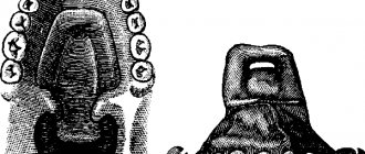
Cleft lip
Rice. 1a
Rice. 1b
Rice. 2a
Rice. 2b
Rice. 2s
Rice. 2d
Rice. 3a
Rice. 3b
Rice. 3s
Rice. 4a
Rice. 4b
Rice. 4s
Rice. 5a
Rice. 5b
Rice. 5s
Rice. 6a
Rice. 6b
Rice. 6s
Rice. 7a
Rice. 7b
Rice. 7s
Rice. 7d
Rice. 7e
Rice. 7f
Rice. 7g
Rice. 7h
Rice. 7i
Rice. 7j
Rice. 7k
Rice. 8a
Rice. 8b
Rice. 8s
Rice. 8d
Rice. 9a
Fig 9b
Rice. 9s
Rice. 9d
Rice. 10a
Rice. 10b
Rice. 10s
Rice. 11a
Rice. 11b
Rice. 12a
Rice. 12b
Rice. 12s
Rice. 13a
Rice. 13b
Rice. 13s
Rice. 14a
Rice. 14b
Rice. 14s
Rice. 15a
Rice. 15b
Rice. 15s
Rice. 15d
Rice. 15th
Rice. 15f
Rice. 16a
Rice. 16b
Rice. 16s
Rice. 16d
Congenital clefts of the upper lip and palate are among the most common defects in humans and account for 12–30% of all deformities. This pathology ranks second, second only to cardiovascular pathology, among all congenital malformations of the body. In the structure of lesions in the craniofascial and maxillofacial areas, it accounts for up to 90%. According to the literature, the incidence of births of children with congenital cleft palate varies in different regions of our planet from 1 in 300 to 1 in 2000. Statistical studies conducted in recent years indicate an increase in their number. The severity of these conditions is determined not only by external facial disfigurement, pronounced functional disorders of the dentofacial apparatus and ENT organs (breathing, nutrition, speech, facial expressions, hearing), social impairment of children in the family, preschool and school groups, but also by a number of somatic disorders, leading to impaired growth and development of the body.
Etiology of congenital clefts of the upper lip and palate
- I. Exogenous causes
Physical factors: mechanical, thermal, radiation
Chemical factors: hypoxia, malnutrition, hormonal imbalances, teratogenic poisons
Biological factors: viruses, bacteria and their toxins, protozoa
Mental factors
- II. Endogenous causes
Heredity
Biological inferiority of germ cells
Influence of parental age
Classification of cleft lip and palate
1. By nature and prevalence:
- single or double sided
- complete or incomplete
- hidden
- By localization:
- upper lip
- upper lip and alveolar process in/jaw
- upper lip, alveolar process of the jaw and hard palate
- upper lip, alveolar process in the jaw, hard and soft palate
- alveolar process in the jaw, hard and soft palate
- hard and soft palate
- soft palate
The problem of rehabilitation of patients with clefts of the upper lip, hard and soft palate continues to be relevant to this day, despite the large number of developments devoted to its various aspects. In the rehabilitation of this category of patients, one of the key points that directly affects its outcome is the surgical method of treatment. Recently, the timing of cheilorhinoplasty has been more clearly defined. Most leading centers for the treatment of such patients recommend performing it for unilateral nonunion starting at 2–3 months, and for bilateral nonunion at 5–8 months. The indicated timing is due to the fact that by this age physiological jaundice is eliminated, the immunity acquired from the mother at the time of birth, lost after 2 weeks of age, is replenished, albeit minimally, and parts of the upper lip are minimally formed.
As for the timing of uranostaphyloplasty, these issues are not always possible to solve unambiguously due to the variety of variants of this defect and the complex dependencies of its cause-and-effect pathogenetic aspects. In the last thirty years, there has been a clear tendency of domestic and foreign surgeons to perform surgical interventions at an earlier date. This circumstance is caused by the following points: the suffering of children in the period from 2-3 to 6-7 years of age with palatal defects due to deformations of the dentition, speech incomprehensible to the interlocutor and subsequent social inconveniences associated with this; an increase in background diseases every year, which causes a delay in medical rehabilitation. A number of experts consider it necessary to first perform veloplasty (suturing of the soft palate) at the age of 6–9 months, then, at the age of 12–18 months, uranoplasty, which is often associated with the eruption of the upper temporary teeth necessary for fixing the artificially manufactured protective palatal plate on wound healing period, justifying this by the fact that early suturing of the soft palate promotes the development of the muscles of the soft palate, which in turn improves speech function in this category of patients. For the most part, we can agree with this opinion.
The goal of uranoplasty is to restore the anatomical features of all parts of the palate and create the prerequisites for achieving adequate closing function of the velopharyngeal ring. After various types of interventions, postoperative defects often occur in the area of the anterior, middle and other parts of the hard palate or at the border of the hard and soft palate. According to various literature sources, their number reaches more than 75%. The main reasons for such complications should be considered improper planning and non-compliance with the surgical technique; use of local tissues for plastic surgery in cases of medium or large defect sizes, when it is not possible to bring mucoperiosteal flaps together without tension; necrosis of mucoperiosteal flaps due to disruption of their nutrition; failure of sutures due to inflammatory infiltrates or suppuration of wounds associated with hematomas or the occurrence of acute infectious diseases in the immediate postoperative period; improper postoperative management of patients.
Our own technique for eliminating congenital cleft palate.
In order to eliminate the formation of postoperative defects in various parts of the hard palate, as well as to simplify the technique of uranoplasty by eliminating the application of eversion sutures to the mucous membrane of the bottom of the nasal passages in this category of patients, a technology for surgical treatment using tissue implants made of titanium nickelide has been developed
(RF Patents No. 2239374 , 2240062, 2254078, 2284772, 2286730, 2289336, 2352275)
.
The edges of the cleft are refreshed, in the area of the lateral sections of the hard palate the mucous membrane and periosteum are dissected according to Langenbeck and incisions are made in the anterior and middle sections of the hard palate to form additional mucoperiosteal flaps, using one of the well-known techniques (Zausaev V.I., 1953; Dubov M. D., 1960; Kabakov B. D., 1964; Muratov I. V., Kotov G. A., 1998). In patients with bilateral clefts in the anterior and middle sections, mucoperiosteal flaps are mobilized on the right and left sides of the defect and subsequently mutually moved, taking into account the overlap of the cleft, formed by the mucous membrane and periosteum of the premaxillary bone, palatine and alveolar processes of the upper jaws. Mucoperiosteal flaps are separated from the palatine and alveolar processes of the upper jaws, as well as the horizontal plates of the palatine bones. The neurovascular bundles are removed from the large palatine canals after resection of the posterior walls of the latter and their preparation from flaps is performed. Next, the flaps are separated from the nasal mucosa in the area of the border of the hard and soft palate forward, the mucous membrane and submucosal layer are dissected in the retromolar zones to the lingual surface of the alveolar part of the lower jaw and the hooks of the pterygoid processes of the main bone are exposed. The flaps from the latter are separated in the layer of interfascial space from the inner surface of the inner plate of the main bone to the attachment of m. pharingopalatini, without changing the place of attachment of m. tensor veli palatini, and if there is insufficient tissue mobility, the upper pole of the tendons of these muscles is crossed. The flaps of the soft palate are sutured together in three layers. Wounds in the areas of the pterygomandibular spaces, taking into account the retransposition of the palate, are tightly sutured with interrupted sutures. Titanium nickelide fabric is placed between the mucoperiosteal flaps and the bony part of the hard palate so that the width of this implant corresponds to the total width of the mucoperiosteal flaps. In the anterior and middle sections of the hard palate, nickelide-titanium fabric is installed below the bony part of the cleft, the other part of the tissue is fixed to the border of the mucous membrane of the palate and nose with a nickelide-titanium thread 40-50 microns thick, followed by isolation from the oral cavity with tipping mucoperiosteal flaps , adjacent to the tissue, areas of which are de-epithelialized. Mucoperiosteal flaps are fixed with interrupted sutures. The surgical wound is isolated from external influences using an iodoform bandage; additional fixation of the mucoperiosteal flaps is carried out with a protective dental palatal plate.
Depending on the indications, paracentesis of the eardrums is performed, followed by evacuation of mucous or mucopurulent contents from the tympanic cavities and shunting of the latter. In cases of reduced volume of the lower nasal meatus caused by hypertrophy of the inferior turbinate, to prevent difficulty in nasal breathing, laser radiation from an Nd-Yag laser with a wavelength of 0 is applied to the medial surface of the anterior end of the inferior turbinate using the tip of a laser quartz light guide with a diameter of 0.5 mm. 89 microns with a power of 20 W in continuous mode, with the advancement of the light guide in the submucosal layer of the inferior turbinate parallel to the bone skeleton and mucous membrane along the entire length of the inferior turbinate at a speed of 2 cm per second, then a hemostatic tampon is inserted into the nasal cavity. Deviation of the nasal septum is eliminated by resection of the area protruding to the side or by dissection at the base, in the middle and upper part of the quadrangular cartilage. Hypertrophic changes in the pharyngeal tonsil are excised completely or only in the projection of the mouths of the auditory tubes (in order to preserve the function of the velopharyngeal seal). (RF Pat. No. 2368337; authors: A. A. Radkevich, S. G. Vakhrushev, V. A. Ivanov, V. I. Kolga).
Replacement of iodoform tamponade and treatment of the wound is carried out at intervals of 24 hours; sutures from the oral cavity are removed on the 10th day. The vault of the palate, if necessary, is formed by layering the thermoplastic mass “Stens” or others onto the protective palatal plate for a month at weekly intervals.
The advantages of the proposed technology over previously known ones are that the installed titanium nickelide fabric between the mucoperiosteal flaps, the bony part of the hard palate and the bottom of the nasal cavity, due to biochemical and biomechanical compatibility with body tissues, fluid retention properties, ensures restoration of the mucous membrane membranes of the bottom of the nasal passage, reliably isolates the oral and nasal cavities in cases of divergence of mucoperiosteal sutures, necrosis of part of the flaps. In these situations, the missing tissue is replaced along the implant material by marginal epithelization of the oral and nasal mucosa on the exposed areas of the implant. The connective tissue from the recipient areas will grow through the cellular structure of the implant to form a single connective tissue regenerate in the area of the former defect. The absence of the need to apply reversible sutures to the mucous membrane of the bottom of the nasal cavity significantly reduces the time characteristics of the operation, prevents hypertrophic cicatricial deformation and narrowing of the lower nasal passage. The immediate elimination of pathological conditions from the organs of the nasal cavity ensures the normalization of nasal breathing and prevents the development of rhinosinusitis. Segmental adenotomy makes it possible to normalize the functional characteristics of the auditory tubes without leading to functional insufficiency of the velopharyngeal seal.
Clefts of the alveolar processes of the upper jaws.
A defect in the alveolar process of the upper jaw is a common form of manifestation of congenital pathology - cleft lip and palate; according to the literature, this combination accounts for 70-80%. Some authors believe that in almost all forms of cleft lip there is an alveolar ridge cleft, which can be hidden, incomplete (at the level of only the apical base) or complete. In all forms of cleft of the alveolar process of the upper jaw, the apical base of the upper jaw is underdeveloped or has a bone defect significantly larger than in the area of the crest of the alveolar process. The presence of a defect in the area of the basal part of the alveolar process leads to instability of the results of surgical and orthodontic treatment. In addition, attention is drawn to the fact that in the group of children with congenital malformations of the maxillofacial localization the most severe forms of clefts have increased, including bilateral clefts of the upper lip, hard and soft palate, accompanied by clefts of the alveolar process of the upper jaw, in which pronounced protrusion is most often observed premaxillary bone and medial displacement of the lateral fragments, which creates unfavorable conditions for the healing of the surgical wound, since under conditions of pronounced tissue tension, local hypoxia is observed, creating a threat of suture dehiscence.
Complete social rehabilitation of such patients is possible only if full surgical treatment is carried out at an early stage of their development. Instability of the position of two or three bone fragments of the upper jaw leads to the development of severe deformations of the facial skeleton and reduces the stability of the teeth located at the edges of the alveolar defect. Uncorrected defect of the alveolar process and oronasal anastomosis are the reasons for a number of unsatisfactory functional and cosmetic results of treatment of patients with congenital clefts of the upper lip and palate. The constant reflux of oral contents into the nasal cavity contributes to the development of chronic rhinitis, maxillary sinusitis, and is also a complicating factor in reconstructive cheilorhinoplasty. The correct shape and size of the basal part of the upper jaw is a necessary condition for obtaining stable results of orthodontic treatment, formation of the position of the teeth and bite. The asymmetrical contours of the upper lip and the displacement of the base of the nasal wing on the side of the cleft, caused by the presence of a bone defect in the alveolar process and the deformation of the anterior outer surface of the upper jaw and the edges of the pyriform foramen, do not allow for effective reconstructive surgery for deformities of the upper lip and nose. Additional air leakage through the oronasal anastomosis, unstable and dystopic position of the teeth of the upper jaw have a negative impact on the speech function of patients. In connection with the above, elimination of the defect of the alveolar process of the upper jaw in patients with clefts, which can be achieved by performing osteoplastic surgery for these types of congenital defects, creates a normal basis of the alveolar process, preventing or reducing the possibility of developing orthodontic pathology, and contributes to the elimination of general somatic changes body, creates conditions for the eruption of impacted teeth. The continuity of the alveolar process of the upper jaw improves the conditions for the eruption and formation of the anterior teeth, the anterior part of the upper jaw, and optimizes the correct growth and development of the upper lip and nose. Many surgeons observed the displacement of tooth buds and their eruption through the thickness of the autograft.
Technique for eliminating congenital defects of the alveolar processes of the upper jaws.
Vestibular mucoperiosteal flaps are cut out and mobilized in the projection of the cleft, taking into account their use to cover the defect of the oral mucosa and periosteum, pathological tissues are excised between the bony edges of the fragments of the upper jaw, blind sutures are applied to the mucous membrane and the skin part of the bottom of the nasal passage in the projection of the cleft. , a bone autograft is collected, which is combined with finely granulated porous titanium nickelide with a particle size of 1–500 microns, in a ratio of 3:1 and placed in the area of the bone defect by dense tamponade until complete replacement, under the mucous membrane of the bottom of the nasal cavity and on the anterior side Nickelide-titanium tissue is installed in the bone autograft, taking into account the overlap of the cleft, the mucoperiosteal flaps are moved until the defect is completely covered, the wound is sutured tightly. In cases where it is necessary to reconstruct a defect in the mucous membrane and periosteum on the palatal side, nickelide-titanium tissue is installed on the posterointernal side of the autograft under the palatal mucoperiosteal flaps, taking into account the overlap of the bone defect, after their formation, mobilization and movement to completely eliminate the defect. The wound is sutured tightly.
Surgical treatment of cleft palate
Maxillofacial surgeons perform the first simple operation in infancy - usually before 2 years. We are talking about eliminating a defect in the soft palate, or veloplasty. Thanks to it, blood supply to tissues and development of the muscles of the soft palate improves. Moreover, even the bone plates of the hard palate also begin to grow after surgery [2].
However, veloplasty is sufficient only for cleft soft palate - the mildest form of pathology. If the cleft palate also affects the hard palate, the main stage of treatment is a much more complex operation - the so-called uranoplasty. Such interventions have a history of almost 200 years, and during this time several effective techniques have been developed. Essentially, they all boil down to three main tasks:
- close the cleft palate;
- lengthen the soft palate;
- narrow the middle part of the pharynx.
The main differences lie in the surgical protocol: it can be radical, when all corrections are performed in one intervention, or gentle, in which the surgeon’s actions are divided into two stages [4].
Gentle uranoplasty was developed primarily in order to perform operations as early as possible. Therefore, it is faster to return the child to normal life and begin rehabilitation. However, there is still no consensus among specialists about the optimal period for surgical intervention. Some doctors believe that it is best to eliminate the cleft at 2–4 years of age. Others, on the contrary, argue that it is not advisable to operate before 4 years of age [4].
Regardless of the operation protocol, preparation for it plays the most important role. Uranoplasty is a complex intervention (including when performed in childhood), and to minimize risks, all actions are carried out with special care:
- restorative treatment;
- sanitation of the oral cavity and pharynx;
- examination of all organs and systems of the body;
- swab from the throat and nose for pathogenic microflora;
- testing the protective palatal plate and, if necessary, correcting deficiencies;
- preoperative speech therapy training, which greatly facilitates the work of a speech therapist after uranoplasty [4].
The maxillofacial surgeon is the main coordinator in the rehabilitation of the child. Then ENT doctors, orthopedists, dentists , audiologists, and neonatologists (in the early stages) are involved, who guide the child according to the specifics of the corresponding pathologies.
Then we get team play and an integrated approach to rehabilitation. Grichanyuk D. A., Ph.D., Associate Professor of the Department of Maxillofacial Surgery of BelMAPO, member of the American and European Association of Craniomaxillofacial Surgeons [1]
Risks of the procedure
When performing an operation to restore the soft palate, complications such as complete dehiscence of the suture, slit-like or perforated defects, malocclusion (oblique or mesial bite), immobility of the palate, rough scars, and tissue necrosis are possible. In case of improper care or poor quality of surgery, infectious complications are possible.
When eliminating snoring through conventional surgery, laser therapy or cryotherapy, complications such as loss of function of the soft palate are possible, which may include impaired swallowing and nasal breathing, cicatricial stenosis of the pharynx, nasal speech, and lack of obturator function of the palate. The most modern and safe method of treating snoring is somnoplasty.
Orthodontic treatment of cleft palate
In addition to surgery, the assistance of an orthodontist . Medical measures are carried out from the first days after the birth of children with cleft palates until adulthood, and, if necessary, throughout adult life.
Pre-teething period
The primary task is to provide nutrition to the baby so that milk does not enter the mouth and nose during feeding. The problem is solved with the help of specially designed nipples and obturators.
Already at 1–4 months, they begin to correct the shape of the upper jaw using a removable apparatus with a sliding screw. Subsequently, it is replaced with a retention plate that closes the cleft. Before veloplasty, the orthodontist makes a new protective plate for the hard palate.
Period of temporary occlusion
Before uranoplasty, the orthodontist must help the surgeon narrow the cleft of the hard palate. For this purpose, a plate with special devices - pelota - is used. They put pressure on the mucous membrane along the edges of the cleft, irritating it and thereby stimulating the growth of bone tissue.
From 5–6 years of age, the doctor needs to ensure that the jaws develop evenly: restrain the growth of the lower jaw and optimize the growth of the upper one. The first problem is solved by a cap with a chin sling, the second by orthodontic devices for correcting anomalies (activators and regulators).
Mixed dentition period
Before uranoplasty, the orthodontist makes the next protective plate, which is necessary during the recovery stage. After 1–1.5 months, it is replaced with a removable denture with missing teeth.
Replacing baby teeth with permanent ones is the most important time for an orthodontist. Morphological defects worsen, some teeth begin to tilt, overcrowding and crossbite appear. The doctor must diagnose and minimize all these negative changes in a timely manner. At the same time, measures for balanced jaw development are continued.
Period of permanent dentition
At this stage, multibonding systems ( braces ) begin to be included in the process of correcting the bite. Fixed structures help expand narrowed areas of the dentition and correct the position of individual teeth. The upper jaw is also retained: by this time it can already be expanded sufficiently, but the new position is still unstable and requires support. After completing hardware treatment, the teenager should undergo examinations by an orthodontist once every six months [2].
A cleft palate is a serious pathology, but with the coordinated and professional actions of specialists, the defect and its consequences can be completely eliminated. In the future, the person lives a full life, without experiencing discomfort while eating or communicating.
List of sources
- “Cleft palate and cleft lip”, interview with D. A. Grichanyuk, maxillofacial surgeon, Ph.D. // URL: https://www.the-village.me/village/city/people/271423-hirurg (accessed 09/02/2020).
- Special issues of orthodontics. Educational and methodological manual / Tokarevich I. V., Gorbatsevich N. A., Moskaleva I. V., Gorbatsevich D. V., Chernyavskaya M. V., Vasilenko E. P., Kolomiets E. G., Korneeva A. S., Minsk: BSMU, 2012. // URL: https://www.bsmu.by/downloads/kafedri/k_ortodont/3.pdf (access date 09/02/2020).
- Alexandrova L.I. Comprehensive assessment of the results of early preoperative orthopedic therapy taking into account dynamic indicators of the quality of life of children with congenital cleft lip and palate: dissertation. Ph.D. honey. Sciences: 01/14/14 - dentistry. Perm, 2022. // URL: https://www.psma.ru/index.php?option=com_mtree&task=att_download&link_id=218&cf_id=24 (date accessed 09/02/2020).
- Surgical methods of treatment of children with congenital cleft lip and palate: textbook / Chuikin S.V., Davletshin N.A., Averyanov S.V., Chuikin O.S., Ufa: State Educational Institution of Higher Professional Education "Bashkir State Medical University of Roszdrav" , 2011. // URL: https://library.bashgmu.ru/elibdoc/elib491.pdf (accessed September 2, 2020).
The operation is over. What's next?
The baby is prescribed treatment aimed at preventing the occurrence of rough postoperative scars of the upper lip. Scars worsen the aesthetics of the face, limit the mobility of the upper lip, which causes impaired articulation and deformation of the dentition. Physical therapy will help prevent such scars.
If the cleft lip is deep and extends into the nasal cavity, then during the postoperative period the child should wear a “nasal liner” (endonasal retainer). This is a device that creates the correct shape of the child’s nostril on the side of the cleft. The retainer is a conical tube formed along the outer edge, in the shape of a healthy nostril. The retainer is made by the orthodontist before surgery.
Children with congenital clefts need regular examinations by an otolaryngologist because they are three to four times more susceptible to ENT diseases due to abnormal communication between the mouth and nasal cavity.
Regular visits to the dentist will ensure timely sanitation of the mouth. This is an important condition for successful rehabilitation.
An important stage in the rehabilitation of a child with a cleft is sessions with a speech therapist.
Olga Legostaeva, speech therapist at the Rehabilitation Center for Speech Development at the Institute of Congenital Diseases of the Maxillofacial Region, Professor Gonchakov, explains exactly what kind of help speech therapists can provide:
“90% of children with facial clefts need speech therapy help. Sometimes such children are diagnosed with “rhinophonia” - the so-called “nasality”, the pronunciation of sounds “in the nose”. Sometimes - “rhinolalia” (voice disturbance, nasalization plus specific pharyngeal pronunciation, some sounds are replaced by exhalation through the nose, speech becomes completely incomprehensible). Sometimes, if a cleft is one of the signs in the structure of a particular syndrome (Pierre Robin syndrome, etc.), delayed speech development is possible. But even in this case, the prognosis can be favorable.
An important point is that working with those children who had facial clefts differs from working with other disorders. Often children come to us whose parents complain: “We have been working with the child for two or three years, no results. - What are you doing? - Speech therapy massage. But in this case it is not necessary to do a massage. If the child is not breathing properly, the massage will be of no use.
The most effective in this case are breathing exercises and exercises for the formation of the velopharyngeal ring.
Breathing exercises are needed to teach your child to breathe using the diaphragm and abdominal muscles. Our children with clefts usually use their collarbones and shoulders to breathe. If you teach a child to use the abdominal muscles, take a deep breath through the nose and exhale strongly through the mouth, and open the mouth correctly, correct breathing will be formed. Then it will be easy to put sounds.
The second set of exercises is necessary in order to develop the muscles of the velopharyngeal ring. Due to physiology, our children cannot close their throats, so when they phonate, the air goes into their nose. But such simple exercises as coughing, yawning, swallowing liquid in small portions, arranged in a certain system, help develop the velopharyngeal ring and create the conditions for good sound. Vocal phonopedia perfectly develops the palate.
This work is not quick, it is long, painstaking work that requires the constant participation of parents. Parents are always present at our classes; they not only write down the lessons in a notebook, but also learn how to correctly present tasks to the child and ensure that he completes them correctly.
You should start classes as early as possible. We don’t have the concept of “started studying too early.” If a child does not speak after surgery at two to two and a half years old, this is a reason to see a speech therapist. If he is three years old and speaks “in his nose,” this is also a reason to see a speech therapist.
In my experience, three-year-olds are much easier to deal with than five-year-olds. Five-year-olds have quite a long speaking experience. And in order to correct their speech defects, they have to be “broken.” Three year olds are more flexible. Therefore, I recommend starting to practice as early as possible. Usually, if we start teaching a child at three years old, by the age of five or six he no longer has problems with pronunciation. At an early age, many speech problems can be corrected.
It is only important to understand that the rehabilitation of children with facial clefts must be comprehensive. A facial cleft is not only an anatomical defect, it is a defect that impairs breathing, hearing, and speech. Therefore, when working with such children, a speech therapist alone is not enough. In our center, for example, there is a team that includes a speech therapist, a phonologist and a psychologist.”
A phonopedist is a speech pathologist who restores physiologically correct sound production.
Natalya Borisovna Silkina is a phonopedist; for three years she worked at the Rehabilitation Center for Speech Development at the Institute of Congenital Diseases of the Maxillofacial Region, Professor Gonchakov.
“90% of children with facial clefts hear worse than ordinary children,” says Natalya Borisovna. — Their hearing loss occurs due to the fact that they do not hear all frequencies. This defect can be eliminated with the help of exercises to develop phonemic hearing (starting from two years), and with the help of vocals.
It is best if the mother sings with the baby. Always. At trainings, we teach mothers to sing continuously, almost around the clock. Only in this case do the kids begin to sing on their own. The fact is that such children are not able to perceive those songs that sound from electronic media. The speed is too high. For them it is background, noise.
Moreover, it is better to talk to them slowly and loudly, pronouncing the words clearly: they will not hear enough. Therefore, when I practice with them in the center, I pronounce everything and sing it five times slower.
Older children can be taken to a vocal studio. Vocal exercises stretch tissues and relax the apparatus. The palate is the same muscle, only its sensitivity is reduced due to the suture. Vocal exercises allow you to gently work this muscle. But ideally, vocals should be studied with a phonopedist who knows the features of rhinolalia and rhinophony.”
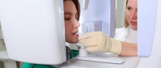
How does the procedure work?
Palatoplasty to remove clefts is performed using a standard set of surgical instruments. This technology is called Z-plasty or Furlow plastic. It consists of sequentially isolating and stitching triangular flaps of the mucous membranes of the oral and nasal cavities, taking into account all the anatomical features of the structure of these structures. Veloplasty is usually accompanied by other types of surgical operations aimed at the correct formation of the hard palate and lips.
A surgical solution to snoring problems involves the surgeon excision of part of the soft palate and removal of the uvula, but more advanced methods are now being used to solve this problem.
Using a laser after preliminary anesthesia, the doctor makes small wounds, which, as they heal, lift the soft palate. The essence of cryotherapy is exactly the same, except that the operation is performed using a cryoapplicator. Radio wave therapy is the most gentle and effective method. All these procedures usually do not require hospitalization and are carried out within 10-15 minutes. The rehabilitation period is individual.