A panoramic photograph of the teeth (orthopantomogram or OTP) is an x-ray of the upper and lower jaw. Allows you to examine most dental problems in detail, conduct high-quality diagnostics and prescribe treatment.
A panoramic dental photograph shows not only the teeth, but also the jaw joints, paranasal sinuses and facial bones. All this makes it indispensable for orthodontic, surgical and reconstructive treatment. You can obtain OPTG using an orthopantomograph.
Indications for panoramic dental imaging
The patient's need for a panoramic photograph of the teeth will be determined by the dentist. It is necessary to do it for a number of diseases of the oral cavity. Among them:
- inflammation of bone tissue;
- bite pathologies;
- tooth abscess;
- joint dysfunction;
- infectious diseases of the jaw;
- periodontal diseases;
- mechanical injuries.
OPTG is also necessarily prescribed when planning orthodontic correction of the dentition. A panoramic photograph allows you to see parts of the teeth that are located under the gums. With its help, the orthodontist can correctly draw up a treatment plan, predict its timing and select teeth that need to be removed (if necessary).
A panoramic photograph must be taken after sustaining jaw injuries (dislocations, fractures, etc.). Its main function is the diagnosis of dental diseases that cannot be determined solely by external signs. For this reason, OPTG is prescribed in the presence of symptoms indicating deep inflammatory and purulent processes.
The doctor gives a referral for a panoramic dental photograph after a visual examination of the oral cavity and collection of anamnesis. This takes into account the presence of pain (especially after canal filling), the nature of the discharge, dysfunction of the jaw joint and other symptoms.
A panoramic photograph is the most informative diagnostic procedure in pediatric dentistry. It is necessary for delayed teething or deviations in their formation. The picture clearly shows the rudiments of baby teeth already in the first months of a child’s life. With the help of OPTG, it is possible to predict bite pathologies and prescribe timely treatment.
With the help of OPTG, it is possible to diagnose diseases of the ENT organs, in particular, diseases of the maxillary sinuses and nasal passages. In this case, a panoramic image is prescribed for dizziness, fever, headache, and breathing discomfort.
Classification
In orthodontic practice, various assessment criteria are used to determine which dental bite is correct and how to correct existing anomalies. The ideal situation, in which nothing needs to be corrected, is characterized by the following indications:
- Semi-elliptical shape of the upper and parabolic shape of the lower arc.
- Small - no more than a third - overlap of the vestibular surface of the mandibular elements.
- Contact of antagonists during closure and absence of obvious gaps.
Compliance on all points is quite rare, but in most cases the deviations are minimal and do not require medical intervention. Often the cause of imbalance is not congenital defects, but bad habits, some of which are formed at an early age.
Types of correct bite
In accordance with the generally accepted classification, there are four main types of occlusal relationships, characterized by the absence of problems and not having a negative effect on the jaw region.
Orthognathic
The optimal, from an aesthetic point of view, is a condition in which all elements of the dentition have an even shape, are located in the correct manner, without gaps or deviations from the midline.
Straight
A common phenomenon, the main feature of which is the lack of overlap - the cutting edges of the incisors meet along a line, which causes a specific type of smile, but is not considered as a pathology.
Biprognathic
Another type of even dentition without threes and diastemas, the distinctive feature of which is a slight deviation of the vertical growth vector of the units. With this development, the crowns are slightly tilted forward.
Progenic
In a normal state it is not accompanied by abnormal manifestations. The identifying feature is a slight frontal protrusion of the lower units.
What diseases can be diagnosed by a panoramic dental photograph?
A panoramic photograph of teeth is an effective method for x-ray diagnosis of a number of dental diseases. Among them:
- benign and malignant neoplasms;
- fractures, cracks and dislocations;
- deep caries;
- bite pathologies;
- periodontitis;
- improper teething;
- cancer of the maxillary sinuses, etc.
Causes of tooth enamel destruction
There are a number of factors that exacerbate the rate of enamel damage. These include:
- the anatomical structure of the tooth, when a large amount of food accumulates in the dental cavities and spaces between the teeth;
- insufficient saturation of enamel with fluoride;
- genetic predisposition;
- weak immunity, various diseases of the body;
- composition of salivary fluid - a small amount of saliva, viscous in composition, causes the accumulation of bacteria on the surface of the tooth and in the dental spaces, and consequently, the formation of dental plaque;
- insufficient oral hygiene, improper brushing of teeth;
- eating mostly soft food (soft food weakens the protective functions of enamel, while hard food helps to naturally strengthen teeth);
- lack of vitamins and beneficial microelements in the human body;
- The bristles of a toothbrush used for oral hygiene are too hard.
Restoration of tooth enamel is required for those who eat large amounts of citrus fruits (oranges, lemons, grapefruits), which significantly weaken tooth enamel. The acid contained in these fruits tends to dissolve the protective surface of the tooth. Therefore, after eating citrus fruits, be sure to rinse your mouth with water.
Preparing for a panoramic dental photograph
The procedure does not require special preparation. You can consume drinks, food and medicine - this will not affect the x-ray in any way. Immediately before the procedure, metal jewelry must be removed from the body. It is not recommended to drink alcohol.
When visiting a dentist, you must take panoramic and x-ray photographs from the last few years, as well as a medical history. With their help, the doctor will be able to track visible changes in the patient’s health status or compare treatment results.
X-ray radiation poses a possible risk to pregnant women. According to some studies, it can negatively affect the intrauterine development of the fetus. Pregnant women should tell their dentist about their pregnancy. In this case, carrying out OPTG depends on the ratio of possible risks and benefits.
Restoring tooth enamel
In modern dentistry, tooth enamel restoration is carried out using several methods. The doctor makes a decision on what is most suitable for you, based on the overall clinical picture: the health of the teeth and gums, the presence of caries, the degree of enamel destruction, the presence of bad habits.
Restoring tooth enamel using fluoridation
This is the most famous and widely used method of restoring tooth enamel. Fluoridation involves feeding the enamel with useful microelements by applying a special gel or varnish to the surface of the teeth. The fluorides contained in the solution enhance the protective function of the enamel and suppress the metabolism of bacteria, making the enamel much more resistant to acids.
In our Dentistry clinic on Shchelkovskaya, the dental fluoridation procedure is provided to all patients who want to restore and protect tooth enamel. The procedure is carried out by appointment by calling 8 (495) 033-00-63 or using the online registration form. During your consultation with a dentist, you will receive an opinion on the condition of your mouth and teeth and recommendations for maintaining the health of your enamel.
Fluoridation can be carried out in two ways. The first is that a fluoride-containing composition is applied directly to the tooth enamel. Most often, the composition has the form of a gel and is well accepted by the tooth enamel itself. The second method is to make special impressions of the patient’s dentition. The medicinal composition is applied to an impression, which is then applied to the patient’s teeth. After the fluoridation procedure, the impressions are removed. In order for the treatment to be most effective, in the second case it is recommended to repeat the procedure several times.
Recovery method - remineralization
This method of restoring tooth enamel is similar in its beneficial properties to fluoridation, but in other respects they differ. When remineralizing the tooth surface, a more concentrated composition is used. In addition to fluoride, the protective substance also contains calcium and a large number of minerals.
Remineralization occurs in the following way: the medicinal substance is applied directly to the surface of the teeth, and it is absorbed. Absorption of the composition takes a long time, but this ensures complete penetration of beneficial minerals into the crystal lattice of the tooth.
Restoring enamel with filling
Filling damaged tooth enamel is slightly different from filling a tooth with ordinary caries. In the case of restoration of tooth enamel, filling occurs in layers, the filling material evenly covers the damaged surface of the teeth, and also eliminates cracks in the enamel.
Veneers protect the enamel from further destruction
Installing veneers is a way to restore enamel if its destruction is deep and other methods are ineffective. Veneers are thin pieces of porcelain placed on the surface of teeth (most often the front teeth of the upper and lower jaw). Although veneers are made of durable material, they are subject to wear and tear (at best, veneers will need to be replaced after about fifteen years).
The incomparable advantage of veneers is their ability to eliminate all visible dental defects. The color of the manufactured veneers is no different from the natural color of the tooth surface, and sometimes even better. Veneers hide gaps between teeth, pigment spots, mask the incorrect position of a tooth in a socket, and make chips and cracks invisible.
How is a panoramic dental x-ray performed?
Panoramic photography of teeth is the specialty of a dental radiologist. It is carried out using an orthopantomograph - a modern device that exposes the body to a minimal amount of x-ray radiation.
To take a photo, you need to stand or sit near the device. Next, the patient must wear a protective apron that protects the chest from radiation. When the patient is ready, the doctor starts the orthopantomograph. The entire procedure lasts no more than 15 minutes and is completely painless.
During the process you must remain stationary. After the panoramic photo is taken, you need to wait a few more minutes. During this time, the specialist will prepare the image and provide it to the patient. The entire procedure lasts no more than 15 minutes, including the time required for preparation and waiting.
Structure of a wisdom tooth –
As we said above, the crown part of a wisdom tooth differs little from the crowns of other large molars (molars). In this case, it is more difficult to answer the question - how many roots does a wisdom tooth have? there may be two, three, four, or even five. On the other hand, there are eighth teeth with one large root. But if you take a closer look at such a root, you can usually see that it consists of several tightly fused roots at once (Fig. 3-4).
How is a wisdom tooth different from other teeth?
- firstly, wisdom teeth, as a rule, have strongly curved roots.
- secondly, curved roots mean that such teeth will have highly curved root canals and, accordingly, this makes them difficult when filling is necessary (for pulpitis and periodontitis).
What does a wisdom tooth look like: photo
In the photo, notice that some roots curl into a boot shape or several roots spread widely in different directions. This shape and position of the roots makes wisdom teeth extremely unpleasant for dental surgeons, because the curved roots often break off when the tooth is extracted, and then they have to be literally “plucked out” from the bone tissue. Read and see our article about how they are removed: → “Removal of wisdom teeth”
Additional specialists and procedures
A patient who has received an appointment for a panoramic dental x-ray may need the help of the following specialists:
- dental surgeon;
- maxillofacial surgeon;
- oncologist;
- otolaryngologist;
- periodontist;
- orthodontist, etc.
OPTG is an important, but not the final stage of diagnosis. To confirm the diagnosis and develop effective therapy, in most cases a number of additional studies are prescribed. If tumors are present, a tissue biopsy is performed. A laboratory examination of pathological discharge is also performed.
If an orthopantomogram indicates problems with the canals or subgingival part of the tooth, it is necessary to open the tooth (filling) and further treatment. In some cases, tooth extraction may be indicated (if it cannot be treated).
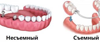
Treatment for missing teeth
The specificity of the anatomical structure of the jaw apparatus is that even complete edentia does not guarantee the formation of correct occlusion after implantation. The result of prosthetics is largely determined by the degree of development of defects and anomalies that formed even before the loss or removal of elements. If the row ratio was within acceptable limits, it is enough for the doctor to take into account the necessary adjustments when developing a replacement structure, but the presence of problems with bone tissue will require the use of more complex restoration techniques.
How to check and find out if the bite is correct when making a jaw prosthesis without teeth? For this purpose, special wax rollers are used, placed in the oral cavity. The measuring instruments are a ruler arc and an intraoral plate. After collecting the necessary readings, first a plaster prototype is created, and then the final structure.
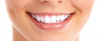
What is an ideal smile?
Probably, every person has his own idea of a beautiful smile. But in dentistry this concept has clear parameters. When a dentist makes a Hollywood smile, he strives for these criteria:
- Geometry. On a person’s face, you can draw several conventional lines that form the eyes, lips, edges of the incisors and fangs. For a beautiful smile, the parallelism of these lines to each other is important.
- Teeth are physiologically correct size. Ideal teeth don't just have different sizes. The largest teeth are the incisors. Then the size of the teeth gradually decreases. With this size, the smile looks attractive.
- Correct bite. Antagonist teeth must touch each other at physiologically correct points.
- Upper lip. When smiling, it only exposes the teeth, but not the gum line.
It is customary to call a Hollywood smile a smile that meets all these criteria. She's not just beautiful. It makes you more attractive, younger.
Gum disease: symptoms and treatment
Gum diseases in adults and children occur in the same way. Below we will analyze each disease - with a description of its characteristic symptoms and photographs of the patients’ teeth and gums. The information will allow you to easily distinguish one disease from another, and understand how much professional treatment you will need in different situations.
The author of the article has more than 10 years of experience as a periodontist, and therefore our recommendations really work (state-issued documents on advanced training in the Periodontology program can be viewed in the editorial section).
Prevention of tooth enamel destruction
As you know, prevention is always better than cure. This also applies to cases of tooth enamel destruction. Keeping your enamel strong and healthy is not difficult if you follow a number of recommendations.
Use a brush with soft or medium bristles to brush your teeth. Choose toothpastes that contain calcium and fluoride. Do not be lazy to maintain complete oral hygiene, that is, brush your teeth twice a day.
Use dental floss after eating - it perfectly cleanses the dental spaces of food deposits (and it is the cracks between the teeth, not even visible to the eye, that are potential spreaders of bacteria). If possible, use a mouth rinse after each meal - most of them contain microelements that serve to strengthen the tooth surface.
Eat foods fortified with calcium. These are fish, dairy products, cheeses, eggs. Eat more foods that are a source of vitamin C (pepper, rose hips, black currants, cabbage, garlic), as well as vitamin D (salmon, tuna, fish oil, potatoes, oatmeal).
When you brush your teeth, try to leave the paste on the tooth surface for at least three minutes. During this time, the medicinal substances included in the composition penetrate into the structure of the teeth, nourishing the enamel.