It is believed that at the age of 6-7 years the first milk teeth (lower central incisors) should fall out, and at the same time the first permanent molars grow. But what if the pattern breaks down? Can the upper incisors fall out first or, for example, first the lateral and then the central incisors? What could this mean, and should parents pay special attention to this?
What if my teeth fell out too early? Or did the molars start growing before the incisors fell out? Is it normal for molars to start growing at 4-5 years of age? With what it can be connected? When should a situation alert parents?
Why are the first teeth called baby teeth?
There are several opinions on this matter. Doctors attribute the authorship of the term to Hippocrates, who believed that these teeth are formed from mother's milk. Philologist N.N. Vashkevich states that “the term is a tracing from the Latin lactose “milk”. But the tracing paper is false, it is a misunderstood Arabic liwaqt - “for a while,” “temporary.”
One way or another, the first teeth actually actively “feed” on breast milk, since it is from it that the child absorbs the main building material of teeth and bones – calcium. And although baby teeth erupt, as a rule, at 6-7 months, their formation in the child’s jaws occurs long before that.
It is worth noting that for the health and strength of the first teeth, it is mother’s milk (not cow’s) that is necessary, since the nutrients from it are maximally absorbed by the baby’s body. Therefore, the universal recipe “breastfeed your baby” will help in this matter.
When do baby teeth appear?
The rudiments of baby teeth appear in the embryo at about 5-7 weeks of pregnancy. By the time a child is born, the crowns of 10 temporary and 8 permanent teeth have already been formed in his jaws. The timing of the eruption of baby teeth is quite arbitrary. The average formula is as follows: the child’s age in months minus 6. That is, the first 2 teeth (usually the lower middle incisors) appear at 6-7 months, the next 2 (upper middle incisors) at 8-9 months. Next, the upper lateral incisors usually come out, then the lower lateral incisors, then the front molars, canines, and back molars. Thus, by the age of 2-2.5 years, the child should have all 20 milk teeth. These are ideal timing and ideal sequence; deviations from them are quite common. Teeth may begin to appear as early as 4 months, or may linger for up to 8-9 months. In rare cases, a baby is born with teeth already erupted.
If your little one doesn’t “meet the deadline,” don’t be alarmed. This does not mean that the child is developmentally delayed. You should also not be proud of the early appearance of teeth - it does not indicate the child’s superpowers. Early or late appearance of teeth may be a hereditary factor. In case of a strong deviation from the schedule - the appearance of teeth before 4 months or their absence after 9 months - just show the baby to the pediatric dentist. In general, the first examination by a pediatric dentist should be scheduled at least when the child is one year old. The doctor will see how the teething process is progressing and talk with you about the baby’s oral hygiene. The baby will get acquainted with the environment of the office, with the doctor, will get the first positive experience of visiting the dentist, because nothing unpleasant awaits him during this visit, and funny pictures on the walls of the clinic, toys, a chair in which he can ride with his mother - all this will certainly have an impact on him. him a good impression. From now on, you should take your child to the pediatric dentist at least twice a year.
When does teeth change?
In order to monitor the order of changing teeth and be able to identify pathology if necessary, you should remember the time frame:
- Children's incisors change at the age of 6-8 years.
- Fangs fall out and new ones appear at 9-12 years of age.
- Molars begin to change at 9-10, and can change up to 12 years.
- Large molars erupt from the age of 6 to 11-13 years, and the last ones (“eights”, so-called “wisdom teeth”) can appear at 20-30 years.
And if you notice a change in incisors and fangs immediately - the child is happy to show everyone his charming toothless smile, then with molars everything is not so simple.
How to distinguish a permanent molar from a baby tooth
You can distinguish between temporary and permanent teeth, first of all, by color. Milk enamel is white and even has a bluish tint.
These are the teeth that are called snow-white. But the permanent ones are always a little darker and have more of a yellowish or grayish tint.
Also, temporary teeth are smaller, and permanent teeth
are larger
; molars and premolars have a large chewing surface and several large tubercles on it. The teeth give the impression of being impressive and strong.
The formula of the dentition in children whose teeth have already been replaced will be similar to that of an adult - 8 incisors, 4 canines, 8 premolars and 8 molars. That is, on one side of the jaw there will be 2 small molars and 2 large molars. To understand this formula and learn to distinguish a permanent molar from a baby tooth, we recommend comparing them in the photo.
My baby is teething - how can I help him?
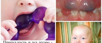
The child's profuse salivation will tell you that the first tooth is on the way. 1-2 months before the first tooth erupts, the baby’s saliva begins to flow so actively that it is already difficult to do without aprons and bibs.
All your older relatives will probably tell you about the unpleasant side effects of teething. However, there are many misconceptions here.
The very first teeth usually come out painlessly. What most often happens is this: while spoon-feeding a child, the mother hears the sound of metal on the edge of a tooth - that is, she detects an event that has already happened, without even noticing anything unusual in the child’s behavior.
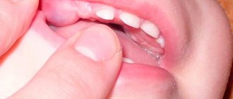
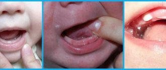
The emergence of canines and molars may be more difficult. The baby may be capricious, refuse to eat, sleep poorly, and put everything in his mouth. You should take care of inflamed gums - regularly treat them with special gels, offer your child to chew a cooling ring (cold relieves pain well).
However, do not believe that an increase in temperature is associated with teething. Fever and catarrhal symptoms are caused by an infection that is “caught” by the child’s body, weakened by malnutrition and lack of sleep. That is why, during the period of teething, it is better to protect the baby from communicating with strangers. Digestive disorders, an upset stomach of a child at the time of teething, is associated with his desire to chew and suck everything he can reach, just to relieve discomfort in the gums. This is how pathogenic microbes enter his mouth. Try to surround your child with clean objects, wash his hands and toys more often. Regularly let your baby chew small pieces of solid food - dried bread, bagel, apple slice, etc. This will help the teething of those teeth that are already “on the way”, improve blood supply, and therefore nutrition of the gums, develop the chewing reflex, and help the formation of the speech apparatus.
PsyAndNeuro.ru
Early trauma is adverse events (violence, parental deprivation) that occur in childhood and affect later life. Early trauma is thought to increase the risk of developing mental disorders. However, not all people exposed to adverse childhood experiences develop a mental disorder.
There are sensitive periods in psychomotor development during which the brain is especially plastic. If the time of exposure to unfavorable factors coincides with these periods, then the risk of developing a mental disorder increases. On the other hand, the acquisition of new experiences (including unfavorable ones) during such periods has a protective effect. There is no consensus on this issue; scientific research can support both points of view.
To date, there are no reliable methods for assessing the strength of early trauma. Objective research methods, such as identifying DNA methylation disorders and changes in the functioning of the amygdala, are invasive, expensive and time-consuming.
In March 2022, an article by Kathryn A. Davis and co-authors was published in the journal Biological Psychiatry, dedicated to the search for new methods of assessing early trauma. The researchers suggested that teeth could be used for this purpose, which are a kind of “time capsule” that remains unchanged over the years and is imprinted by the environment. The authors of the article summarized the achievements of dentistry, anthropology and archeology, and studied the results of archaeological studies of the human population and primate populations. The collected data were compared with the latest information about the etiology of mental disorders and the role of early trauma in their development.
There are at least five reasons why teeth were chosen as a marker of early trauma.
Firstly, dental development continues during age-related crises. Mineralization of “baby” teeth begins at the 4th month of intrauterine development and ends by 2–3 years. The formation of permanent second molars lasts from 3 to 14–16 years, and permanent third molars, or “wisdom teeth,” complete their formation by 18–25 years. These periods coincide with sensitive periods of formation of the nervous system.
Secondly, teeth retain “traces” of environmental influences (like rings on trees). Odontoblasts and ameloblasts produce proteins that promote the mineralization of dentin and enamel. Traces of this mineralization are visible during the complete formation of the crown in the form of striated exhaustion, Retzius stripes. It is possible that adverse events occurring during the formation of teeth can affect this process and will be “visible” on the enamel.
Thirdly, during the period of tooth formation, exposure to stress factors such as lack of nutrition, illness, poisoning with toxic substances leads to disturbances in the structure of dentin and enamel hypoplasia. According to some studies, similar changes are found both in living people and in archaeological excavations.
Fourthly, presumably, teeth can “retain” the memory of the effects of stress factors, and the main role in this process is played by the so-called neonatal line - a dark stripe in the enamel of baby teeth. There are several studies that have found a correlation between an increase in its width and stress exposure during the perinatal period. Similar studies were conducted on primates, where separation from the mother, death of a sibling, recovery period after surgery, and change of habitat were used as stress factors. A neonatal stress line was also found in the enamel of primates. The idea that teeth store a biological memory of early trauma is supported by studies demonstrating the effect of stress on the condition of hair and nails, which, like tooth enamel, are formed from ectoderm.
Fifthly, the phases of the “life” of teeth coincide with periods of development of major depressive disorder. Thus, the first teeth begin to fall out at the age of 6 years, in the period preceding puberty, in which, as is known, the risk of developing depression increases. Starting at age 13—the age of possible onset of depressive disorders—teeth can be removed for orthodontic reasons. In late adolescence, third molar extraction may occur. All these teeth can be sent for examination to special laboratories to identify “traces” of early trauma. A similar phase pattern can be observed in relation to other mental disorders.
Based on previous research in the fields of psychiatry, dentistry, archeology and anthropology, the authors of the new paper formed the TEETH (Teeth Encoding Experiences and Transforming Health) model. This model is based on the fact that various stress factors acting in childhood affect the development processes of all organs and systems, including the process of dental development (see diagram).
As a result of the study, three postulates were formulated.
First, impaired brain development and dental growth may be associated with early trauma. The fact is that stress factors acting during the development of the nervous system can leave their “mark” in the structure of the brain. Since the brain and tooth enamel are formed from the ectoderm, it is possible that adversity in childhood leaves an imprint on their structure. For example, enamel defects are observed in Down syndrome and cerebral palsy. Moreover, the authors of the article found a study demonstrating that children with low socioeconomic status and decreased salivary cortisol levels have thinner enamel than their advantaged counterparts.
Secondly, exposure to adverse environmental factors at the time of teeth formation can leave a long-term imprint on them, which can later be seen and objectively measured. Pre- and postnatal damage disrupts both the structure (decreased cortical thickness) and function of the brain (decreased activity of the amygdala). The response to stress changes, which is likely due to changes in cortisol levels. Researchers suggest that stress-induced defects in tooth formation are detected in macro signs of damage, such as changes in tooth size, and micro signs, such as changes in the structure and chemical composition of teeth, manifested in the formation of stress lines in the enamel. Since we know the stages of dental development, these signs can be used to trace the time of exposure to a stress factor: macro signs can indicate the presence of early trauma with an accuracy of three to five years, and micro signs with an accuracy of one week.
Thirdly, disturbances in the development of the nervous system can lead to mental disorders. Previously, scientists in their research relied on studying the brain and the level of stress neurotransmitters as markers of the risk of developing psychopathological symptoms. The authors of the new study found at least eight articles describing the effects of various environmental factors on teeth.
To date, the TEETH model has not been tested on a large number of respondents. Despite this, if we continue research in this direction, then perhaps in the future we will have a simple, non-invasive way to diagnose the risk of developing mental disorders.
Author of the translation: Wirth K.O.
Source: Kathryn A. Davis, Rebecca V. Mountain, Olivia R. Pickett, Pamela K. Den Besten, Felicitas B. Bidlack, and Erin C. Dunn. Teeth as Potential New Tools to Measure Early-Life Adversity and Subsequent Mental Health Risk: An Interdisciplinary Review and Conceptual Model. Biological Psychiatry.
When should you start brushing your child's teeth?
Now pediatric dentists are inclined to believe that systematic cleansing of a child’s oral cavity should begin... from the first days of his life. After feeding, you need to take clean gauze or a bandage, wrap it around your finger, moisten it with boiled water and rub it over the newborn’s gums. This is how you can avoid such major troubles as, for example, oral candidiasis (thrush).
Cleaning the first teeth can be done with a cotton swab or fingertip. You should start using toothpaste and brushing at 12-14 months. Almost all children's brushes are now made from soft artificial bristles, but still be careful and check the brush you like: what age it is designed for, and whether it is soft enough. Give preference to products from well-known manufacturers. Toothpaste also differs in composition and taste depending on the age of the children for whom it is intended. The child should be explained that toothpaste should be spat out, even though it is sweet. However, keep in mind: nothing bad will happen if a child swallows a certain amount of paste at first: manufacturers are aware of this tendency of children and make children's pastes safe for the body.
Of course, it is necessary to involve the child in the dental care process as much as possible: show how to use a brush correctly, brush your teeth in the presence of the child, thereby demonstrating how important this procedure is. You can involve your baby in the process of choosing toothpaste and brush, especially since the brush needs to be changed every 3 months. A slightly older child can be asked to choose brushes for the whole family. It is necessary to develop in your child the skill of regular brushing of teeth 2 times a day. More attention should be paid to brushing your teeth at night. And yet, you can let go of the situation and leave the child to his own devices in matters of oral hygiene only when the child turns 10 years old.
Diseases of baby teeth
The most common troubles are caries (including bottle caries), pulpitis, periodontitis.
You should be alerted to any changes in the color of the enamel, spots, dots (both dark and white) on the child’s teeth, redness or swelling of the gums. But you don’t have to be afraid of uneven teeth at first: when chewing solid food, the baby teeth will move a little and gradually take the right place.
The causes of diseases in primary teeth are not very different from the causes of problems with permanent teeth. This:
- insufficient care for them (lack of proper hygiene),
- eating disorders,
- undermining of the immune system due to other diseases and taking certain medications.
It is a mistake to think that you can't take too much care of your baby teeth because they will fall out anyway. A diseased tooth in the mouth is a breeding ground for pathogenic bacteria, which not only destroy other teeth, but also negatively affect digestion and ENT organs. A prematurely lost baby tooth is also a problem, as it does not allow the baby to fully chew food or articulate sounds well. In addition, neighboring teeth try to occupy the vacated space - they move. And when a permanent tooth begins to grow here, it simply does not have enough space in the dentition and will have to grow sideways. That’s why it’s imperative to keep an eye on your baby teeth and treat them on time!
We ourselves, out of ignorance, introduce some pathogenic bacteria into the baby’s oral cavity. Suffice it to remember my grandmother’s method of disinfecting a fallen pacifier: lick it and put it in the baby’s mouth. How many times have we done this? Did you feed the child with your spoon? Did you take turns biting an apple or a bun with him? Imagine for a moment the state of your teeth and the newly formed, not fully formed, delicate teeth of the child. What danger did you expose them to?
Foreign microflora is especially harmful to a child under 2 years of age, while temporary teeth are maturing in his mouth and their enamel is very vulnerable.
Leftover food also contributes to the rapid proliferation of bacteria in the oral cavity. A by-product of their activity is acid, which eats away the enamel of a baby tooth, leaving its delicate base - dentin - without protection. Then it’s a matter of little things: microorganisms penetrate the dentin and destroy it. This is how caries occurs. Outwardly, a diseased tooth may look normal for quite some time: a small black dot (the site of the lesion) is not too noticeable. But inside it can already be severely damaged, since caries of baby teeth, due to the softness of their tissues, develops much faster than permanent teeth.
There is also the so-called “bottle caries” - this is a brownish plaque on the front teeth. It is very resistant and cannot be cleaned with a brush. Such teeth rot quickly. This caries is called “bottle caries” because it is a consequence of feeding the baby from a bottle with sweet drinks and juices at night and throughout the night. Carbohydrates are known to be the best food for bacteria. Receiving such wonderful nutrition, pathogenic bacteria rapidly multiply and take over the entire territory available to them. It will be very difficult to force them to give up their positions. The development of “bottle caries” is also facilitated by the fact that at night the natural protector and cleaner of teeth – saliva – is almost not produced. While you are sleeping peacefully, your baby’s fragile little teeth are under powerful attack and are not protected by anything! Is it worth it to buy yourself a vacation at such a price?
Dangerous complications of caries are pulpitis and periodontitis.
Pulpitis is an inflammation of the pulp (the core of the tooth, filled with vessels and nerve fibers). In children, the pulp is practically insensitive, so they may not feel the usual sign of pulpitis - sharp pain. As a result, pulpitis of baby teeth may not be noticed even by an attentive parent, and the role of preventive examinations by a dentist is irreplaceable here.
If bacteria pass through the root of a baby tooth and enter the gum tissue, inflammation begins - periodontitis. Severe pain and fever are already guaranteed here. The gums become red and swollen. Periodontitis is a serious disease, and a child should never be exposed to it.
By visiting your dentist in a timely manner, you can avoid all these troubles. Caries is very easy to treat. Here, however, the first question comes to mind: where exactly to turn. It turns out that the approach to the treatment of baby teeth in different clinics is very different.
Traditionally, caries of primary teeth is treated “in a hurry”: a small child usually does not really want to endure while the doctor picks around his mouth, so the doctor tries to somehow clean the damaged area and fill it with quick-hardening material. As a result, the affected teeth remain untreated and may soon become inflamed again or completely collapse. Sometimes you even have to put crowns on baby teeth.
“The Nutcracker” uses all the possibilities of modern medicine to provide comprehensive care to the smallest patients: from composite materials to “laughing gas” and Sevoran anesthesia. We are convinced that the treatment of primary teeth should be of high quality, and the child’s impressions of the clinic should be the most pleasant.
Characteristics of temporary, replacement and permanent dentition. Part 1
Over the course of a person's life, temporary and then permanent teeth emerge successively in the oral cavity. Knowledge of the anatomical structure, timing of teething, characteristics of growth and development of the jaws at different stages of the formation of the maxillofacial system, physiological occlusions is important for doctors of all specialties. This general morphological direction is basic and determines the patterns of development of the dentofacial system and reveals individual characteristics.
Timing and basic patterns of eruption of primary teeth
A newborn child's jaws contain the rudiments of 20 temporary and 16 permanent teeth of various stages of development. The lower jaw is located behind the upper jaw, and between the alveolar processes in the anterior section there is a sagittal gap of up to 10 - 14 mm. During the thoracic period, the intramaxillary formation and mineralization of temporary teeth continues, and from the 6th month the process of teething begins and a temporary bite is formed. At the same time, mineralization of permanent teeth begins. The average age of teething is the age at which the tooth erupts in 50% of children. It has been established that in a zone with a high fluoride content in drinking water, the eruption of lateral incisors, canines, and second molars begins 2-3 months later compared to a zone with a low fluoride content in drinking water. The sequence of eruption of temporary teeth on each jaw occurs in the following order: I – II – IV – III – V, and in general in the occlusion as follows: Iн – Iв – Iв – Iн – Ivн – Ivв – IIIв – IIIн – Vн – Vв. The presented order of eruption is the most typical, however, options are also possible when the child’s lateral incisors erupt first, and soon after them the central ones. Teeth usually erupt sequentially, in pairs, symmetrically. Violation of paired eruption is observed with malformations of teeth and jaws. It is considered normal when, by the end of the 1st year of a child’s life, 8 incisors have erupted, then the first molars, canines and second molars (Table 1). Table 1. Timing of mineralization, eruption and formation of teeth
However, recently, due to acceleration processes, many children begin to erupt at an earlier time - 4 or 5 months of life, so some authors propose average periods of eruption, which were determined by them as a result of their own clinical studies (Table 2).
Table 2. Anatomy of primary teeth.
Temporary teeth differ from permanent teeth in size, shape, and color (Table 3).
Table 3. Comparative characteristics of temporary and permanent teeth
Among the temporary teeth there are: incisors – 8; fangs – 4; molars – 8. The teeth are not homogeneous in their structure. This depends on the group affiliation of the tooth, on the described surface of the tooth. For convenience of description, surfaces are distinguished: vestibular, lingual, medial, distal, occlusal. The group of temporary incisors are single-rooted teeth. There are 4 incisors on each jaw: 2 medial (central) and 2 lateral (lateral), left and right, respectively. What is common in the structure of the incisors is the shape of the crown, flattened in the vestibular-lingual direction near the cutting edge. The root has a cone shape.
Rice. 1
Anatomical shape of primary teeth of the right upper jaw
Rice. 2
Anatomical shape of primary teeth of the right lower jaw.
The group of temporary canines are single-rooted teeth. There are 2 fangs on each jaw: right and left. What is common in the anatomy of these teeth is the presence of a crown that is pointed on all surfaces and the longest cone-shaped root. Group of temporary molars. These are teeth with a multi-tubercular chewing surface and several roots. Primary molars are the largest teeth in the primary dentition. The child has 8 molars: 4 molars of the upper jaw - first and second (right and left); and 4 molars of the lower jaw - first and second (right and left).
There are several systems for recording the dental formula of primary teeth.
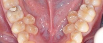
There are three main periods in the temporary dentition: 1st period (from birth to 2.5 - 3 years) - formation of the temporary dentition; 2nd period (from 3 to 4 years) – formed temporary bite; 3rd period (from 4.5 to 5 – 6 years) – reduction (abrasion, wear) of the temporary occlusion. The period of formation of temporary occlusion. With normal development of teeth and jaws, the process of eruption of temporary teeth begins at 6–8 months (Fig. 3), lasting up to 2.5–3 years. As teeth erupt and chewing function develops, involution of those organs that previously provided the act of sucking is observed. With the eruption of the second primary molars, the first rise in the height of the bite occurs. During this period, the alveolar process actively develops, the basal part of the lower jaw thickens, its branches grow, the outlines of the mandibular canal change, the size of the mandibular angle decreases, and the relief and architecture of the lower jaw become more complex. By 2.5 - 3 years, the temporary bite is considered formed. This period lasts up to 4 years and is a stable state of the dental system. Violations in the primary occlusion can lead to various complications, both local and general. They affect the growth and development of the jaws, the eruption of permanent teeth, reduce chewing ability, and lead to changes in the shape of the hard palate. Rice. 3
The period of formed temporary occlusion has the following dental characteristics: 1. There are 20 temporary teeth, they have a pronounced anatomical shape, 2. The teeth within the dentition have close contact. The contact point is point-like, but with an increase in physiological mobility, by the end of the period of formed temporary occlusion, the contact point becomes planar. 3. the upper and lower rows of teeth have the shape of a semicircle and their midpoints coincide with each other; 4. The teeth of the upper jaw in the frontal region overlap the teeth of the lower jaw. This is explained by the greater width of the upper dental arch compared to the lower one; 5. each tooth of the upper jaw articulates with two lower ones, with the exception of the second molar, which articulates only with its antagonist - the second lower molar; 6. The distal approximal surfaces of the second temporary molars are in the same vertical plane. By the end of the eruption of primary teeth, the first stage of physiological rise in the height of the bite takes place, which begins with the establishment of contact between the first temporary molars and ends with complete eruption and correct articulatory relationships of the second temporary molars. The next period of formation of the dental system is the period of reduction of the temporary occlusion, lasting from 4.5 to 6 years. A fully formed temporary bite during this period is unstable and undergoes a number of changes. It is also called the period preceding the change in the temporary bite, or the period of “wear and tear” of the temporary bite.
Fig.4 Fig. 5
Rice. 4. Reduction of temporary occlusion (models of the jaws): a) front view, b) rear view.
Rice. 5 Correct ratio
As a result of the growth of the jaw bones in preparation for the replacement of temporary teeth with permanent (larger ones), this period is characterized by the formation of physiological gaps between the teeth (diastemas - between the central incisors and three - between the remaining teeth). The abrasion of the cusps of the chewing teeth and the cutting edges of the frontal teeth gradually increases. The physiological mobility of individual teeth to be replaced increases. The dentition lengthens. Due to the abrasion of the chewing surfaces of the lateral teeth, a medial (towards the midline) shift of the lower jaw occurs and a “direct sliding bite” is formed. In this case, the incisors contact their cutting edges, and a medial step appears in the lateral area, formed by the distal surfaces of the second temporary molars (Tsilinsky’s symptom). This relationship of teeth ensures the subsequent correct position and relationship of the first permanent molars. The spaces of primates (diastema and trema) are a reserve site for the medial displacement of the lower dentition and the subsequent unimpeded placement of permanent teeth. Wearing of teeth leads to a decrease in the height of the crowns, and a straight “sliding” bite is formed. The differentiation of the elements of the temporomandibular joints is completed, the formation of third molars, the development and mineralization of premolars and second molars continues. Towards the end of the period of “wear and tear” of the temporary dentition, the eruption of the first permanent molars begins.
Timing and basic patterns of eruption of permanent teeth
The development of permanent teeth generally resembles the development of primary teeth. Permanent molars do not have temporary predecessors, which is why they are called additional molars. All other permanent teeth are replacement teeth. During the eruption of permanent replacement teeth, destruction and loss of temporary teeth occurs, which also includes progressive resorption of the roots of temporary teeth and their alveoli (Fig. 6).
Rice. 6
Due to the pressure of the permanent tooth on the alveolus of the temporary tooth, differentiation of osteoclasts begins, which are actively involved in the processes of bone tissue resorption. The localization of the zones of physiological resorption of the roots of temporary teeth varies depending on the group affiliation of the tooth: in single-rooted teeth it is located in the area of the apex of the tooth on the lingual side, and in multi-rooted teeth - in the zone of root bifructation. The timing of the eruption of permanent teeth with the correct development of the child coincides with the time of loss of temporary teeth (Table 4). The process of temporary tooth loss occurs synchronously with the process of permanent tooth eruption. Clinically, after the loss of a temporary tooth, cusps or part of the cutting edge of the erupting permanent teeth are detected.
Table 4. Timing of eruption of permanent teeth.
The eruption of permanent teeth begins with the first permanent molar at 6 years of age. Then, sequentially at the age of 6–8 years, the central and lateral incisors erupt. At 9–10 years of age, the first premolars erupt, followed most often by the canines (10–11 years) and the second premolars (11–12 years). At 12–13 years of age, the second permanent molars erupt. Thus, by the age of 12–13 years, all temporary teeth are replaced by permanent ones. The final formation of roots is completed by 15 years. Replacement teeth have a special anatomical structure that facilitates their eruption - a conductive canal, which contains a conductive cord. The anlage of such a permanent tooth is initially located in a common bone alveolus with its temporary predecessor. It is subsequently completely surrounded by alveolar bone, with the exception of a small canal containing the remains of the dental plate and connective tissue. Together, these structures help guide the movement of the permanent tooth as it erupts.
Periods of mixed dentition
So, with the eruption of the first permanent molar, the mixed dentition begins. The mixed dentition represents a higher degree of development and differentiation of the masticatory apparatus. It is characterized by the presence of temporary and permanent teeth, which lasts from 6 to 12-14 years. The mixed bite is of particular interest to orthodontists, because at this time the most intensive growth of the jaw bones occurs, metabolic processes in bone tissue are at a high level. Therefore, timely identification of etiological factors is especially effective during the day, as is the treatment of dental anomalies themselves. However, information about the development of mixed dentition is ambiguous. So, for example, A.D. Osadchiy (1967) distinguishes two periods in the mixed dentition: 6 – 8 years – early mixed dentition and 9 – 12 years – late mixed dentition. I.L. Zlotnik (1952) also distinguishes these two periods, but with an age difference of 6 - 9 and 10 - 12 years, respectively.
Rice. 7
This allocation of periods is associated with the presence in the dentition during the period of early mixed dentition of the first permanent molar, four permanent incisors on the upper and lower jaw. And premolars and canines are in the period of late mixed dentition. At 9 years of age, the growth of the jaw bones slows down, but there is a noticeable growth of the alveolar process, associated with the eruption of permanent canines and premolars and the formation of the roots of the incisors and first molar (F.Ya. Khoroshilkina, 1999). This division takes into account the growth rate of the jaw bones and alveolar process and the level of intensity of metabolic processes in them. The eruption of the first permanent teeth (first molars) provides a second physiological rise in the height of the bite, and sagittal and transversal occlusal curves are formed. The third period of increasing the height of the bite begins at 12 years of age with the eruption of the second molar. It is accompanied by active growth of the dentoalveolar arches, which lasts from 13.5 to 15 years. During the development of the chewing apparatus in children, the mixed dentition is the most labile. The simultaneous presence in the oral cavity of temporary teeth that have lost stability due to root resorption, and permanent teeth that are at various stages of eruption and root formation, leads to a significant decrease in chewing function, leading to uneven training of the masticatory muscles, abnormal growth of the jaw bones and often the formation of anomalies dental system. During this period, both self-regulation of existing anomalies and the formation of new ones are noted. Due to the unstable condition of individual parts of the dental system and the entire masticatory apparatus as a whole, as well as the increased growth of the jaws during this period, it is necessary to use it to perform corrective orthodontic interventions. Article continues here
Authors: V.I. Kutsevlyak, A.V. Samsonov, S.V. Altunina, Yu.V. Tkachenko
What should you do to keep your baby teeth healthy?
Let’s summarize all those simple and, in general, obvious rules that will help us preserve the baby’s healthy baby teeth given by nature throughout the entire period of their functioning and not lead to the moment when the child’s baby tooth hurts or its premature loss occurs.
- Avoid unnecessary use of medications during pregnancy and breastfeeding to avoid damaging developing teeth.
- Eat right during pregnancy.
- Feed your baby breast milk.
- Introduce solid foods into your child's diet in a timely manner.
- Do not share food with your child (especially before the age of two), and do not lick his pacifier for the purpose of “disinfection.”
- Don't let your child fall asleep with a bottle of sugary drink.
- Carefully monitor your child’s oral hygiene from the first days of life.
- Have your child undergo regular preventive examinations with a pediatric dentist.
What is tooth root resection, indications and contraindications for surgery
Resection of the apex of the tooth root or apexectomy is a surgical procedure during which part of the tooth root is removed. When performing a tooth resection, only the inflamed and infected part of the root is removed, healthy tissue is not affected. To speed up the process of restoration of tooth tissue after root resection, a special bone material is placed in place of the removed fragment.
Tooth resection must be performed by a competent and experienced dental surgeon, since this operation is a complex surgical procedure. Due to the complexity and the fact that the surgeon will have to cut through the gum and work with the jaw bone to gain access to the root of the tooth, root resection surgery is always performed under anesthesia - local or general.
In our dental clinic in Moscow - “Firadent”, patients are offered the service of performing an operation to resection the apex of the tooth root under sedation, that is, in a state of light medicated sleep. You will sleep, and the doctor will do all the work and cure your bad tooth. Sedation allows you to completely eliminate stress and pain during dental treatment!
Naturally, such a complex operation as resection of the apex of a tooth root is not carried out just like that; there must be certain indications for the intervention. Tooth root resection is prescribed for:
- The need to remove pathological neoplasms on teeth with installed core inlays and pins;
- Resection of the tooth under the crown is relevant. This way, you can save the tooth, provide it with high-quality treatment, and at the same time not remove the installed prosthesis;
- Inability to carry out high-quality filling of dental canals (for example, in case of their abnormal obstruction) or to unfill tooth canals that were treated previously.
It is the resection of the tooth root that allows you to save the tooth from removal due to cysts and other large tumors. However, there is a fairly extensive list of contraindications for this operation, which are important to consider when planning treatment.