Cleaning the tooth canals is a procedure for removing microparticles of the inflamed pulp from the roots, i.e. affected tissues. This is a particularly labor-intensive process, since the final effect of caries treatment depends on the quality of the work.
Clinical case: a patient has caries that has developed into pulpitis. When opening the tooth, the dentist diagnosed an infection and a severe inflammatory process; it was recommended to remove dead pulp tissue from the roots. If the work is not performed perfectly, an infection gets into the roots, a filling is placed on top, and the pathological process already develops under a thick layer of filling material. The toothache does not subside, the patient goes to the doctor again or endures the pain, quenching it with medications, and eventually loses the tooth. That is why, in case of caries, pulpitis and other problems, it is important to contact trusted doctors who perform high-quality root cleaning. It is advisable for the dentist to work with a microscope - multiple magnification allows you to monitor the process of cleaning the canals from remnants of pulp tissue.
One tooth may have two or more tubules, which are localized in the layers of the periodontium. The main task of the canals is to be a connecting link between the crown pulp and the nerves.
More than 10 years ago, deep caries and advanced pulpitis were the reasons for removing a unit, but now, thanks to effective cleaning and treatment of the canals, it is possible not only to preserve the crown, but also to restore its integrity to an ideal result.
When is tooth canal cleaning necessary?
Here are three common cases when a patient is required to have canal cleaning:
- Before installing the prosthesis on the tooth, the unit is depulped and ground down so that a crown can subsequently be installed on it;
- When diagnosing pulpitis (inflammation of pulp tissue). Usually pulpitis is a consequence of advanced caries and infection. The problem is accompanied by acute pain; it is painful for the patient to chew on the side of the pathological tooth, which indicates severe inflammation inside the tooth;
- If a person has been diagnosed with periodontitis - inflammation of the tissues around the tooth. Usually the problem is a consequence of untreated caries and a dynamically spreading infection.
Internal tissues and pulp become inflamed due to the penetration of pathogenic microorganisms. They can also get inside due to mechanical injuries (chipping off a piece of a tooth, splitting a crown due to a blow or fall, tooth dislocation), gum disease.
Note! Pain is not always the only signal to remove the pulp and clean the tubules of dead granules. Very often, cell death occurs without pronounced symptoms, when the tooth stops hurting, and the inflammatory process is already actively developing inside it. A visit to the dentist and a complete examination of the oral cavity will help you understand that there is a purulent-inflammatory process in the tooth.
It is important to thoroughly clean the canals from pulp residues, since pathogenic microflora will still provoke purulent-inflammatory processes. Therefore, it is advisable to remove inflamed tissue not only from the canals, but also from the crown part of the tooth.
In particularly complex and advanced cases, partial pulp resection is prescribed. If the dentist does not consider this method, unfortunately, the tooth is removed.
Dentistry for those who love to smile
+7
Make an appointment
Possible complications of ignoring pain
The sensations that may occur in a pulpless tooth clearly indicate the presence of pathology, which must be eliminated as quickly as possible. Otherwise, infection will lead to quite serious consequences:
- complete destruction and/or loss of the tooth;
- acute or chronic gingivitis (gum disease);
- development of periodontitis (inflammation of the tissue surrounding the root, bone tissue of the alveoli and gums);
- chronic inflammation of the gums;
- the appearance of a cyst.
Ignoring the inflammatory process and delaying contact with the dentist can cause the development of systemic infections, which will spread throughout the body through the bloodstream from the periosteum.
Preparing for channel cleaning
To assess the condition and structure of the root system, the patient is prescribed a preliminary diagnosis:
- CT scan;
- radiography;
- radiography;
After this, the dentist chooses a cleaning tactic and a special tool for the job.
Before opening the coronal part, local anesthesia is administered, since in a state of inflammation, internal structures can provoke acute pain. In this case, the dentist administers the anesthetic using the following methods:
- point or infiltration, that is, when injections are made into the projection of the apexes. An injection into the cancellous bone using the same infiltration method will also numb the area well;
- The conduction technique involves anesthesia of the nerve branch that surrounds the neighboring units. Anesthesia also affects some of the soft tissues, so after the injection and treatment, the patient may continue to experience unpleasant sensations in the immobilized part of the mouth for a long time.
Granulomas on the roots, we treat the tooth canals
In the article “Available about root canal treatment,” I discussed in theory what proper endodontic treatment should look like, and what happens when it is not performed entirely in line with modern requirements. And today I propose to support the theory with practice and analyze one of the interesting clinical cases from my own practice.
So, a young man approached me with a referral from an orthodontist and a desire to save a tooth, or rather, what was left of it. This is how he looked at the time of our meeting on an x-ray.
This is the upper 6th tooth (designated by dentists as 16, there is a lot of detail about the numbering of teeth in dentistry). His background is as follows: a couple of years ago he was treated for pulpitis. Everything was as usual - they removed the nerve, somehow cleaned the canals, filled them and put in a big filling. As you can now see, very little time passed and a natural “accident” happened: the tooth and the filling broke. What’s bad is that one of the walls broke below the gum level. I will explain further why this is bad. As a result, we have 2 main problems that threaten the tooth with removal - a deep level of chipping of the palatal wall (indicated in the picture with a red line) and poorly treated canals (the unpassed part of the canal is indicated by a white dotted line), one of which has radiological signs of chronic inflammation - granulomas (its outlines are indicated by a black dotted line).
But despite these problems, the patient had a strong desire to preserve his own tooth by any means available. What was ultimately done for this?
In order to better understand and evaluate, we must say what shortcomings we had to correct... and what you can see for yourself in the picture of your teeth:
- barely noticeable white stripes on the x-ray - this is the filling material in the canals. Such thin, interrupted “threads” that do not reach the top of the root indicate that the canal was poorly expanded, and therefore not sufficiently treated with solutions to remove organic residues from it
- darkening around the tips of the roots (in this case around one root) indicates the presence of a chronic inflammatory process on this root. This is precisely caused, as a rule, by errors in the processing of the canals during previous treatment.
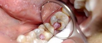
So, during the re-treatment, it was discovered that in addition to the 3 previously treated canals in this tooth, 2 (!!!) additional canals were missed at once. Those. they were not discovered by the doctor and were simply closed with all the remains of the pulp, which acted as a food storehouse for the microflora. It is on this root that the inflammatory focus is visible on x-ray. In addition, the so-called "step". In dentist jargon, this unpleasant thing means that the doctor has deviated from the natural course of the canal and lost it, resting on the hard tissues of the root. As a result, the apical part of one of the previously discovered canals also remained underprocessed.
The picture on the left shows 3 instruments inserted into 3 different canals in the same root. On the right picture, the black dotted line indicates the outline of the granuloma, the blue line outlines the outline of the root, the white dotted line indicates the untraversed part of one of the 3 canals, the red arrow shows the part of the canal in which a “step” was made during the previous treatment
Ultimately, these errors were corrected, two undetected channels were completely processed, and the step in the third was bypassed by finding the correct path. In total, after all the instrumental preparatory measures, it turned out that there were 5 separate canals in this tooth.
This control image taken before filling shows all 5 individual canals...
This is a photo of a tooth cavity, in which 5 separate orifices of all five canals are visible. Channels 1,2,5 are the main ones. It was they who were found and processed during the previous treatment, although not perfectly. Quite often, there are additional canals in the upper molars (3,4). Their mouths are initially very small, sometimes barely noticeable, and quite often these channels remain undetected. This is one of the common causes of complications after endodontic treatment...
This is quite a rare case. Still, usually in the upper molars there are 3-4 canals, very rarely less than 3. But no less rarely, more than 4. This is just that rare case, and therefore interesting.
The result of 2 visits (1 hour + 1.5 hours) was the filling of all canals.
Now all 5 canals are sealed exactly to the top of the root. All that remains is to wait for the inflammatory process to disappear.
The same thing, but the view from the tooth cavity. The canals are tightly sealed with gutta-percha*. Next comes tooth restoration.
Further, the remaining root due to serious damage will be restored with a stump insert* and a crown. True, before that, the surgeon will still have to tinker with him in order to solve the second problem of this sufferer after the canals (I talked about it at the beginning). We are talking about a deep chip of the palatine wall. The fact is that for high-quality restoration it is very important that the edges of the tooth rise at least 1-2 mm along the entire perimeter above the level of the gum (it is allowed that in some places the edge of the tooth coincides with the level of the gum). This will make the restoration airtight and preserve the remains of the tooth for many years to come. In this case, the wall was chipped on the palatal side below the gum, which means we will have to remove some part of it in order to transform the subgingival defect into a supragingival one. This manipulation is called “surgical lengthening of the clinical tooth crown.” I will also talk about this, though in other posts.
This completes the story of saving one single tooth.
Finally, I want to make a couple of small but important additions to this clinical case.
Firstly, very often in such situations the patient faces a dilemma: keep his tooth, fight for it until the last drop of... the sweat of your dentist, or remove it with subsequent prosthetics? In such cases, the last decision, dear friends, remains with you, the patients. Repeated canal treatment, correcting mistakes already made by the previous dentist (and, perhaps, your own) always remains a difficult task, and therefore no one can ever give a 100% guarantee on the final success of all rescue measures. What factors does the doctor take into account?
Objective:
— the condition of the tooth (and not only the canals)
- opportunities for its subsequent QUALITY restoration (you can perform a feat in the canals, but then, without properly assessing the degree of tooth destruction, make an inadequate restoration, and in the end still lose it)
— the importance of preserving a specific tooth (it seems that all teeth are important, you might say, but there are still situations in which the heroic saving of a tooth makes absolutely no sense, for example, this often happens with wisdom teeth)
Subjective:
- the equipment of your workplace (the presence of a rubber dam, an apex locator, a large number of various files *, x-rays, ultrasound, magnifying equipment - in general, everything that I have already written about earlier and without which “picking in the canals” is impossible). Why do I include completely objective things here? Yes, because, unfortunately, many doctors believe that it is possible to work “high quality” without something from this list, or even without everything at once. Sensitivity of fingers and experience, they say, you can’t drink away, as they say. This is self-deception.
- skills, knowledge, experience - well, everything is clear here... it’s very difficult to soberly evaluate your loved one. And in conditions of high competition, it is still somehow not customary for us to send patients somewhere to colleagues who are more equipped and “advanced” in some narrow issues. Proximity to the body of one's pocket may outweigh the patient's interests.
Based on this, the doctor should try to give an honest, objective prognosis for the success and feasibility of treatment. And remind you that only surgeons using forceps can achieve 100% results. If you are then ready to invest time and money in treatment without guarantees, go to battle. If you do not accept even 1% of possible failure after mutual efforts and your waste of money, you move to the surgeon’s office. In addition, it should be noted that from a financial point of view, canal retreatment followed by prosthetics usually costs the patient about the same as tooth extraction followed by implantation. At the same time, tooth extraction gives a more predictable result. The success of complex endodontic treatment over a 5-year period, even with the most remarkable specialist, does not exceed 70-80%. For implantation, the same figures are approximately 95%. So in the matter of “pulling” hopeless teeth, it is always worth soberly assessing the risks.
And the second addition... The modern protocol for the treatment of chronic inflammatory processes around the root (what is often called a granuloma or cyst ) involves opening and closing the canals as little as possible. What am I talking about? Some time ago it was “fashionable” in cases like the one I analyzed to put “medicine” (calcium preparations) into the channels and “marinate” it there for a long time. At the same time, change it several times with some frequency (and each dentist had his own favorite scheme - some once a week, some once a month, and some in accordance with the phases of the moon... just kidding). At the same time, it was believed that this should be done for 6-12 months and monitor whether the cyst “goes away.” So... This is already a very outdated approach, which has many disadvantages, the main one of which is the absolute pointlessness of keeping calcium preparations in the root canals for a long time. Therefore, such an approach, at a minimum, will not make you better (if you don’t consider wasting your money and your time a big loss), at most, it can do harm (for example, a temporary filling during a long wait by the sea for good weather will begin to “leak” or even fall out, leaving microbes the opportunity to again settle in the tooth canals and immediately making all previous treatment meaningless). Even in cases of exacerbation of inflammation, in the presence of swelling, pain, fistula, discharge from the canal... the modern treatment protocol remains the same - open the canals, thoroughly clean and wash (in case of exacerbation, this stage takes a longer time), immediately seal with permanent materials, make a good sealed restoration, and then sequential monitoring after 3.6 months. All this in 1-2-3 visits within a minimum time. No many months of walking with calcium, no numerous replacements, no walking with an open canal in the tooth, rinsing it and plugging it with cotton wool while eating, no leeches - all this is yesterday and the day before yesterday. If you are offered something like this, you can safely turn towards the exit, you can’t go wrong...
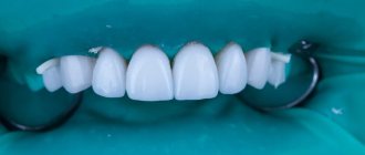
Will root canal cleaning save a tooth that is more than 50% destroyed?
Yes, quite, but instead of installing a composite filling, it is recommended to install a microprosthesis or a ceramic crown. The only difference is in the final stage of the operation.
Clinical case: the patient was diagnosed with advanced caries, which developed into pulpitis. The tooth is badly damaged, but the root is intact. The dentist removes necrotic tissue, qualitatively treats the canal passages, cleans them under a microscope, and then places a microprosthesis or a full-fledged crown.