Molars are divided into canines, incisors, premolars, and molars. The first three types of teeth erupt in place of similar milk teeth. In turn, molars do not have predecessors and appear behind the temporary ones, therefore their second name is accessory.
Each jaw has 16 teeth - four incisors, two canines, four premolars and six molars. In some cases, third molars, better known as wisdom teeth, do not erupt because they do not have buds in the jaw - then instead of 16, a person has 14 teeth above and below. Experts explain this phenomenon by a reduction, or simplification, of the dental system caused by changes in dietary style.
Classification
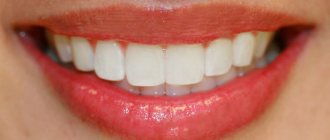
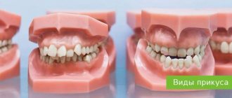
In orthodontic practice, various assessment criteria are used to determine which dental bite is correct and how to correct existing anomalies. The ideal situation, in which nothing needs to be corrected, is characterized by the following indications:
- Semi-elliptical shape of the upper and parabolic shape of the lower arc.
- Small - no more than a third - overlap of the vestibular surface of the mandibular elements.
- Contact of antagonists during closure and absence of obvious gaps.
Compliance on all points is quite rare, but in most cases the deviations are minimal and do not require medical intervention. Often the cause of imbalance is not congenital defects, but bad habits, some of which are formed at an early age.
Types of correct bite
In accordance with the generally accepted classification, there are four main types of occlusal relationships, characterized by the absence of problems and not having a negative effect on the jaw region.
Orthognathic
The optimal, from an aesthetic point of view, is a condition in which all elements of the dentition have an even shape, are located in the correct manner, without gaps or deviations from the midline.
Straight
A common phenomenon, the main feature of which is the lack of overlap - the cutting edges of the incisors meet along a line, which causes a specific type of smile, but is not considered as a pathology.
Biprognathic
Another type of even dentition without threes and diastemas, the distinctive feature of which is a slight deviation of the vertical growth vector of the units. With this development, the crowns are slightly tilted forward.
Progenic
In a normal state it is not accompanied by abnormal manifestations. The identifying feature is a slight frontal protrusion of the lower units.
Anatomical features of the upper teeth
- The central incisor (the largest of all eight) has a chisel-shaped crown with a convex surface and one cone-shaped root. The only root canal in 75% of cases is straight.
- The lateral incisor with the same chisel-shaped crown and the same convex surface has a characteristic depression in the enamel - a “blind fossa”. There is one root canal, but more often it is deviated to the side.
- The canines on the upper dentition are often larger than those on the bottom. They are characterized by a crown pointed on all sides and the longest cone-shaped root. Canines have a single root canal, which can be straight (45%), distally deviated (30%) or vestibular deviated (12%).
- The first premolar with a prism-like crown is characterized by a convex lingual surface. On the chewing side there are tubercles, and between them there is a fissure. In the upper dentition, these teeth are always larger than in the lower one. The root has extended longitudinal grooves, which divide it in 60% of cases into two parts - buccal and palatal. There are often also two root canals.
- The second premolar with the same prism-shaped crown has predominantly one straight cone-shaped root with expanded lateral surfaces. Sometimes the root bifurcates closer to the top. There are often two root canals.
- The first molar is the largest tooth in the dentition. The rectangular crown has a diamond-shaped chewing surface with four cusps and an H-shaped fissure between them. There are usually three root canals, but sometimes there are 4 (25%) or 5 (1%).
- The second molar with a classic cube-shaped crown has 4 cusps and an X-like fissure on the chewing side. The tooth has 3 roots and three (87%) or four (13%) canals.
Which teeth bite is not considered correct?
The presence of deviations from the norm is at least the basis for a dental diagnosis, based on the results of which a decision is made on the need for treatment. The best option is orthodontic correction in childhood, since the dentofacial apparatus, which is in the stage of formation, is more easily amenable to the targeted influence of a special machine and takes on the desired structure without surgical intervention. The most commonly diagnosed abnormal forms have certain signs that allow us to understand whether a person’s bite is correct or not, what is going wrong, and how this deviation can affect the further development of the jaw:
- Distal - diagnosed with excessive development of the upper jaw, noticeably protruding forward and distorting facial features.
- Mesial is a reverse pathological condition in which the mandibular row protrudes, almost completely covering the incisors and canines.
- Cross - characterized by the presence of free areas between the teeth, as well as a “scissor” intersection of elements in a random order.
- Deep - significant, more than 30%, overlap of the lower section.
- Open - contact during closure is observed only between individual chewing units, while in the incisal region there are pronounced gaps, creating the impression of a constantly slightly open mouth.
Any pathological manifestation requires timely medical intervention. Not all patients know what a correct bite in a person is and what it is needed for - but this indicator affects the quality of respiratory and speech function, ensures the conditions for normal food processing and the functioning of the gastrointestinal tract. By contacting Dentika dentistry, you can undergo a comprehensive diagnosis, receive recommendations from qualified specialists and, if necessary, resort to orthodontic correction services.
Anatomical features of the lower teeth
- The central incisor is the smallest tooth in the “adult” bite and the smallest among the incisors. The root is quite short, in 65% of cases there is one root canal, less often - two.
- The chisel-shaped lateral incisor is always larger than the central one. One or less often two root canals – both narrow. “Adult” incisors on the lower jaw are less susceptible to damage than others, so dentists rarely turn to them for the treatment of caries in children and adults.
- Fang - similar in structure to the upper one, but is smaller in size. In 96% of cases it has a single root canal of normal structure.
- The first premolar has a rounded crown in cross-section and two characteristic tubercles on the chewing side. One root is slightly flattened.
- The second premolar is very similar in crown shape to the canine, and is always larger than the adjacent premolar. The surfaces of the single root are smooth and slightly shiny. Two roots occur in only 3% of cases or less.
- The first molar with a cubic crown has five cusps on top of the crown, separated by an F-like fissure. In 88% of cases, three root canals are formed.
- The second molar is smaller than the first, but completely replicates its anatomical features. Root canals are curved and have poor patency. In 85% of cases, the tooth has three canals, in 10% - four.
The anatomical characteristics of teeth are similar in all people, but each person may have individual characteristics, for example, the absence of wisdom teeth or an increased number of root canals in a particular tooth.
Treatment for missing teeth
The specificity of the anatomical structure of the jaw apparatus is that even complete edentia does not guarantee the formation of correct occlusion after implantation. The result of prosthetics is largely determined by the degree of development of defects and anomalies that formed even before the loss or removal of elements. If the row ratio was within acceptable limits, it is enough for the doctor to take into account the necessary adjustments when developing a replacement structure, but the presence of problems with bone tissue will require the use of more complex restoration techniques.
How to check and find out if the bite is correct when making a jaw prosthesis without teeth? For this purpose, special wax rollers are used, placed in the oral cavity. The measuring instruments are a ruler arc and an intraoral plate. After collecting the necessary readings, first a plaster prototype is created, and then the final structure.
The structure of the periodontium under a microscope –
In Figure 8 below you can see collagen fibers penetrating the root cementum. Please note that the terminal sections of the fibers (located in the cement of the root or in the bone wall of the alveoli) are called Sharpey's fibers. Next, Fig. 8 shows a histological preparation of the periodontium of a tooth, where 1 – bundles of collagen fibers, 2 – the main amorphous substance, 3 – periodontal vessels.
Electron microscopy data made it possible to find out that periodontal fibers penetrate into the cement of the tooth root to a depth of only 3 to 5 μ.t, and into the bone wall of the alveoli - no more than 20 μ.t. It also became known that although periodontal fibers consist predominantly of mature type 1 collagen, it also contains some immature elastic fibers (oxytalan). These fibers are only 2-3 mm long, and they are not located perpendicularly like all steel fibers, but parallel to the surface of the tooth root. Oxytalan fibers cross the dento-alveolar fibers at right angles, and their role is to redistribute blood flow in the periodontium under conditions of chewing load.
Collagen fibers occupy only 40% of the periodontal volume, and the remaining 60% is nothing more than the main amorphous substance (which, in turn, consists of as much as 70% water). In addition to water, there is also a large number of different cellular elements present here - first of all, we are interested in fibroblasts, which are located along the collagen fibers. Fibroblasts can also differentiate into fibrocytes or myofibroblasts during the cell cycle.
Another group of cellular elements includes cementocytes and cementoblasts. The latter are located on the surface of the cementum of the tooth root, and their function is to build replacement cement. There is also a small group of cellular elements, which include osteoblasts, osteoclasts, and odontoclasts. The following are also found in small quantities in the periodontium: lymphocytes, plasma cells, mast cells, eosinophils and neutrophilic leukocytes.
Periodontal histology: video
Below in video 1 you can see the histology of tooth tissue in stunning resolution. Video 2 is the best lecture on periodontal histology you can ever hear. The video is in English, but if you wish, you can turn on subtitles, and then select translation from English into Russian in the settings.
The importance of routine observations in normal occlusion
In children with an orthognathic or straight bite, the arrangement of dental units in a row may change due to untimely (early or late) change of primary teeth. During a preventive examination, the orthodontist will identify the presence of abnormalities in the rudiment itself. The following types of preventive measures are carried out:
- Clinical examination (allows us to identify existing anomalies and risk factors).
- Formation of groups of patients for further clinical observation.
- Drawing up a plan of preventive and therapeutic measures (pediatricians of all profiles participate).
In adult patients with normal dentition, malocclusion can develop under the influence of external and internal unfavorable factors. The main reasons that increase the risk of developing dental anomalies in adults are:
- carious process;
- loss of teeth;
- various types of prosthetics;
- prosthetics errors;
- bad habits.
During the examination, the dentist will identify the presence of predisposing factors, after which he will develop a scheme for carrying out preventive measures to maintain a normal bite. Selection is carried out on an individual basis, taking into account the condition of the dental system and the physiological characteristics of the patient’s body.
Size of modern human teeth
Over more than two million years, the size of teeth gradually decreased due to the transition to eating soft and processed foods. The first transformations affected the fangs - in primates they were larger and more strongly advanced relative to the rest of the row. The interdental spaces have disappeared, the size of the front teeth has become smaller. Refusal of raw meat led to a decrease in the chewing load and a narrowing of the jaw, as a result of which there was no room left for the “eights”. Now wisdom teeth are considered atavism and are often subject to removal.
Causes of pathological bite
The formation of occlusion is a long and complex process that begins at the stage of intrauterine development, continues in early childhood and ends after the end of puberty. Pathological occlusion is the result of long-term influence of a number of unfavorable external or internal factors at different periods of a person’s life:
- Genetic factor. A genetically determined anomaly is formed when there is a congenital discrepancy in the size of the teeth and dentofacial bones (crowding of teeth or the presence of spaces between teeth) or a hereditarily determined abnormal shape and size of the lower and upper jaws.
- The influence of negative factors on the body of a pregnant woman. The formation of the dental apparatus in the fetus can be influenced by bad habits, poor nutrition and diseases suffered by a woman during pregnancy.
- Birth injury. During childbirth, the baby may receive injuries leading to displacement of the temporomandibular joint.
- Negative factors affecting the baby’s body in the first years of life. The reason for the formation of a distal bite may be the incorrect selection of nipples or the late introduction of solid foods into the diet.
- Bad habits and behavioral factors in early childhood. The habit of sucking a pacifier or finger, or sleeping with the head thrown back can cause a child to develop a deep bite.
- Factors influencing the formation of anomalies in later life. These include carious lesions of the teeth, ENT pathologies, deviated nasal septum, and diseases of the skeletal system.
Treatment of abnormal bite should begin as early as possible. Early diagnosis and therapy make it possible to restore the normal functioning of the dentofacial apparatus more effectively. Doctors at the Consilium-Dent use specially developed diagnostic tests and tables. They are used both for direct examination of the oral cavity and for diagnosis using dental casts.
The need for a particular therapeutic measure is determined by a specialist based on the diagnostic results on an individual basis. You cannot resort to self-medication in this situation. This can lead to activation of the pathological process.
Treatment of malocclusions
Before prescribing treatment procedures, the orthodontist assesses the condition of the oral cavity, carefully examines the depth of the problem and the presence of concomitant pathologies, taking into account the patient’s age. Treatment of dental anomalies is carried out using conservative and radical methods:
- Braces are the most popular method of bite correction. This orthodontic method has the widest range of applications and is suitable for solving any problem of malocclusion. Treatment with this method is long and includes preparatory, main and retention (recovery) periods.
- Aligners. Removable structures are used to treat the initial stages of deviation. With their help, a gentle correction of the dentoalveolar apparatus is carried out, changing one set of mouthguards to another.
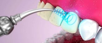
- Application of veneers, onlays, crowns. Weak areas of the dentition that are susceptible to caries are protected by using orthopedic structures. Chipped areas of enamel are restored using ceramic onlays and veneers.
- Surgical intervention is performed to treat late stages of dental anomalies (mainly of a hereditary nature). During the operation, the position of the jaws is changed, their size is reduced or increased.
Laser therapy is performed as an auxiliary treatment to help speed up regeneration and reduce the risk of complications.
Internal anatomy of the tooth
What structure do teeth have in an adult, what parts do they consist of, and what can cause so much pain? If you look at the crown from the outside and mentally “climb” inside, the tooth consists of several layers.
- Enamel. This is the outer layer of the tooth and the hardest part in the human body, consisting of mineral prisms. Its thickness is minimal in the neck area and becomes as strong as possible on the “working” surface of the tooth.
On the outside, the enamel is covered with pellicle - a special organic film, without which acids have a negative effect on the teeth, dissolving enamel minerals.
- Dentine. This is the structural basis of the tooth, a hard, durable tissue that consists of a collagenous base substance that forms the tubules. Odontoblast processes are located inside the dentinal tubules.
Dentin is the primary tissue of the tooth.
In lower vertebrates (fish, amphibians), teeth consist only of dentin. In higher vertebrates, starting with reptiles, enamel and cement appear in the teeth “Anatomy of human teeth” Dr. med. Gaivoronsky I. V.
- Pulp. It consists of loose connective tissue in which nerves and blood vessels pass.
- Root. The part of the tooth, normally not visible to the eye, is located in the alveolar socket of the jaw. On the outside, the root is covered with a protective layer - cement, which is a complex of lime salts, collagen fibers and special cells called cementocytes. The structure of cement is similar to bone tissue, but it does not contain blood vessels or living cells, so it does not grow or renew itself.
- There are many connective tissue fibers around the root of the tooth. They are called periodontium or periodontal ligament. These are the “ropes” that hold the tooth root in the bony alveolus.
How are tooth sizes determined?
To determine the size of a person’s permanent teeth, the calculation tables of Wetzel and V.L. Ustimenko with average standards and permissible deviations are used. However, an experienced dentist is able to independently identify the anomaly during a visual examination or by applying the formula for the ratio of the height and width of the natural crown. During diagnosis, the specialist takes into account the patient’s face shape and height. For example, with a wide jaw, the size of teeth exceeding the norm is not a pathology.
To determine the size of the root canal, there are also tables that indicate the average distance from its apex to the cutting edge or cusp of the tooth surface. The longest is the root of the canines - about 26 millimeters, the root canal of the incisors is 21 - 23 millimeters, and the size of the roots of the chewing zone and premolar teeth ranges from 19 to 22 millimeters.
Odontogenesis: the process of tooth formation
The crowns of the front milk teeth are formed in the prenatal period; they lie in the dental sacs inside the jaw. When a child is born, the formation processes continue, the roots of the teeth grow, and the interalveolar septa separate in the gums.
The tooth takes up more and more space, because of this, as well as under the influence of anatomical and physiological changes, part of the gum atrophies, and the crown of the tooth comes to the surface. When this happens depends on:
- the child's health status;
- nutrition;
- heredity.
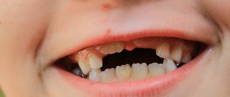
In general, the eruption of primary teeth begins at 6-8 months with the lower central incisors, and the lateral incisors soon appear. After a few months, the first molars become visible, then the canines and second molars. On average, the eruption process ends in 20-24 months. The appearance of teeth at an earlier date is less common, and a delay in onset of up to 10-12 months is much more common [1].
If there are obvious violations of the order or very delayed teething, the child must be examined to exclude diseases or pathologies of the structure of the teeth. It is important to understand that the development of baby teeth affects the oral cavity and facial skeleton, posture and even the psycho-emotional indicators of the child. From the age of 6-7 years, baby teeth gradually fall out and are replaced by permanent teeth [1]
Functions of periodontium –
- Retaining function - it consists in holding the tooth in the alveolus, and the dento-alveolar fibers of the periodontium are primarily responsible for this.
- Shock absorption and distribution function - intercellular substance and periodontal fibers allow you to evenly distribute the chewing load from the tooth to the alveolar tissue.
- Protective function - the connective tissue and cellular components of the periodontium represent the so-called “histohematic barrier”, which ensures the structural and antigenic homeostasis of both the periodontium itself and the surrounding tissues. The implementation of the protective function is mediated by both specific and nonspecific protective factors.
- Plastic function - ensures the preservation of the structure of the periodontium, as well as the reparation of both the periodontium itself and adjacent tissues (for example, the bone plate of the alveolus, as well as the cement of the tooth root). The periodontium is very rich in cellular elements, including osteoblasts, which are responsible for the formation of bone tissue on the surface of the alveoli, as well as cementoblasts, on which the production of replacement cement for the tooth root will depend.
- Trophic and sensory functions are provided by a well-developed vascular and nervous network (a large number of receptors). These functions are closely related to those listed above.
Dental formula
Dental patients look with curiosity at the notes on the examination card, on which numbers, corners, and sometimes letters are written. We are talking about a system of numbering teeth without naming them: that is, a diagram written on paper, where each place corresponds to a tooth.
Thus, one of the popular formulas for adults in Russia consists of four blocks [1]
| 87654321 | 12345678 |
| 87654321 | 12345678 |
The teeth are numbered from front to back as they are seen by the dentist examining the oral cavity. Using this diagram of teeth, he describes their general condition using symbols:
- dash—tooth missing;
- the number is circled - treatment is required;
- the figure remains unchanged - a healthy tooth.
Roman numerals are used to designate baby teeth.
| V IV III II I | I II III IV V |
| V IV III II I | I II III IV V |
There are other recording methods, but their general essence is the same - to clearly show the condition of the dentition, so that all specialists involved in the treatment of the patient can immediately see problem teeth and build the correct tactics of help.
Blood supply to the periodontium –
The sources of blood supply to the periodontium are the superior and inferior alveolar arteries. In turn, smaller “dental arteries” branch off from them, which already penetrate the apical openings at the apexes of the roots of the teeth. Before penetrating the apical foramen, its alveolar and periodontal branches are separated from the dental artery. The bone wall of the alveoli throughout its entire length is penetrated by a system of perforated canaliculi, through which smaller arterioles penetrate from the alveolar branch of the dental artery to the periodontium.
Periodontal blood supply diagram –
The vascular network of periodontal tissues of adjacent teeth is combined into a single system, which allows for collateral blood flow. An important point is that periodontal vessels can be connected to intrapulpal vessels - through additional openings on the lateral surface of the tooth root. These may be ways for the infection to spread.
The lymphatic system is formed by capillaries that blindly begin in the intercellular substance of periodontal tissue, and is rather poorly developed. From the periodontium of the teeth of the upper jaw, the outflow of lymph occurs into the parotid lymph nodes, and from the teeth of the lower jaw - into the submandibular and sublingual lymph nodes. This is what explains the increase in certain groups of lymph nodes, for example, during exacerbations of chronic periodontitis.
Innervation of the periodontium –
It is carried out from the side of the trigeminal nerve, the afferent and efferent fibers of which form a plexus in the periodontal tissues. The endings of these fibers are pain receptors and mechanoreceptors. In most teeth, the maximum concentration of receptors is concentrated in the area of the root apices, but in the periodontium of the incisors, the receptors are evenly distributed throughout the periodontium. Sympathetic nerve fibers responsible for regulating blood flow are also found in the periodontium.
Macrodentia
The tooth size exceeds the norm by more than 2 millimeters. Pathology occurs due to the fusion of two rudiments or the main and supernumerary teeth during the period of formation. The cause of macrodentia can be endocrine diseases, metabolic disorders or heredity. There are five types of pathology:
- localized - one or two teeth are significantly larger than the rest;
- generalized - the entire dentition differs in size from the other;
- isolated - enlargement of one medial incisor;
- absolute - the size of the teeth of both jaws exceeds the norm;
- relative - excessive growth of the upper or lower incisors.
Histological structure of the tooth
The special hardness of teeth is due to their structure. The outside of the crown is covered with enamel, the basis of which is hydroxyapatite crystals. 96% of the enamel consists of inorganic substances, which is why it is so hard (which, however, does not mean that experiments can be carried out on teeth, testing their strength). But dentin, which is located under the enamel, has much less inorganic substances - about 70%. It is softer, and therefore is destroyed by caries more than enamel. Inside the tooth there is a cavity filled with a neurovascular bundle, or pulp. The entire rich spectrum of pain during the deepening of the carious process is associated with it. In addition to nerve fibers, the pulp also contains a large number of blood vessels, through which infection from the affected tooth spreads throughout the body. This is why it is so dangerous to leave dental diseases untreated.
Microdentia
The size of teeth in microdentia is less than anatomical standards. The list of reasons for deviation includes exposure to radiation, premature removal of a baby tooth, narrow jaw, and infectious diseases. There are several types of anomalies:
- isolated - a single violation affecting the lateral incisors;
- relative - the teeth are of normal size, but look smaller due to the enlarged jaw, as a result of which interdental spaces and diastema are formed;
- generalized - the defect covers a group of teeth.