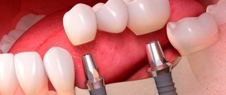
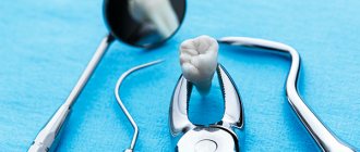
The first molars of the chewing group of teeth (6s) are large 2-3-root teeth, the main function of which is the primary mechanical processing of food. They are subject to high chewing loads. Due to their complex structure and location in the row, it is difficult to remove bacterial plaque around the molars, which leads to the development of caries and destruction of dental tissue. The loss of the first molars is fraught with impaired chewing function, displacement of the dentition, impaired occlusion, and other serious problems. Since the upper 6s are located almost close to the maxillary sinuses, the implantation of the 6s in the upper and lower jaws is different.
- When used:
Absence of 1 tooth - Type of anesthesia:
Local anesthesia - Procedure time:
From 15 minutes - Treatment period:
Up to 4 months - Healing period:
Up to 7 days - Age restrictions:
From 18 years old
Types of tooth extraction
In medical practice, all removal procedures are divided into:
Easy removal
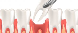
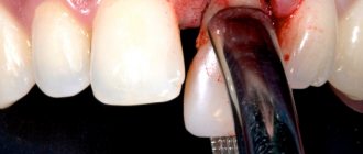
The doctor manages to extract the entire tooth using special forceps; its duration is up to 5 minutes. This is how single-rooted teeth or teeth affected by periodontitis are removed.
Difficult removal
To extract a tooth, the doctor uses auxiliary tools (most often an elevator), and the entire extraction process takes up to 15 minutes. It is most often performed when a part of the root or crown is broken during the removal of multi-rooted teeth.
Atypical removal
It has no clear boundaries in terms of time and extraction method and implies a complex and multi-stage procedure. For this, the doctor can use a bur, an elevator, forceps, a chisel, surgical sutures and needles to close the wound. Often such removal requires from 30 minutes to 2 hours.
This division is reflected both in the cost of the removal itself (due to the various labor costs of the doctor) and in the rehabilitation period:
Simple removal results in minimal damage to surrounding tissue. At the same time, squeezing them with an instrument (and stopping full blood circulation in this area) lasts no more than 5 minutes, and does not in any way affect the subsequent healing of the wound. If you follow the recommendations, the risk of complications is minimal.
Complex removal involves the use of more aggressive instruments and prolonged compression of the walls of the bone alveolus. After it, there may be difficulties with the formation of a blood clot and the development of alveolitis of the “dry socket” type, which is supervised by taking anti-inflammatory drugs and applying turunda with iodoform.
Atypical removal is a surgical intervention in which the doctor violates the integrity of the mucosa, periosteum, or a certain part of the bone. Pressure on tissue can last up to 2 hours, which greatly affects tissue microcirculation. Sometimes a hemostatic sponge is used to stop socket bleeding during such removal. The wound is sutured, which ensures the tightness of the hole and protects it from the development of alveolitis, but it is important to maintain a hygienic regime to avoid infection on the soft tissue.
The doctor’s further tactics, his prescriptions and recommendations for the recovery period depend on how the tooth was removed.
What is the difference between implantation on the upper and lower jaw?
Features of the operation from below
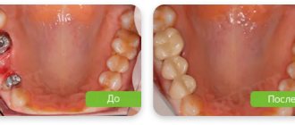
Implantation of the 6th tooth from below is performed using a two-stage method with delayed loading. The implant is implanted using the patchwork method. The crown is installed on the abutment after the artificial root has healed (after 2-4 months). Features of implantation are associated with the anatomical structure of the mandibular structures:
- High jaw bone density - the implant takes root faster, and good primary stability is achieved.
- Bone tissue is stronger and therefore decreases more slowly than in the upper jaw.
- The volume of the mandibular bone is anatomically higher - there are more opportunities to replace the lower tooth without tissue expansion.
- Absence of sinuses - there is no risk of injuring the membrane of the maxillary sinus during implantation.
How is tooth extraction performed?
Before removal, the doctor conducts a survey to find out if the patient is allergic to anesthetics and medications. The doctor also asks the patient about the presence of trophic diseases (diabetes mellitus, atherosclerosis) and pathologies of the cardiovascular system (hypertension, arrhythmia, etc.). This is necessary for the correct selection of anesthesia.
Then the doctor administers anesthesia (usually conductive torus for the lower jaw and infiltration for the upper jaw) and waits for the effect to occur.
Simple removal consists of 5 steps:
- applying forceps to the tooth;
- moving them so that the cheeks are under the gum;
- tool fixation;
- luxation (swinging) - necessary to detach the periodontal ligament, which holds the tooth in the bone;
- traction (luxation) - removal of a tooth from the alveolus. This is done by tilting the tooth in the direction of least resistance (where the alveolar wall is thinner) and simultaneously pulling the tooth outward.
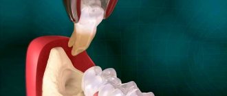
For complex extractions, the doctor uses an elevator - a chisel-shaped instrument with a pointed end, which is carefully inserted into the hole and, with its help, pushes the tooth out from the top of the root.
In the process of atypical removal, a surgical field is formed: the doctor dissects the mucous membrane, peels off the flap with the periosteum and removes the bone substance located above the tooth. The tooth itself is removed using an elevator, but often during the removal of wisdom teeth, the doctor is forced to saw it into pieces with a drill, extracting each root and crown separately.
Is it necessary to place an implant if there is no six?
The upper and lower sixes, despite their strength, are more susceptible to mechanical stress and exposure to the bacterial environment than other dental units. Molars are one of the first teeth a person loses. The dental system is designed in such a way that almost immediately after the loss of a tooth, the bone tissue in this place begins to decrease. The absence of the sixth tooth is accompanied by the following problems:
- the abrasion of the frontal group increases;
- the dentition shifts - neighboring units move towards the missing one;
- antagonists move forward - on the opposite jaw the second molar leans forward;
- tilting of the teeth contributes to the appearance of a wedge-shaped defect, exposure of the roots and displacement of the dental axis;
- the chewing load, which previously fell on 6, is redistributed to other units (adjacent premolars, molars);
- occlusion and chewing function are impaired;
- When the occlusion is violated, excessive chewing pressure is created, accelerating bone loss in the area of the missing tooth.
Considering the importance of the six as a supporting chewing unit, you need to immediately think about how to replace it. Implantation is considered a physiological, long-lasting way to replace teeth. Traditional prosthetic methods do not prevent bone loss. Implant-supported dentures are the only way to restore full functionality of the dentition.
Recommendations after removal
Regardless of the method by which the tooth was removed, in the first 2 days the patient must observe a special diet, activity and hygiene. This is necessary to preserve the blood clot in the wound, which helps maintain the sterility of bone tissue and promotes its regeneration.
In the first 48 hours after removal you cannot:
|
Failure to comply with these recommendations most often causes the clot to fall out of the socket and the development of alveolitis or bleeding.
After tooth extraction it is recommended:
- make oral baths from chamomile and sage decoctions (the decoction prepared according to the instructions is taken into the mouth and held there for 10 minutes, without rinsing or moving the tongue) 2-3 times a day;
- take non-steroidal anti-inflammatory drugs (especially after atypical removal, when the tissue is severely damaged and swelling occurs) - nimesulide, diclofenac sodium, ibuprofen, paracetamol, etc. (it is important that the medicine does not reduce blood clotting).
Sometimes patients need preliminary preparation for removal. People taking aspirin and other medications that impair blood clotting should stop taking them a week before the procedure. For those who suffer from increased excitability and anxiety, you can start taking valerian or motherwort 1-2 weeks before removal, having previously agreed with your dentist and your therapist.
Even if the removal was painless and quick, you should not ignore the doctor’s advice. The right approach to rehabilitation will help you avoid complications and recover faster.
How long does it take for a wound to heal?
Normally, the wound surface heals in 4-5 weeks. In older patients, healing is delayed due to decreased tissue repair and reduced blood circulation.
The rate of healing depends on the extent of the procedure and the roots. In the case of complex removal of a molar with 3-4 roots, the process of tissue regeneration is delayed up to 20-25 days.
Restoration of gum tissue goes through the following stages:
- in the first days, the wound is closed by a blood clot (thrombus), formed as a result of stopping the bleeding;
- 3-4 days after removal, the thrombus resolves, and new tissue appears on the surface of the wound in the form of a thin film;
- bone tissue forms on the sides of the socket already at 3 weeks;
- after 3-6 months, the hole heals and has no differences with the jaw bone.
The speed of wound healing after removal depends on the characteristics of the body and the age of the patient. If necessary, the doctor corrects the tissue regeneration process with medications and physiotherapeutic methods.
Prices for tooth extraction
| Name | Price |
| Consultation | For free |
| Use of bone material | 4,000 rub. |
| Simple tooth extraction | 2,700 rub. |
| Removal of periodontitis tooth | 1,000 rub. |
| Tooth extraction is complicated | 3,200 rub. |
| Removal of a dystopic wisdom tooth | 4,000 rub. |
| Removing a tooth fragment | 500 rub. |
| Removal of exostosis in the area of 1 tooth | 2,500 rub. |
| Removal of cysts and benign formations | 3,500 rub. |
| Treatment of alveolitis | 1,200 rub. |
| Lengthening the clinical crown of a tooth | 5,000 rub. |
| Free flap gum grafting | 8,000 rub. |
| Removing stones from the ducts of the salivary glands | 3,500 rub. |
| Splinting for a jaw fracture | 15,000 rub. |
| Socket curettage | 200 rub. |
| Filling the hole with a medicinal substance (Alvogil, al-vostaz, hemostatic sponge) | 200 rub. |
| Resection of the apex of the tooth root | 4,500 rub. |
| Removing a foreign body from the canal | 1,500 rub. |
Is express implantation possible?
The upper jaw bears less of the chewing load. Sometimes it is possible to simultaneously implant the 6th upper tooth immediately after its removal. The hole is filled with osteoplastic material and an artificial root is immediately installed. But this method is possible if a number of conditions are met:
- sufficient volume and good quality of bone;
- no inflammation;
- atraumatic tooth root removal without damaging bone and soft tissue;
- integrity of the bed walls.
The lower jaw experiences a large mechanical load, so it is better to choose the classic protocol and wait until the hole heals and the bone tissue is restored.
With a single restoration using the express method, the adaptation crown is “removed” from the bite before re-prosthetics with a permanent structure. We do not recommend immediate loading of the implant with a crown for the chewing group, especially for the 6th lower tooth - the lower jaw bears a large mechanical load. Even when the crown is removed from the bite, there is still a possibility that the artificial root may be injured or dislodged during chewing, which will lead to its rejection.
If the patient insists on immediate installation of a crown, the clinic does not give any guarantees.
Guarantees
MCDI ROOTT is one of the few metropolitan clinics where the patient is provided with a lifetime warranty on implantation systems. Warranty conditions are specified in the contract. To maintain the guarantee, the patient complies with the following conditions:
- systematically undergoes dental examinations and diagnostics;
- carefully adheres to the doctor’s recommendations regarding oral hygiene (entered in the Patient’s service record);
- undergoes annual maintenance.
If the doctor’s recommendations are ignored, the examination schedule is violated, or the patient decides to continue treatment in another dentistry, the warranty obligations are canceled.
If the artificial root is rejected during osseointegration before installing a permanent crown, the clinic will reinstall the implant free of charge, provided there are no contraindications to re-intervention. The possibility of reoperation is determined based on the results of examination and diagnosis.
Methods
There are two ways to pull out a lower tooth:
- Simple. It is relevant if the coronal part has been preserved in sufficient volume (the dentist has something to grasp with forceps).
- Surgical. It is used if access to the tooth is difficult or its upper part is severely damaged, as well as when molars, canines and incisors have not fully erupted. In such cases, in order to minimize the risk of developing severe postoperative inflammation, professional hygiene is first carried out and plaque accumulations are removed, and the patient is prescribed antibiotics a few days before the procedure.
Before extirpation, local anesthesia is administered. It makes the working area insensitive to manipulation and helps the patient easily endure all procedures.
External changes
Due to missing teeth, facial muscles weaken, facial tissues begin to sag, which visually increases a person’s age and adds 7-10 years to it. The chin becomes sharper, the lower jaw protrudes above the upper jaw, the corners of the lips and the tip of the nose droop. This changes the overall facial expression, which constantly appears gloomy and dissatisfied to others. From a close distance, the absence of a tooth is clearly visible, even if it is not part of the smile.
Alternative to implantation
An alternative option is to install a bridge based on the patient’s teeth. A bridge is a construction of 3 artificial crowns. The middle one serves to compensate for the defect, the lateral ones - to fix the prosthesis on previously ground, pulpless abutment teeth (5.7). This budget option for prosthetics has a number of disadvantages:
- the chewing load is distributed exclusively on the supporting units, and not evenly on the entire jaw;
- it is not able to stop bone atrophy, bone tissue decreases even faster, the gums sag, a gap forms between the crown and the gum, where bacterial plaque begins to accumulate;
- The supporting teeth are severely injured, quickly destroyed, and then subject to prosthetics.
