I. K. Lutskaya, Doctor of Medical Sciences, Professor BelMAPO (Minsk)
Modern endodontics in most cases guarantees high efficiency in the treatment of pulpitis and periodontitis. However, violation of the algorithm of interventions or clinical protocols can contribute to the development of errors and complications.
Regular clinical and radiological examinations are extremely important to assess the quality of endodontic treatment.
According to the European Society of Endodontics, assessment of the results of root canal treatment should be carried out within 1 year after treatment and thereafter as necessary. The high quality of therapy is evidenced by the following results: absence of pain, swelling and other symptoms, absence of changes in the sinuses, preservation of tooth function and x-ray confirmation of the presence of a normal periodontal gap around the root. Uneven expansion can be considered as the outcome of the disease - scar tissue changes.
The causes of complications after root canal filling may be errors made at the stages of endodontic treatment.
1. At the preparatory stage:
- Root canal infection.
- Lack of adequate access to the root canal orifice.
- Perforation of the bottom and walls of the tooth cavity.
2. During mechanical treatment of the root canal:
- Obturation of the root canal lumen with dentine filings.
- Formation of an apical ledge when the canal is bent (“Zipping”).
- Excessive lateral expansion of the middle third of the canal along the internal curvature of the root (“Stripping”).
- Perforation of the root walls.
- Destruction of anatomical (physiological) narrowing.
- Fracture of the instrument in the canal.
3. During the process of filling the root canal:
- Heterogeneous, insufficient filling of the canal lumen.
- Removal of filling material beyond the apical foramen.
- Longitudinal root fracture.
Root canal infection
Penetration of microorganisms into the root canal can occur due to merciless preparation with pressure on the coronal pulp, careless amputation and removal of tissue from the orifice. The development and proliferation of microbes is possible due to the repeated use of tools, including burs and excavators. Infection of the root canal increases the risk of post-filling complications such as painful percussion, lack of positive dynamics after treatment of pulpitis or periodontitis. In preventing this complication, great importance is attached to careful isolation of the surgical field, since microflora can penetrate into the canal along with oral fluid. It is optimal to use protective equipment such as rubber dam and its analogues (Fig. 1). Before instrumental treatment, it is advisable to completely excise carious dentin from the walls of the carious cavity in order to prevent infection from entering the root canal.
Rice. 1. Treatment of pulpitis using a rubber dam.
Errors in creating access to root canal orifices
The reasons for this situation are insufficient preparation of the carious cavity, incomplete excision of the roof of the pulp chamber, and lack of control over the insertion of the endodontic instrument (Fig. 2). The consequence is the following complications. Overhanging edges of the cavity do not allow completely removing the remaining pulp from the tooth cavity, which inevitably leads to the appearance of pigmentation and worsens the aesthetic parameters of the tooth.
Rice. 2. Incomplete opening of the tooth cavity.
Due to poor visibility, it is not always possible to identify all existing root canal orifices, which precludes treatment and filling of undetected canals (Fig. 3).
Rice. 3. Poor quality treatment of the cavity walls.
The apparent “saving” of hard tooth tissues in the process of cavity formation can lead to poor-quality endodontic treatment.
At the same time, excessive, excessive tissue removal causes a decrease in the tooth’s resistance to mechanical stress.
A measure to prevent such an error is the formation of correct access, which is characterized by the absence of overhanging edges and the straightness of the cavity walls, which should be smooth, without roughness or notches.
Periodontitis
If the carious process is complicated by pulpitis, which is not treated, inflammation of the tissues surrounding the causative dental unit develops. Symptoms of periodontitis are bleeding of the gingival margins during hygienic sanitation of teeth and an unpleasant odor emanating from the oral cavity. Possible pain from thermal irritants.
Periodontitis is classified into acute and chronic. In an acute process, severe pain occurs, which intensifies with load on the causative tooth. Pain symptoms can also arise from the action of thermal irritants. The gums around the causative tooth become swollen, hyperemic, an abscess often appears, which spontaneously opens and purulent contents come out through it, after which the patient’s condition improves. Patients note that the causative tooth becomes higher than other dental units in the jaw.
Treatment of acute periodontitis consists of opening the pulp chamber, through the resulting defect the tooth is cleansed of purulent contents or pulp decay. To make this process go faster, dentists at Dr. Granov’s clinic recommend rinsing the mouth with a soda-saline solution. After 2–3 days, the inflammatory process stops and the tooth can be filled. If the patient’s condition does not improve, then an incision is made into the mucous membrane and periosteum in the area of the periodontitis tooth, and a rubber drainage is placed into the resulting incision. If this does not give a positive result, then the tooth is removed.
If the acute form of periodontitis is not treated, then after 7–14 days the process becomes chronic. Symptoms of chronic periodontitis:
- Excessive tooth mobility.
- Gaps appear between the teeth.
Injury to the root pulp
When treating pulpitis using the amputation method, injury to the root part of the pulp is possible in the absence of adequate access to the mouths of the canals (Fig. 4).
Rice. 4. Hypertrophied gums obstruct the view of the cavity.
Excessive pressure on the bur or excavator will cause bleeding from the canal due to rupture of the neurovascular bundle. The application of a therapeutic pad over the mouth of the canal under pressure contributes to the disruption of blood circulation and the functioning of the root pulp (Fig. 5). In any case, trauma to the root pulp increases the risk of ineffective treatment of pulpitis with a biological method.
Rice. 5. Therapeutic lining over the mouths of the canals.
This complication can be avoided by careful preparation of the carious cavity with complete excision of the altered dentin and subsequent careful removal of the roof of the pulp chamber.
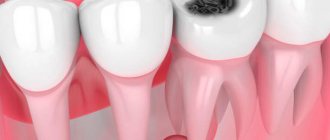
Symptoms of caries
Diagnosis of the disease should only be carried out in the dentist's office. But the person himself can determine the presence of this pathology by very characteristic symptoms:
- the appearance of dark spots on the teeth: most often they appear on the chewing surface, but often inflammation develops between the teeth (but it is usually not possible for the patient to see this - special equipment will be required),
- there is a roughness of the surface of the teeth,
- When eating food, aching pain appears, teeth react to sweet and sour, as well as cold and hot. But the unpleasant sensations subside immediately as soon as contact with the irritant ceases - this is how caries, even deep ones, differs from pulpitis - inflammation of the dental nerve,
- Breath odor may become unpleasant,
- a carious cavity can even be felt with the tongue; food debris often gets clogged into it, which causes discomfort or even swelling of the tooth - for example, if a small gap appears under a filling.
Perforation of the bottom and walls of the tooth cavity
May occur during the search for root canal orifices and their expansion; with poor visibility of the bottom of the tooth cavity as a result of inadequate formation of access to the root canals.
The presence of softened pigmented dentin, intense staining of hard tooth tissues after previous treatment (resorcinol-formalin method, silvering) also greatly complicate the search for root canal orifices (Fig. 6).
Rice. 6. Dentin pigmentation and paste residues at the bottom of the cavity.
In some cases, the causes of perforation are the following factors: insufficient or, on the contrary, excessive expansion of the tooth cavity; carrying out endodontic treatment through an artificial crown. Insufficient knowledge of anatomical features, such as displacement of the tooth axis and a decrease in the height of the crown due to its significant abrasion, contributes to the commission of errors.
Preventive measures for perforation of the walls of the tooth cavity are rational excision of hard tissues, adequate pressure on the bur during the preparation process, its correct direction and precise control of the depth of insertion of the rotating instrument.
Incomplete removal of the root pulp is allowed in cases where adequate access to the canal mouths is not provided or the latter are inaccessible due to the location of denticles in them. The reason may be insufficient expansion of the canal mouths or incorrect determination of the working length. The anatomical features of the structure of the roots can also become a factor in the poor patency of the canal for instruments. Violation of the work technique, for example, removal of tissue with a pulp extractor with rupture of the neurovascular bundle, incomplete removal of the root pulp, leads to bleeding from the canal, which prevents further endodontic interventions.
Obstruction of the canal lumen by dentine filings is manifested by the impossibility of reintroducing a small endodontic instrument to the entire working length. The reason is the accumulation of dentinal filings in the lumen of the canal and their compaction. An attempt to re-pass the canal with force may entail pushing the products of mechanical treatment of the root canal (endolubricants, dentinal filings, pulp residues, etc.) beyond the apical foramen, which can cause pain after endodontic treatment.
This complication is prevented by carefully passing the canal to the apical narrowing with small instruments after every second step, as well as washing the canal lumen with solutions.
The formation of apical enlargement (Zipping effect) most often occurs in curved canals. During canal treatment, slipping of the instrument tip during rotation leads to the so-called “Zipping” effect. The reason is the use of large, inflexible files, which cannot follow the shape of the canal. The canal lumen may be blocked by dentine filings. The risk of creating an apical expansion significantly increases when working with files that have an aggressive apex.
Excessive longitudinal expansion of the canal in the middle third along the internal curvature (Stripping) occurs during mechanical treatment of curved root canals. The reasons may be the following: the use of rigid, inflexible files; mechanical treatment without taking into account the thickness of the canal walls, as well as underestimation of the degree of root curvature.
Due to excessive removal of dentin in the area of the internal curvature of the root, not only does the tooth’s resistance to mechanical stress decrease, but there is also a real risk of longitudinal perforation of the root canal wall.
Destruction of the anatomical (physiological) narrowing occurs when the working length is incorrectly determined. Another reason is a slight decrease in the working length of the channel during straightening. If further processing of the canal is carried out to the previous working length, destruction of the physiological narrowing is inevitable.
Prevention of this complication consists in accurately determining the working length and its correction during mechanical treatment of a curved root canal.
Perforations of the root canal walls occur most often during instrumental treatment of curved roots.
Perforations of the mouth and middle thirds are formed mainly when the filling material is removed from the canal in the process of creating a bed for the anchor pin, as well as when the latter is screwed into the canal.
Apical perforations can occur when using insufficiently flexible rotating instruments in difficult, curved canals. A similar complication is possible from the application of excessive pressure during machining with hand tools, when trying to forcefully pass the canal. The cause of lateral perforation is the passage of a curved canal with an endodontic instrument with an aggressive tip without prior bending.
Measures to prevent various types of perforations are good access to the mouths of the root canals, analysis of the configuration of the root canals according to radiographs (Fig. 7). During mechanical treatment, obstruction of the canal lumen with dentinal filings should be avoided; pre-bend the tool; use an anticurvature technique for passing the canal.
Rice. 7. Excessive preparation and perforation of the wall of the first molar.
Instrument fracture in the root canal
The risk of instrument fracture is very high in case of file deformation (bending, unwinding of turns) and most often occurs when passing and expanding narrow, curved, previously sealed canals (Fig. 8). The main reasons for this complication may be the lack of adequate access to the mouth of the root canal; violation of the sequence of use of endodontic instruments; use of instruments without taking into account indications; non-compliance with operating mode and rotation speed; application of significant force during manual or machine endodontic treatment; metal fatigue caused by repeated use of the tool.
Rice. 8a. Introduction of a curved file.
Rice. 8b. Broken instrument in the root canal.
Prevention of tool breakage consists in strict adherence to the operating mode and use of the tool according to indications. The sequence of use of tools must be taken into account. During machining, the use of endolubricants is recommended.
Incomplete and insufficient obturation of the root canal is mainly due to incorrect determination of the working length, incomplete passage of the canal (Fig. 9), the use of the technique of one gutta-percha or silver pin in canals that have an oval, dumbbell-shaped, slit-like (irregular) shape that does not correspond to the shape of the pin, and also using liquid-mixed paste for filling (using a channel filler). As a result, shrinkage is inevitable, as well as dissolution of the paste some time after filling.
Rice. 9a. Obturation of root canals: high quality.
Rice. 9b. Obturation of root canals: incomplete.
The removal of filling material beyond the apical foramen is often observed after excessive mechanical treatment of the root canal. The result is destruction of the physiological apical constriction. It can also be disrupted due to a chronic inflammatory process in the tissues of the apical periodontium. In addition, there is a real possibility of removing material beyond the apex when using a machine channel filler. The risk of complications increases sharply when filling a root canal without taking into account the working length (Fig. 10).
Rice. 10. Removal of a significant volume of sealer beyond the apex.
The removal of the filling material beyond the apical foramen is observed in the case of using a large amount of sealer, as well as as a result of excess pressure during the condensation of the filling material in the root canal.
Pushing the gutta-percha pin beyond the apex may be a consequence of incorrect determination of the working length and/or incorrect selection of the size of the main pin (Fig. 11).
Rice. 11. Removing the gutta-percha pin beyond the root apex.
Removal of gutta-percha beyond the root apex is possible during the process of lateral condensation of gutta-percha (Fig. 12).
Rice. 12. Lateral condensation of pins.
Preventive measures: control of working length at all stages of endodontic treatment; competent formation of the root canal; maintaining the integrity of the anatomical (physiological) narrowing.
If removing a small amount of sealer beyond the apical foramen may not cause problems, since it is quickly resorbed, then gutta-percha removed beyond the apex, which itself is biologically inert, can maintain inflammation in the apical periodontal tissues for a long time, being a mechanical irritant.
Longitudinal root is possible during the process of lateral condensation of gutta-percha pins and is a consequence of excessive thinning of the walls of the root canal during mechanical treatment. In addition, a longitudinal root fracture can be observed with strong lateral pressure on the sprider during the condensation of gutta-percha pins.
Prevention measures include assessing the condition of the hard tissues of the tooth root, their thickness, as well as improving manual skills and applying adequate efforts in the process of condensation of gutta-percha pins.
Treatment methods
Cure caries, especially at the initial stage, is not difficult - this involves removing the inflamed tissue and then filling it. In the presence of deep lesions, binoculars and a microscope can be used, which allow you to increase the working space and remove even the smallest inflamed cells, while preserving healthy tissue. Depending on the situation, it may also be necessary to restore the coronal part if it has been severely damaged.
Fissure sealing A method that allows you to stop the carious process. Essentially, this is the prevention of inflammation. Fissures or depressions (grooves) on the surface of the enamel are filled with a special sealant, which evens them out and thereby prevents the accumulation of bacteria and plaque.
Price:
from 2500 rubles more details about the solution
Treatment of caries If the doctor notes caries at the spot stage, then the main procedure is remineralization (restoration of the required mineral content) using a special solution. If the caries is superficial, medium or deep, then the resulting cavity will need to be treated, followed by filling using composite materials. When the carious process penetrates into the deep layers, it is advisable to use medicated pads containing calcium hydroxide - this way additional protection is created to preserve the dental nerve.
Price:
2500 rubles more details about the solution
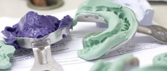
Installing an inlay In a number of situations, when the tooth is quite badly damaged and a large filling will not hold, you can develop an individual inlay made of ceramic or zirconium dioxide. It will accurately follow all the curves of the tooth and be securely fixed inside the cavity. At the same time, it will last many times longer than a large filling.
Price:
from 20,000 rubles more about the solution