Errors and complications associated with creating endodontic access
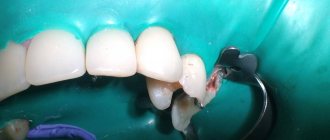
Insufficient removal of the arch of the dental cavity (Fig. 1).
Rice. 1. Insufficient removal of the arch of the tooth cavity
Causes:
- insufficient knowledge of the topographical features of the tooth cavity;
- non-compliance with the rules for opening the tooth cavity.
Prevention:
- knowledge of the topographical features of the tooth structure;
- following the algorithm for opening the tooth cavity.
Endodontic treatment begins with the complete removal of carious tissue and poor-quality restorations. The opening of the tooth cavity is carried out with a spherical diamond bur. To expand the cavity, it is better to use cylindrical burs or mechanical wellhead files. The most rational approaches to the dental cavity are considered coronal-apical and mesiodistal. They make it possible to minimize the number of errors at the initial stage of processing and not to lose the correct direction of the tool in the channel. To check the free access of the instrument into the canal, the “hanging” instrument rule is used: the instrument must enter in a straight line without bending at the mouth.
Perforation of the bottom or walls of the tooth cavity is one of the most common complications during mechanical treatment of the tooth cavity and root canals.
The problem of tooth perforation appeared with the introduction into practice of drills in 1871 and special instruments for mechanical expansion of root canals (Gates Glidden, Beutelrok, Drill reamer, etc.). Even then, there was an increase in the number of complications. The factors leading to the occurrence of perforations were pointed out by the dentist M. Morgenstern in 1901: if “... careless handling of root drills when using an electric drill, it is easy to drill the root.” Currently, dentists make the same mistakes as they did a hundred years ago.
Perforation of the bottom of the tooth cavity is caused by excessive preparation with a bur during the search for the mouths of the root canals. This happens especially often when re-treating a tooth previously treated with the resorcinol-formalin method, since the search for the mouths of the canals is complicated by the changed color and structure of the dentin of the bottom of the tooth cavity.
The prerequisites for the occurrence of perforations of the bottom and walls of the tooth cavity are:
- displacement of the tooth axis in the lingual or buccal direction;
- reduction in the height of the tooth crown due to significant abrasion of the chewing surface or deposition of a large amount of replacement dentin;
- endodontic treatment of a tooth through an artificial crown.
Clinically, perforations of the bottom or walls of the tooth manifest themselves in the form of a characteristic “sinking” of the instrument, bleeding and a sharp pain in the patient during treatment without anesthesia. Touching the site of a fresh perforation with a probe also causes acute pain.
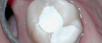
Perforation of the tooth crown at the level of the neck, walls of the crown cavity, bottom of the crown cavity and in the bifurcation area (Fig. 2, 3):
Causes:
- poor knowledge of topographical features of teeth;
- rough preparation without taking into account the position of the tooth and its working length;
- excessive expansion of the root canal orifices, sometimes as a result of an attempt to detect the orifice of a sclerotic canal;
- incorrect choice of tool and violation of the methodology for its use;
- an attempt to prepare curved root canals.
Prevention:
- indication of root canal orifices using dyes;
- compliance with the principles of preparation taking into account the topography of the teeth;
- compliance with the rules for using endodontic instruments;
- knowledge of topographical features of teeth;
- avoiding forced passage of narrow and obliterated root canals and irrational use of machine instruments;
- periodic x-ray monitoring during the passage of root canals.
Rice.
2. Perforation of the tooth cavity Fig. 3. Perforation of the bottom of the tooth cavity. Fracture of the vestibular or lingual wall of the tooth.
Causes:
- wrong direction of bur;
- excessive pressure on the boron.
Prevention: careful use of a boron.
Features of dental canal treatment
Differences in the structure and functions of the “representatives” of the dentition largely determine the nature of the treatment approach. Treatment of canals for different groups of teeth has its own nuances.
Front tooth canal treatment
The front teeth most often have one canal. They are often curved and difficult to pass through for instruments. In order to preserve aesthetics, the opening of the cavity of these teeth is carried out from the vestibule of the oral cavity. Due to their frontal location, it is important to prevent tooth enamel from darkening, so filling materials containing dyes are not used.
Treatment of wisdom tooth canals
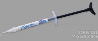
Wisdom teeth can have more than 5 canals; they often have a branched structure, which is not always revealed by x-ray examination. These factors, along with the marginal location of the “eights”, significantly complicate the high-quality treatment of root canals. At the final stage of treatment, various filling pastes are traditionally used.
Root canal treatment of temporary teeth
Endodontic treatment of tooth root canals is usually used only at the stage of root stabilization. When choosing the optimal treatment tactics, it is necessary to take into account the specific structure of temporary teeth. The small thickness of the canal walls, the insignificant degree of dentin mineralization and the relatively large size of the apical foramen are the main reasons for special caution during instrumentation. Zinc oxide eugenol and iodoform pastes, as well as materials based on calcium hydroxide, are usually used as filling agents. They are not toxic to the permanent tooth germ and are able to dissolve along with the temporary root.
Errors in identifying root canal orifices
Causes:
- insufficient knowledge of the topography of root canals;
- Features of endodontic anatomy.
To detect the mouths of root canals, a sharp angled probe, optical devices, and dyes are used. It is often difficult to find additional canals at the bottom of the tooth cavity, but sometimes the entrance to the canal is not identified due to ignorance of the anatomy. The fourth canal in teeth 16, 26, 17, 27 is usually located in the center of the main mesiobuccal canal and is sometimes connected to the main canal. The second canal in teeth 31, 41, 32, 42 is localized on the lingual side of the main canal. To detect it, it is necessary to expand the entrance to the cavity towards the neck of the tooth. In lower jaw teeth 34, 44 with two canals, one canal is located on the buccal side, the other on the lingual side. In teeth with three canals, one canal is located on the lingual side, the other two on the buccal side. If the additional channel remains undetected, the treatment prognosis worsens. The fourth canal in teeth 36 and 46 is located next to the main distal canal; it can be identified by the oval opening of the distal canal, elongated in the lingual-buccal direction.
If your tooth hurts after root canal cleaning
Toothache is an acceptable consequence of endodontic treatment; provided that all doctor’s instructions are carefully followed, the pain should gradually go away within three days.
If the pain does not go away within three days (and even earlier, if the intensity of the pain increases), a repeat visit to the clinic is necessary. This is possible, for example, with periodontitis, when even high-quality canal treatment is not enough and further treatment is necessary.
Important! Only a doctor can prescribe adequate treatment (medical or surgical) after diagnosis; self-medication is extremely dangerous!
In conclusion, here are some tips for choosing a clinic for endodontic treatment. When choosing dental services, pay attention to the following points:
- Quality of the initial consultation: this should be a thorough examination of the oral cavity and the most detailed interview of the patient, who should be given the opportunity to learn about the state of health. Already at this stage, the doctor must demonstrate his ability to create a comfortable environment and win over the patient.
- The quality of the proposed diagnosis: the patient should be offered an X-ray examination; canal treatment should be preceded by a computed tomography scan, which allows one to accurately determine the number of canals to be treated in the tooth.
- Use of latex plates in practice (read about the purpose of rubber dam above).
Lower first molar
Typically this tooth has two roots, a mesial and a distal. The latter is smaller and usually rounder than the medial one. Among Mongoloids, there is a variant with an additional distal-lingual root with a frequency of 6 to 43.6% (Walker, 1988).
Lower first molar. (By Harty).
This double-rooted tooth usually has three canals, the average length of the tooth is 21 mm. Two canals are located in the medial root. In 40 - 45% of cases, there is only one apical foramen in the medial root. The single distal canal is usually larger and more oval than the medial canals, and in 60% of cases opens on the distal surface of the root, close to the anatomical apex.
The attention of specialists was attracted by the work of Skidmore and Bjorndal (1971), who showed that in the distal canal there are two canals in more than 25% of cases. In Mongoloids, due to the tendency to double the distal root, the frequency of occurrence of two canals in this root is even higher - about half (Walker, 1988).
There have been reports of cases involving five channels.
Lower first molar with five canals. (By Harty).
The pulp chamber is wider at the medial than at the distal wall and has five pulp horns. The lingual horns are taller and pointed. The bottom is rounded with a convexity towards the chewing surface and lies immediately below the level of the cervix. The mouths of the canals are funnel-shaped, and the medial canals are narrower than the distal ones.
Of the two medial canals, mediobuccal and mediolingual, the first listed is the most difficult to pass due to its tortuosity. It leaves the pulp chamber in a medial direction, which changes to a distal direction in the middle third of the root. The mesiolingual canal is slightly wider and usually straight, although it may curve medially in the apical third of the root. These two canals can have a dense network of anastomoses among themselves along their entire length.
When an additional distal canal is present, it is more lingual and tends to curve toward the buccal side.
With age, dentin deposition occurs on the roof side, and the canals narrow.
Lower third molar
This tooth is often underdeveloped with numerous and poorly developed cusps. Usually there can be as many canals as there are cusps. The root canals are relatively larger than those of other molars, possibly due to the late development of this tooth.
Despite these disadvantages, it is usually less difficult to fill the roots of a lower than an upper wisdom tooth because access is usually easier due to the mesial inclination of the tooth and because they more often follow normal anatomy, resembling a second molar, and are less likely to be deviated from the norm.
Access in lower molars
Access contours in lower molars.
If there is a second distal canal in the first molar, a more quadrangular approach may be necessary. Care must be taken when removing the roof of the pulp chamber to avoid damaging the bottom. To improve visual control of the canal mouths, access can be expanded. The access walls should extend toward the occlusal surface to resist masticatory forces and prevent displacement of temporary fillings.
If the course of the channels is non-standard, access can be expanded and/or modified.
Thus, standard, universal, tabular methods for determining the working length of dental canals cannot currently satisfy clinicians. Of course, you need to have a more or less correct idea about possible deviations in the morphological characteristics of cavities; the decisive factor is an X-ray examination with the introduction of files into the root canal. In this case, it is advisable not to try to insert the instrument to its full working length, since it is almost impossible to obtain undistorted radiographs.
Lower premolars
These teeth usually have a single root, but sometimes the first premolar may have a bifurcated root in the apical half.
Type I channel predominates. Where there are two canals (usually in the first premolar), there may be type IV/V configurations. Types II/III occur in less than 5% of cases. The highest incidence of two canals is reported to be 10.8% in the second premolar (Zillich and Dowson, 1973).
One report found that two canals in the first premolar were three times more common in African-Americans than in whites (Trope et al., 1986). This option is more common among southern Chinese. In less than 2%, three canals may be present in the first premolar.
The pulp chamber of the lower premolars is wider in the buccolingual direction than in the mesiodistal direction, and has two horns, the buccal one being better developed. The lingual horn is small in the first premolar and larger in the second premolar.
Lower first premolar. (II channel configuration type). (By Harty).
The canals of the lower premolars are similar to the canines of the canine, although they are smaller, but they are also wider in the bucco-lingual direction until the middle third of the root, when they narrow and acquire either a rounded shape or bifurcate.
Lower second premolar. (I channel configuration type). (By Harty).
Lower second molar
In Caucasians, the second molar resembles a small version of the first, with an average length of 20 mm. There are two canals in the medial root, and only one in the distal root. The medial canals tend to merge in the apical third and form a single apical foramen.
Lower second molar. (By Harty).
Studies conducted in 1988 showed a high tendency for root fusion in the Chinese (33-52% of cases). In a longitudinal section, such teeth resemble a horseshoe. Where there is incomplete root separation, incomplete canal separation may occur, which is accompanied by a dense network of anastomoses between the canals and can lead to unpredictable localization of the orifices. One of the locations was called the middle buccal orifice with the middle buccal canal. In Caucasians, this anomaly is recorded in 8% of cases, which is significantly less than in the Chinese.
Lower central and lateral incisors
Lower first incisor. (I channel configuration type).
Both teeth have an average length of 21 mm, although the central incisor is slightly shorter than the lateral incisor. The morphology of dental canals can have one of three configurations.
Lower second incisor. (IV channel configuration type).
Type I - one main canal from the pulp chamber to the apical foramen.
Type II/III - two main canals that merge in the middle or apical third into one canal with one apical foramen.
Type IV - the two main canals remain separate for the entire length of the root and with two apical foramina.
All studies show that type I is the most predominant. Two channels are recorded in 41.4% of cases, and type IV - in 5.5% of cases.
There is evidence that in Mongoloids, two canals are less common in these teeth.
The pulp chamber is a small replica of the upper incisors. There are three pulp horns, not very well defined, and the chamber is wider in the labial-lingual direction. In the version with one channel, it can bend towards the distal and, less commonly, towards the labial side. The canal begins to narrow in the middle third of the root and becomes round. With age, the changes are the same as in the upper incisors and the pulp chamber can be located below the level of the neck of the tooth.
Upper canine
It is the longest tooth in the mouth, averaging 26.5 mm (range 20-38 mm). It is extremely rare to have more than one root canal. The pulp chamber is relatively narrow and has only one horn; it is much wider in the vestibulo-oral section than in the mediodistal section. The root canal is type I and becomes round only in the apical third. The apical constriction is not as pronounced as in the incisors. This fact, and the fact that often the apical part of the root is significantly narrowed, causing the canal to become very narrow at the apex, makes it difficult to determine the length of the canal.
Upper canine
The canal is usually straight, but sometimes at the apex it bends towards the distal (in 32% of cases) and, less often, the lateral side. In 13% of cases, vestibular deviation of the canal was recorded. The frequency of occurrence of lateral (side) canals is about 30%, and additional apical ones - 3%. The apical foramen is located in 70% of cases in the range from 0 to 1 mm in relation to the root apex, and in 30% in the range of 1 - 2 mm.
Access to lower incisors and canines
Essentially, the approach is identical to that in the upper teeth. However, with a pronounced curvature towards the lingual side of the crowns of the incisors and due to very thin (especially in older people) canals, it is sometimes necessary to involve the cutting edge and, sometimes, the labial surface of the tooth into access to avoid bending of the instrument.
The outlines of the access in the lower canine are shown in Fig.
Access contours in the lower incisors.
Access contours in the lower canine.