Pediatrician (working with children of the first year of life)
Murzina
Oksana Yurievna
22 years of experience
Pediatrician of the highest category, member of the Union of Pediatricians of Russia
Make an appointment
Jaundice in newborns (most often physiological) is a condition in which the skin and whites of the eyes turn yellow due to high levels of a substance called bilirubin in the blood. Most babies develop 2-3 days after birth. Bilirubin is a yellow substance resulting from the destruction of hemoglobin during the natural replacement of red blood cells. Under normal conditions, it should be processed quickly in the liver. According to medical statistics, jaundice is diagnosed in 50-60% of cases in newborns born at term, and in 80% of premature infants. Bilirubin in excess amounts has a toxic effect on internal organs.
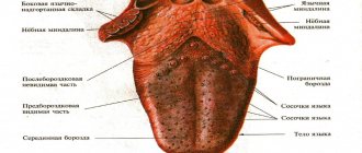
What color should the tongue normally be?
Keep in mind! Such an unremarkable organ as the tongue performs a number of important functions:
- Articulation is responsible for the ability to pronounce sounds.
- Eating food - chewing, moving a bolus of food around the mouth, swallowing.
- Recognition of tastes - the sensation of sour, salty, sweet and bitter.
- Regulation of local immunity with the help of the tonsil in the mucous tissue of the root of the tongue.
Light, whitish deposits on the lingual surface are a type of normal .
You should know! They are a layer of keratinized epithelium (dead tissue from thread-like taste buds).
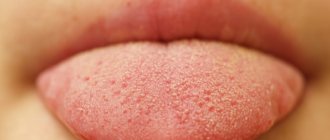
The tongue of a healthy child from 3 to 7 years old should meet the following characteristics:
- Color and structure . The surface is pink, soft, with a smooth groove in the center and visible tubercles of taste buds.
- Raid . A small amount of whitish plaque on the back is acceptable. It can be easily and painlessly removed with a toothbrush or cotton pad.
- Feel . The organ does not hurt, has no swelling or irregularities, and does not interfere with articulation and swallowing.
- Smell . Normally, there is no bad breath.
Important! To avoid possible problems with the oral cavity, teach your child to also clean the tongue surface while brushing his teeth.
This can be done either with a regular toothbrush or with a special scraper spoon.
Plaque on the tongue. Causes
The tongue of a healthy person should have a pale pink color. The formation of a certain amount of light plaque on the surface of the tongue is allowed; it should be loose and the texture should be visible through it. Plaque on the tongue is thin or dense deposits, most often white or grayish in color, which completely or partially cover the surface and thereby change its color.
What it consists of:
- Saliva, epithelium, food debris.
- Bacteria and fungi that feed on the components from the first point.
- Leukocytes that eat fungi and bacteria.
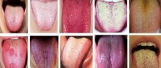
Color and reasons
The causes of plaque on the tongue may vary depending on its color. We will not talk about situations where the tongue changes color due to the use of coloring products - this is not at all dangerous and quickly goes away on its own, but we will touch on different colors of plaque caused by more serious reasons.
White
White color is almost harmless. Many people experience a thin white film in the morning. It is easily removed when brushing your teeth with a toothbrush. This is quite normal - overnight in the absence of oxygen, bacteria actively multiply, which forms plaque on the teeth and mucous membranes.
Grey
Why can plaque on the tongue be gray? Usually this is the next stage of white, that is, a sign of the development of thrush, gastritis or tonsillitis. Sometimes gray deposits also appear with long-term use of antibiotics, due to disruption of the natural flora of the oral cavity.
Yellow
In this case, there are several reasons for the yellow plaque:
• ARVI. Usually accompanied by high fever and general malaise; • Problems with the digestive tract, such as gastritis or ulcers; • Long-term use of antibiotics or vitamins. No treatment is required; • Jaundice. In this case, the lower part of the tongue is affected rather than the upper part, and it is not easy to detect.
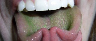
Green
This is a very rare phenomenon and occurs due to exposure to certain types of fungi. Sometimes the cause may be excessive load on the liver, that is, eating large amounts of fatty and fried foods. In very rare cases, green deposits are caused by antibiotics.
Brown
There may be several reasons for a brown coating on the tongue:
- Improper functioning of the gallbladder, formation of stones in it;
- Disturbances in the gastrointestinal tract. Accompanied by sharp pain and diarrhea;
- Alcoholism;
- Liver diseases;
- Regular consumption of dark drinks, especially coffee and strong tea. The teeth also suffer and become yellowish or brown.
Blue
Why is the coating on the tongue such an unusual color, like blue? A sign of anemia, a lack of vitamin B12, iron and folic acid in the body. Sometimes the tongue turns blue in people who smoke for a very long time.
Black
If the tongue is not gray, but rather black, this is a very bad sign. Most often, he talks about serious disturbances in the gastrointestinal tract. Spots and cracks indicate stagnation of bile, that is, problems with the pancreas and liver. At the same time, there is always a bitter taste in the mouth. If the organ is covered with black dots, this is a signal of lead poisoning. If both the mucous membrane and tooth enamel darken, this indicates the proliferation of a chromogenic fungus in the oral cavity.
In conclusion, I would like to advise you to get into the good habit of inspecting your tongue every morning during hygiene procedures. If something seems doubtful to you, consult a doctor for advice. Be attentive to your health!
Using materials from https://topdent.ru/articles/nalet-na-yazyke.html https://netnaleta.ru/u-vzroslyh/o-chem-govorit-nalet-na-yazyke-u-vzroslogo/ https: //yandex.ru/turbo/anzub.ru/s/novosti/nalet-na-yazyke-norma-ili-otklonenie/?utm_source=turbo_turbo https://stom.32top.ru/stat/782/
Why it appears: reasons
The reasons for the formation of brown deposits on the surface of this organ in a child are in many ways similar to the reasons for its appearance in an adult.
However, there are a couple of exceptions due to bad habits.
In adults, the coating on the tongue may become darker due to the abuse of caffeine and cigarettes, which is natural - not applicable to the baby.
In most cases, the coloring of this sensory organ in shades from red to brown is associated with disorders of the gastrointestinal tract.
Note! At the same time, the intensity of the color is directly proportional to the complexity and severity of the disease.
Other reasons include : _
- Diseases of the respiratory system (lungs, bronchi). Deposits appear in the morning after waking up, and after cleaning the oral cavity they appear again.
- Treatment with dyes or antibiotics. On some medicines (Faringosept, Malavit, etc.), the manufacturer himself indicates the likelihood of the appearance of colored plaque as the norm. As a rule, at the end of therapy, the tongue surface acquires its usual, healthy appearance.
- Food poisoning, dysbacteriosis . In this case, the formations are accompanied by a bitter taste, diarrhea or constipation, as well as pain in the lower abdomen. The unhealthy color will disappear only after the underlying disease is eliminated.
- Fungal infection of the mucous membrane (tissue microsis). The disease is characterized by thickening and then darkening of the natural plaque. Without proper treatment, the color will become more and more saturated.
- Dehydration of the body. Most often it is a consequence of disorders of the gastrointestinal tract. To solve the problem, you need to consult with a pediatrician on the selection of medications and provide the body with sufficient fluid.
Stay up to date! Sometimes a brown coating on the tongue may appear due to the consumption of chocolate, cocoa, Coca-Cola and other coloring products.
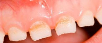
Causes of brown plaque
Most often, a newborn's tongue becomes white or yellow. In the first case, it acquires color due to the formation of thrush, but quickly renews and passes. In the second case, the baby may have problems with the liver, which results in jaundice.
Brown plaque is less common and seems to be a more dangerous phenomenon for the child’s health, especially if it appears on the tongue of an infant.
Brown plaque in teenagers is due to hormonal changes
Doctors name the following reasons for a brown tongue:
- Disturbance of gastrointestinal processes, digestive problems.
- A state of dehydration resulting from elevated temperature or low fluid levels in the body.
- Lack of B vitamins.
- Ingestion of fats, food poisoning.
- Reaction to certain medications.
Coca-Cola turns your tongue brown
There is another reason that is not related to the biological nature of the body. This reason is somewhat banal, but it is not uncommon - eating brown foods:
- chocolate;
- caramels;
- soda;
- dark berries, etc.
A change in the appearance of the tongue occurs during the reaction of dyes with the mucous membrane. After all, parents are not always able to control their children’s consumption of foods with dyes. Buckwheat porridge can also leave a dark tint.
Features of consistency and thickness of plaque
In order not to lose sight of the onset of any serious disease, it is necessary to regularly examine the baby’s oral cavity.
At the same time, you should pay attention not only to the presence of plaque, but also to its consistency, location and thickness .
Your observations will make the pediatrician’s work easier in making a diagnosis.
As a result of many years of medical observations, it was possible to establish the following relationship between plaque on the tongue and the condition of the human body:
- A thin/spotty coating on the tip of the tongue indicates diseases of the heart muscle and respiratory system.
- A thick layer of brown deposits on the root indicates intestinal pathologies.
- A brown stripe in the center of the tongue can be a “signal for help” from the spleen, gall bladder or liver.
Important! Do not try to make a diagnosis and prescribe treatment yourself - this can be dangerous for the baby’s health. Only a doctor can determine the true root cause of the disease.
How is diagnosis carried out?
An accurate diagnosis is established only after a series of the following medical examinations:
- General blood analysis;
- Blood biochemistry;
- Antibody test;
- Scraping from the surface of the tongue;
- Stool and urine analysis.
In some cases , an ultrasound examination of the stomach, intestines or respiratory tract may be required .
Need to know! Depending on the results found, the pediatrician may give a referral to other doctors: a gastroenterologist, an infectious disease specialist, a toxicologist, or a dentist.
Types of plaque
A similar phenomenon happens to everyone: both children and adults. We don’t even think about healthy plaque and don’t pay attention to it. It appears more or less depending on the time of year. In the summer, when the period of berries, fruits, and vegetables begins, the layer on the tongue is quite noticeable, since it has different shades:
- white;
- yellow;
- green;
- even dark blue.
In winter, plaque is not visible on a healthy body.
Yellow coating on the tongue occurs due to liver diseases
Possible treatment
Treatment of brown plaque involves eliminating the root cause , since it is not a separate disease, but only a symptom of the underlying disease.
Only a doctor can make a correct diagnosis and prescribe treatment.
In most cases, he prescribes antibiotics and medications to regulate the functioning of the gastrointestinal tract .
If the course of treatment is successful, the brown plaque disappears on its own.
Clinical picture of ascariasis and enterobiasis in children at the present stage
What are the difficulties in diagnosing helminthiases? What is the treatment for helminthiasis?
Against the background of global environmental changes, the widespread use of immune, antibacterial and other drugs, frequent violations of adaptation processes (adaptation diseases), constant stress, increasing levels of education and culture of the population, many long-known human diseases have changed their clinical picture. Some symptoms practically ceased to occur, while others, on the contrary, began to come to the fore. This also applies to diseases caused by helminths, in particular parasitic roundworms (nematodes). In the temperate zone, the most widespread nematodes are roundworms and pinworms.
Helminths can cause dysfunction of the gastrointestinal tract (GIT), cause allergic (or pseudoallergic) reactions or aggravate them, cause intoxication, and also be a factor that weakens the immune system [2]. Nowadays, cases of massive infestation are rare, when diagnosis does not cause difficulties, and helminth balls cause intestinal obstruction - much more often helminths have become the cause of the development of allergic problems. At the same time, helminthiases are among those diseases that are difficult to diagnose due to objective and subjective difficulties (long periods of absence of oviposition, the possibility of the absence of females among the parasitic individuals, the likelihood of technical errors). Therefore, it is important to know the clinical picture of these diseases in order to be able to prescribe an in-depth examination or effective therapy based on a combination of indirect signs, even without direct evidence of the presence of helminthiasis.
In order to evaluate the clinical picture of nematodes, complaints, anamnesis and examination results of 150 children were analyzed in whom roundworms (116 children), pinworms (27 children), roundworms and pinworms (7 children) were found. Helminths were identified using standard methods: detection of roundworm eggs in feces using a smear method or pinworm eggs in a scraping for enterobiasis, as well as visual detection of roundworms or pinworms in feces, vomit, or during surgical or endoscopic intervention in the abdominal cavity or rectal area [3].
Among the children there were 67 boys and 83 girls. The age distribution is presented in table. 1. Children of primary preschool age (from one to three years old) predominated, their number was 63%. It is at this age that the greatest epidemiological preconditions for the appearance of nematodes occur.
In 150 children with proven infestations of roundworms and/or pinworms, the following clinical manifestations were noted.
107 children (71.3%) had allergic problems: skin rashes, diathesis, atopic dermatitis, neurodermatitis - 99 (66%), of which 18 had periodic problems, two children, in addition to skin rashes, had conjunctivitis ; ten (6.7%) had a proven food allergy to some foods with high levels of specific IgE in the blood serum; Six children (4%) had an asthmatic component or were diagnosed with bronchial asthma.
113 children (75.3%) had gastrointestinal dysfunction: unstable stool, undigested stool, the presence of “green” and mucus in the stool - in 66 (44%); constipation or a tendency to constipation - in 55 (36.7%), 17 children (11.3%) had unstable stool with a tendency to constipation, i.e. periods of normal or liquefied stool alternated with periods of constipation; flatulence, manifested by increased gas formation, bloating, rumbling, belching, occurred in 30 children (20%); nausea, vomiting, and regurgitation were observed in 29 children (19.3%).
In 60 (40%) children who, by age, could complain of abdominal pain, abdominal pain syndrome was noted: most often so-called “flying pains” were noted, which occurred regardless of food, periodically, usually without a specific localization or localized around navel (in the paraumbilical zone). Such pains went away on their own without medications or while taking sorbents and antispasmodics; some children needed to lie down for a while; Pain syndrome of this nature was observed in 54 children (36%). In eight children (5.3%), abdominal pain was constant, often associated with food (occurring after eating), intense, and often required drug treatment and hospitalization to exclude acute surgical pathology; one child underwent an appendectomy for this reason, and roundworms were found in the appendix with catarrhal changes; Two children had abdominal pain syndrome of both types.
66 children (44%) had appetite disturbances: in 54 (36%) appetite was often reduced or selective; in 12 (8%) - increased (the child is constantly hungry). Often, when asked about appetite, parents answered “when and how,” i.e., there were periods of normal appetite and decreased or increased appetite. In assessing this indicator, there is a large amount of subjectivity, since parents assess their children’s appetite differently.
Bruxism (teeth grinding) - a symptom that is often associated with helminthic infestations, but in fact is a nonspecific sign of intoxication of the central nervous system and can accompany any chronic intoxication - was noted in 25 children (16.7%).
Night sleep disturbances were observed in 81 children (54%): restlessness in the evening, difficulty falling asleep - in 15 children (10%); restless night's sleep (screaming, moaning, tossing and turning in sleep, waking up, crying, insomnia, nightmares) - in 76 (50.7%). Some parents of older children found it difficult to characterize the child's nighttime sleep, because the child sleeps in another room. Problems with falling asleep and sleeping at night are an important symptom of helminthic infestations, since it is known that intestinal nematodes (including roundworms and pinworms) are often activated at night.
54 children (36%) had itching and/or redness in the anus (anal escoriation) - in 43 children (28.7%); itching - in 38 (25.3%); both symptoms - in 27 (18%). Anal escoriation and itching are considered symptoms of enterobiasis (pinworms), but out of 54 children who had these symptoms, enterobiasis was proven in 17 children (31.5% of the number of children with these symptoms). In another 37 children (68.5% of the number of children with these symptoms), the presence of ascariasis was proven, but pinworms were not detected either visually or in tests. This may indicate either that in these cases there were pinworms that were not diagnosed, i.e., helminthiasis was mixed, or that anal escoriation and itching are symptoms characteristic not only of enterobiasis, but also of ascariasis.
29 children (19.3%) showed signs of general weakening of the immune system: frequently or long-term ill children (according to the generally accepted classification of Monto J. et al., 87) - 18 children (12%); recurrent stomatitis and gingivitis were observed in 13 children (8.7%); dental caries - in six (4%); recurrent purulent diseases of the skin or mucous membranes - in three (2%).
15 children (10%) had the results of a blood test of their immune status: 13 children had a reduced number of T cells; in all 15 children the number of T-helper cells was reduced, and in six of them it was significantly reduced; in 12 children the helper-suppressor ratio was reduced; in seven children there was a decrease in the level of IgA (secretory immunoglobulin), including in three a significant decrease, while in the remaining eight children the level of IgA in the blood serum was either normal or elevated; Six children had a decrease in the number of lymphocytes, including one child who had severe lympho- and neutropenia. These results confirm the known data that roundworms and pinworms have a depressing effect on immune function, and that people with weakened immune systems are more likely to develop helminthiasis [1].
49 children (32.7%) had various symptoms that can also be associated with the effects of helminths on the body: “geographical” tongue - in 12 children (8%); retardation in physical development, insufficient weight gain or weight loss over a period of time - in 21 children (14%); a painful appearance (blue under the eyes, pallor) was detected during examination in seven children (4.7%); Ten children (6.7%) had bad breath, especially in the morning; increased fatigue, asthenic syndrome - in eight children (5.3%); emotional lability, capriciousness, irritability of the child - in four (2.7%), and this indicator is subjective, as is the parents’ assessment of appetite; hypersalivation, another symptom usually associated with helminthiasis, was observed in three children (2%); various manifestations of hypovitaminosis, including rickets - in five children (3.3%). Often, during examination, changes in the skin in the form of “goose bumps” were revealed.
Most children had more than one symptom. Summarized data on the clinical picture are presented in table. 2.
Therapy in all cases consisted of two stages: first, anthelmintic therapy, and two different drugs were prescribed (decaris and vermox) with an interval of three to five days between them in order to maximally cover the different stages of the life of helminths; some time after anthelmintic therapy - drugs for microbiological and functional correction. Accordingly, the results of anthelmintic therapy, the results of the entire treatment, as well as follow-up within six months after therapy were assessed. In most cases, improvement occurred quickly during treatment. In 36 children, only after anthelmintic therapy, allergic manifestations significantly decreased or completely disappeared, in 39 children, after the first stage of therapy, the functioning of the gastrointestinal tract normalized, in 41 children, abdominal pain immediately stopped, and other symptoms also improved after anthelmintic drugs. This confirms that the symptoms were caused by the presence of helminths.
92 children had no complaints after the entire treatment; about 37 there is no data on changes in condition during and after treatment; in 16 children the effect was incomplete, i.e. some symptoms persisted after the end of treatment; in four children the effect of therapy was unstable, since relapses occurred after the end of treatment; Three children had no effect from therapy.
Literature
1. Blagova N. N. Some immunity factors in patients with ascariasis and enterobiasis during treatment with albendazole.
Author's abstract. dis. ...cand. honey. Sci. St. Petersburg, 1997. 2. Bogoyavlensky Yu. K., Rachkovskaya I. V., Chebyshev N. V. Nematodes and anthelmintics. M., 1994. 3. Laboratory diagnosis of parasitic diseases: https://www.infectology.spb.ru/. Bulletin of infectology and parasitology. Table 1. Age distribution
| Age | Up to 1 year | 1 – 3 years | 37 years | 7 – 12 years | 12 – 15 years |
| Number of children | 9 | 94 | 23 | 18 | 6 |
back
Table 2. Clinical picture of intestinal nematodes
| Complaints | Amount of children | % of total |
| Allergic problems | 107 | 71,3 |
| Gastrointestinal dysfunction | 113 | 75,3 |
| Abdominal pain syndrome | 60 | 40 |
| Appetite disorders | 66 | 44 |
| Bruxism (teeth grinding) | 25 | 16,7 |
| Night sleep disorders | 81 | 54 |
| Anal escoriation and/or itching | 54 | 36 |
| Signs of weakened immunity | 29 | 19,3 |
| Other symptoms | 49 | 32,7 |
back