07.05.2018
A large number of patients are faced with the need to have teeth removed. Injuries, untimely contact with a dentist, errors in previous dental treatment and much more are the main causes of tooth loss.
Unfortunately, patients often put off compensation for the resulting defect after tooth extraction. At the same time, without suspecting that this entails consequences that do not provide the possibility of further restoration of teeth through implantation.
To understand this slightly confusing formulation, we will first try to describe what processes occur in the dental system after tooth extraction.
Types of complications after tooth extraction
Problems that may arise after operations are usually divided into two groups. The first group includes general complications:
- a sharp drop in blood pressure (collapse);
- reflex changes in blood vessels due to insufficient level of pain relief;
- change in consciousness (fainting) due to fear;
- shock, including pain due to inappropriate anesthesia.
The incidence of general complications is low. Usually they are limited to short-term fainting, from which the patient is brought out with the help of ammonia. If the anesthesia is insufficient, the patient experiences pain, which he reports to the doctor - the issue of increasing the concentration of the substance used or choosing a different method of pain relief is decided.
A local complication after tooth extraction is considered to be a pathological condition localized at the intervention site. It does not affect the body as a whole, but causes serious inconvenience to the patient.
Local complications after tooth extraction include:
- bleeding at the site of the extracted tooth;
- paresthesia;
- fracture and dislocation of the jaw;
- leaving particles of the extracted tooth in the gums;
- alveolitis;
- perforation of the maxillary sinus;
- removal of a baby tooth along with the germ of a permanent one;
- root wedging into soft tissue;
- osteomyelitis and other complications of a purulent nature;
- bite pathology;
- traumatic removal of the maxillary tubercle;
- injury to nearby teeth (luxation or fracture);
- root or crown fracture;
- injuries to the gums, tongue and soft tissues;
- aspiration of a tooth or its root.
Each type of complication has its own treatment method. It is important for patients to seek medical help as soon as symptoms appear to help avoid the development of serious pathologies.
Aesthetic consequences
The presence of even one gap in the dentition can change the proportions of the face over time. Age-related aging processes accelerate, become more noticeable, and intensify:
- due to alveolar bone resorption, facial height decreases;
- vertical lines become more noticeable in the area of the lips and chin, making the face look rougher;
- The bite gradually changes, which can cause the proportions of the lips to be disrupted and the chin to change;
- with edentulism in the upper jaw, the nasolabial groove deepens faster and more noticeably, which is why the face looks older;
- It is possible to form a double chin if the attachment of the muscles to the body of the lower jaw is disrupted and their tone decreases.
Causes of complications after tooth extraction
General disorders of the condition usually occur against the background of the patient’s nervous overstrain, which are associated with fears, often formed in childhood. But technologies are changing, and now treatment by dentists is not accompanied by pain or any other unpleasant sensations. An individual method of anesthesia is selected for each patient to ensure that tooth extraction is easy and painless.
Local complications after tooth extraction can be associated with several factors. The most common of them are:
- anatomical features (cause a root or crown fracture, injury to adjacent teeth);
- careless work of the dentist (improper use of forceps leads to fractures of the tooth or its root, an incompletely separated tooth ligament injures the gum during removal, and when the forceps or elevator slide, damage to soft tissues is caused);
- lack of x-ray control (causes tooth remains to remain in the gum);
- severe carious lesions (provokes fractures of the root or dental crown);
- failure to follow the doctor’s recommendations (excessive or poor hygiene after tooth extraction causes alveolitis or insufficient opening of the mouth during surgery can cause injury to the gums or soft tissues).
Anatomical features that provoke complications after tooth extraction
- The roots are thin and long.
- Thick alveolar walls, which are difficult for the doctor to work with.
- High level of root curvature.
- Hypercementosis.
- Dense partitions between roots.
- If there are many roots, the degree of their divergence is taken into account.
Knowing the causes of complications after tooth extraction, you can prevent their occurrence. But if it was not possible to avoid complications after the operation, it is necessary to immediately proceed to treatment; delaying seeking medical help leads to pathologies.
Changes in soft tissues
Attached soft tissues also change when bone volume changes. The gums shrink in volume and become thinner. In the lower jaw, the layer of attached soft tissue may be very thin or almost absent.
Periodontal nutrition is disrupted and blood supply deteriorates. This further accelerates destruction: surface tissues become thin, susceptible to inflammation and irritation. In advanced cases, bedsores form. Destructive processes provoke the development of chronic periodontal diseases. These, in turn, can lead to the loss of adjacent teeth.
If toothless ridges of bone tissue on the jaw have already formed, the tongue can gradually increase in size: it fills the space that was previously occupied. With the loss of one tooth, such manifestations are minimal; if there are several gaps in the dentition, the enlargement of the tongue will be clearly visible.
Treatment of local complications after tooth extraction
The choice of treatment method is based on the causes of the pathology, its nature and the patient’s condition. Age is also taken into account, since not all medications are suitable for children. Therefore, the treatment regimen is developed individually for each patient.
Complications related to bone structures
Most often, a fracture of the tooth root occurs during extraction. In this case, it is usually removed so as not to provoke an inflammatory process. If symptoms of infection have already arisen, and the pathology is discovered a few days after the operation, wait 1-1.5 weeks and perform surgical treatment again. In this case, the patient takes anti-inflammatory drugs to stop the inflammation process.
If during extraction a tooth that was located next to the extracted one is fractured, the doctor evaluates how reasonable and possible it is to save it. With minor damage, it is possible to build up the damaged area. If the fracture is serious, the tooth is removed.
During surgery, cases of dislocation of the tooth closest to the extracted one are not uncommon. For high-grade injuries, replantation is indicated. If the mobility of the tooth is preserved, it is strengthened with a stabilizer in the form of an endodontoendoxal implant. If the tooth tissue is completely dead, it is removed and replaced with an artificial tooth. If the dislocation is incomplete, strengthening is carried out using a splint.
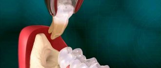
A situation where the root is pushed into the jaw tissue from below is possible when the third molar of a large tooth is removed. If the hard part is palpable, it is removed through an incision made over it. If there are no objective signs of a root being located in any part of the jaw, an x-ray is taken, which is performed in two projections (side and front) - this is how the desired root is detected and removed.
When removed, the root of a tooth may end up in the sinus of the upper jaw. In this case, the root is urgently removed, as it can cause infectious sinusitis. The operation is not performed through the hole so as not to enlarge the hole in the sinus. First, perforation of the bottom is carried out, then an operation to remove the root is made - for this, a burr hole is made in the sinus of the upper jaw from the outside and in front. The operation is performed using endoscopic devices under continuous monitoring.
Trauma to the alveolar process is not considered a serious complication during tooth extraction. This condition does not cause discomfort and is not considered dangerous. It goes away on its own without affecting recovery after surgery. If the fragment has sharp edges, they are smoothed even during tooth extraction. The most commonly injured teeth are the upper and lower third molars.
Pathologies of soft tissues during tooth extraction
In case of injury to soft tissues or gums, the primary task is to stop bleeding: a hemostatic sponge is applied to the wound, and in case of severe damage, the wound is sutured with excision of those areas whose integrity cannot be restored.
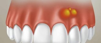
Some patients are concerned about swelling localized on the gum near the extracted tooth. In the first few days after surgery, it is considered physiological. But if the pain persists and the swelling grows, you need to consult a doctor - a purulent complication may develop, which requires opening and draining the resulting cavity.
If a blood clot does not form at the site of the extracted tooth, alveolitis appears. It is provoked by non-compliance with the doctor’s recommendations for oral care (smoking, rinsing in the first few days, failure to perform hygiene procedures, aggressive foods). Sometimes the patient is bothered by pain that is subjectively felt in the temple area. Before starting treatment, the hole is sanitized, treated with an antiseptic solution and a cotton swab with an anti-inflammatory agent is placed in it.
If the rules of asepsis and antisepsis are not followed, osteomyelitis and other purulent complications may occur after tooth extraction. If left untreated, the pathology spreads to neighboring organs and also affects the jaw. To get rid of this condition, the patient undergoes an autopsy of the infectious focus and is prescribed anti-inflammatory drugs.
Other damage after tooth extraction
In some cases, paresthesia occurs due to tooth extraction. It is characterized by a lack of sensation in the cheeks, lips, chin and tongue. The condition lasts about two weeks, then passes. You can speed up recovery by taking B vitamins, ascorbic acid, dibazole and galantamine.
If the patient opens his mouth excessively, the jaw may dislocate. This complication is typical for elderly patients, as well as during surgery on large and small molars of the lower row. When a dislocation usually affects one side, less often both. Patients have this defect corrected on the spot, immediately after detection.
In rare cases, a jaw fracture is detected. It is typical for surgery on the second or third molar. Diagnosis of a fracture is complicated by blurred symptoms, but is clearly visible on x-rays, so x-rays are often used to monitor the success of the operation. A jaw fracture is treated by repositioning the bones and fixing them with splints through the teeth or with the help of intra- and extrafocal bone tissue synthesis.
When removing small or large molars from above, perforation of the maxillary sinus is possible. It is characterized by the presence of a through hole in the hole. The condition may be accompanied by bleeding and discharge of pus. An important stage of treatment is to control the formation of a blood clot in the hole in the absence of inflammation. To speed up healing, an iodoform turunda, a gentamicin sponge or a tampon with an anti-inflammatory and analgesic agent are placed on the hole. To ensure that the applications remain in place, they are fixed with a plastic mouthguard or bandage like a ligature in the shape of a figure 8 on the adjacent teeth. A removable denture can be used to cover the hole.
If clot formation does not occur, an iodoform tampon is secured to the socket to the edges of the gum using silk sutures or a mouthguard. The application should be worn for 5 to 7 days - during this time the wound begins to heal and there is no need for additional coverage. It is important to apply the tampon correctly so that it does not completely fill the hole, otherwise the patient faces sinus inflammation.
If a significant size hole is found at the bottom of the sinus, a blood clot may not form. Then the walls of the resulting hole are processed so as to remove all sharp parts, and then sutured without tension. If there is no positive effect, biological material can be applied to the hole, after which plastic surgery of the defect is performed with soft tissue. To speed up healing, an iodoform tampon is applied to the wound, which is fixed with a plastic plate.
If a root or whole tooth is inhaled, an emergency tracheotomy is performed to prevent the patient from suffocating. When an object passes into the lower parts of the respiratory system, it is removed using a bronchoscope, and for this the patient is transported to a suitable medical facility. If a tooth or part of it has been swallowed, the patient is monitored and the stool is independently monitored. Usually the object comes out naturally without causing significant discomfort.
If after tooth extraction the patient does not insert an implant in time, malocclusion develops over time - this is due to the natural displacement of the teeth towards the vacant space. If the dentition is not restored in time, bite correction with braces will be required, and the installation of a prosthesis will be accompanied by bone grafting.
Complications after tooth extraction in a child
Modern medical standards require that even children’s baby teeth be treated for caries. Until recently, they were simply removed, believing that healthy permanent ones would still grow in their place. But it turned out that this is not so.
Firstly, caries easily passes from baby teeth to permanent ones. Secondly, after the forced removal of baby teeth, a change in the bite often develops. As a result, it took a long time to correct the position of the child’s jaw using plates and braces. Therefore, doctors are increasingly deciding to place temporary implants even in childhood to maintain the correct bite.
The risk of developing malocclusion occurs not only in the postoperative period. There have been cases recorded when, during treatment, the doctor accidentally removed a baby tooth along with the germ of a permanent tooth. This is due to their close location in some children. Correction in this case is only possible with the help of a permanent tooth implant. Therefore, it is important to notice and treat carious lesions in children in a timely manner, rather than delay them and bring the situation to removal.
How to stop the drift?
To prevent destruction, after the operation, restoration of the masticatory organs should be undertaken. To do this, the patient must contact an orthodontist. Braces are often required to correct an overbite. It happens that the hole becomes inflamed, causing the wound area to become infected and may fester. In such situations, cleaning and disinfection are performed. Therapeutic methods also include:
- Installation of removable corrective devices. They include a plastic plate. It is held in the mouth by hooks and wire loops. To straighten and return the units to the correct position, springs and arms are used. To further expand the jaw, an expansion screw is installed on the plate.
- Orthodontic devices that are not removable are represented by systems for correcting jaw closure. This effective technology allows you to correct the chewing organs of adults. The braces are fixed with special glue. A strong bracket is pulled to make the connection. It creates the right amount of force so that the units move in the right direction.
- In difficult situations, radical technologies are used. Treatment may involve face bows, which significantly change the configuration of the skull.
Nowadays, aligners and mouth guards are also used. They are suitable for patient patients. The correction period takes from 12 months to 2.5 years. The aligners are changed every 15 days. The advantage of aligners is the transparency of the material. They can be removed for hygiene purposes or for eating.
Correction of common complications
General violations occur less and less often in dentists’ offices. This is due to the latest technologies in this area, which allow treatment to be carried out painlessly and quickly, as well as the development of asepsis and antiseptics. But some complications still arise, although this does not happen as often as before.
If the patient’s eyes are cloudy due to fear of intervention, use a cotton swab soaked in ammonia. It is brought 7-10 cm from the nose, which brings the person to his senses. If anxiety does not go away, medical sleep is offered as pain relief.
If there is a large loss of blood during the operation, you need to carefully and promptly make up for the deficiency. Otherwise, vascular disorders develop, including hypovolemic shock and cardiac failure. Therefore, treatment should be carried out only in well-equipped clinics.
Low-grade fever is considered a normal reaction of the body to the intervention. But if it goes beyond 38.5 ºС for several days, you need to inform your doctor about this - the symptom may indicate the onset of a complication.
Features of the structure of wisdom teeth
The structure of a wisdom tooth is no different from other molars and consists of a neck, a root system (4–5 pieces) and a crown. In some cases, root development is disrupted and a single conglomerate is formed, that is, a tooth grows with one root.
Its only difference from its neighbors is that recent studies recognize the wisdom tooth as a rudiment that has lost its functional purpose during the evolutionary development of man. Scientists do not consider it as a full-fledged organ, since some part of humanity does not grow it at all.
However, the wisdom tooth has several parameters that distinguish it from the same “seven”:
- lack of milk precursors, which ultimately complicates eruption and impedes growth. There is no room left for it on the dental arch, which is why various developmental pathologies arise;
- the number of roots is greater than that of other molars - up to 5 pieces. In this regard, problems often arise when removing a wisdom tooth;
- significantly curved roots of the “eights” make cleaning and treatment of root canals very difficult;
- poor blood supply compared to other teeth leads to rapid aging, accompanied by fragility and susceptibility of tissues to caries, despite minimal load;
- many pathological conditions, for example, pathologies of the masticatory muscles, malocclusion, pericoronitis, are associated with the growth of “eights”.
Thus, the appearance of third molars has many more negative consequences than positive ones. Many patients are forced to see a dentist even before they can be seen in person.
Prevention of complications after tooth extraction
- Responsible choice of clinic and doctor is one of the most important conditions for successful treatment.
- X-ray monitoring of all performed manipulations allows timely detection and prevention of complications.
- Compliance with all dentist recommendations for oral care after surgery.
- Timely installation of an implant helps to avoid changes in bite.
- A thorough medical history allows you to choose the right anesthesia and calculate its dosage.
- Reducing treatment time, a comfortable environment and a friendly attitude allow you to avoid most common complications.
- Compliance with the rules of asepsis and antisepsis prevents and stops the development of inflammatory and purulent pathologies.
Tooth extraction is a surgical operation; this procedure must be handled responsibly. An informed and careful choice of clinic and doctor will help avoid most complications. Competent patient management and a friendly atmosphere will leave only positive emotions about treatment at the dentist, contrary to prevailing stereotypes.
Sources used in writing the article:
- “Outpatient surgical dentistry”, Bezrukov V.M.
- "Surgical dentistry", T.G. Robustova.
- “Guide to maxillofacial surgery and surgical dentistry”, A.A. Timofeev.
Please rate this article
Stanislav Mezheritsky
Chief physician, surgeon, orthopedist
The author of the article is Stanislav Mezheritsky, a practicing dentist with 19 years of experience, chief physician and one of the founders of the Matisse Dent clinic. The main focus is orthopedic and surgical dentistry. Author of numerous publications, regular participant in specialized seminars.
What determines the stability of treatment?
The preservation of the result in orthodontics is usually called retention. Retentio is a Latin term meaning "preservation", "retention". So, maintaining a beautiful smile does not depend on the money paid by the patient. Even after correction with the coolest expensive ceramic braces or transparent Invisalign aligners, the teeth can move apart very quickly. In some cases, this is due to the fault of the doctor, but much more often the patient himself becomes the source of the problem.
- The teeth will definitely become crooked again if the orthodontist has not identified and eliminated the real cause of the anomaly.
- The doctor is also responsible for the correctness of the treatment program. This means that he must correctly choose the orthodontic design and directions of tooth movement, as well as carefully approach the need to remove those that interfere with effective correction.
- When drawing up a program, a professional will initially focus on a high-quality result, and not on accelerated treatment. With a patient who wants to wear only the “magic” Smartclip self-ligating braces in order to have as few visits to the clinic as possible and correct the bite faster, it is difficult to find a compromise. But without this, it will be impossible to realize his dream of an even, white-toothed smile.
- Finally, to consolidate the result after removing braces, the specialist must offer the patient the optimal retention device. In this case, one should take into account the psychological characteristics of the individual demonstrated during long-term treatment.
This limits the orthodontist's responsibility. If all the rules were followed, and the teeth become crooked again, the patient can only blame himself.