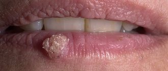
Oral papillomas are benign neoplasms in the oral cavity that grow from epithelial cells. Papillomas are discovered during a dental examination and look like separate growing seals on a small stalk, they are painless and have a white or pale pink color.
This type of neoplasm in the oral cavity is diagnosed most often. About 60% of patients are women aged forty years, about 20% are teenagers of any gender. Often, adults experience the appearance of individual papillomas, while children may experience so-called papillomatosis (multiple papillomas). In half of the cases, papillomas are localized on the mucous membrane of the tongue.
Causes of papilloma in the mouth
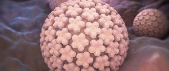
The most common cause of this type of tumor is the human papillomavirus (HPV).
Factors that provoke the appearance of papillomas in the oral cavity are, for example, constant microdamage to the cheeks and tongue. A relatively small damage is enough for viral particles to penetrate inside and trigger the formation of papilloma. In children, the provoking factor is a too short frenulum of the tongue - the lower incisors injure it, creating a gateway for infection.
When analyzing papilloma under a microscope, it can be noted that this neoplasm is a tumor, which consists of many layers of epithelial tissue, which in some places has become significantly keratinized. In some areas, traces of the appearance of a focus of inflammatory infection can be noted.
Complications
In the absence of adequate treatment, the pathology can progress rapidly, which will cause the formation of new lesions. In addition, the risk of developing the following complications increases:
- Hoarse voice.
- Deterioration of speech function.
- Inability to use dentures .
- Impaired respiratory (if papillomas have spread from the gums to the tonsils, larynx).
- Painful discomfort while eating.
- Bleeding .
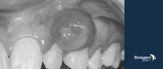
Removal of tooth granuloma
- In addition, the risk of damage to the tumor increases, which causes the development of cancer.
If papilloma appears on the gum, you must immediately contact a dentist, who, after conducting research, will refer the patient to an oncologist or dermatologist.
Classification of oral papillomas
Based on the number and concentration of neoplasms, oral papilloma is differentiated from papillomatosis – a massive accumulation of neoplasms in one place.
According to their origin, papillomas are divided into the following types:
- Traumatic (reactive) papilloma. May appear after traumatic effects of a mechanical, chemical or temperature nature. A distinctive and characteristic feature of reactive type oral papilloma is that their growth stops immediately after the irritant that caused them is eliminated.
- True (neoplastic) papilloma. This type of papilloma begins to develop after the mechanism of cell division, growth, and differentiation is disrupted. In most cases, this type of papillomas appears in the distal part of the cheek, in the area located behind the molars and in the area of the pterygomandibular fold.
- Viral papilloma of the oral cavity. May appear after the patient has been infected with the human papillomavirus. This type of infection occurs through direct contact with a carrier of the virus. When the integrity of the oral mucosa is compromised (for example, due to microtrauma), a path for infection appears.
What is the best way to treat?
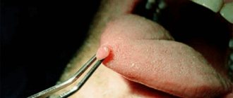
A large number of papillomas certainly causes inconvenience and does not look aesthetically pleasing. However, this is not a reason to use old-fashioned methods for removal with thread, pliers or a blowtorch.
For single small (1x1 mm) formations, celandine juice
- a proven folk remedy.
When there are many papillomas or they are large, this method is not suitable
, as it can last for months and lead to severe inflammation.
Removal using radio wave surgery is much more convenient and effective
The operation does not require special preparation, is practically painless, and after it you can drive a car.
Once I had the opportunity to remove about 70 papillomas from one patient at once. It is difficult to convey all the joy of a person who got rid of them, whom they tormented for several years.
Treatment of oral papillomas
Diagnosis of this disease includes a collection of the patient’s medical history, as well as a thorough histological examination of removed papillomas.
Treatment of papillomas is only surgical. The neoplasm is excised down to the borders of healthy tissue. Techniques such as electrocoagulation, cryosurgery, sclerotherapy and others are rarely used, since as a result of their implementation it is impossible to conduct a histological analysis of the papilloma removed to the base.
If a large accumulation of papillomatous neoplasms is detected, a combined technique is used: a scalpel is used to dissect the largest number of papillomas accumulated in one place, and single papillomas are removed using electrocoagulation.
If oral papillomas have a viral etiology, antiviral and immunomodulatory therapy is prescribed along with surgical intervention to prevent relapses.
Depending on the etiology of the disease, relapses may occur with greater or lesser probability. So, if there is a human papillomavirus in the body, the risk of papillomas returning after surgery is quite high.
Papillomas in the throat - treatment of laryngeal papillomatosis
Let's take a closer look at how complete treatment of laryngeal papillomatosis in children and adults occurs, its full cycle, in the Yesong center, in Korea.
Data from the study:
1 OPERATION
“The intratrochlear excision method is the surgical removal of masses of papillomas with the capture of a 2 mm layer of the mucous membrane, including submucosal glands, using minimal cold instruments for children and some adults, with complete removal of the papilloma, especially if the papilloma has re-formed at the site of the scar from a previous operation and CO2 laser applications. Papillomas are excised to a healthy mucous membrane, all infected parts of the mucous membrane are removed.
IMPORTANT: Since papillomas cells can grow deep into the tissue, destroying the membrane barrier, complete removal of the submucosal gland in the affected area is necessary to reduce the risk of relapse.
Next, the surface of the larynx is treated with a 585 nm pulsed color dye laser ( PDL laser) or KTP laser to prevent scarring and stenosis formation. In the study, the use of cold instrumentation followed by treatment with a PDL laser instead of a CO2 laser resulted in a significant reduction in laryngeal scarring. Minimizing the formation of scars and scarring of the vocal folds after surgery is the basis for effective voice restoration in the subsequent recovery period.
After laser treatment, Cidofovir into the affected area to prevent future relapses.
The operation is completed with the “microflap” technique - suturing the vocal cords in order to minimize the adhesive process and possible stenosis of the larynx.”
Laryngoscope 126: June 2016 Kim and Baizhumanova: Recurrent Respiratory Papillomatosis and Its Treatments. Page 1360-1361
2 OPERATION
On the 4th day after the first operation, a second throat operation is performed . After the first operation, fibrin is formed on the operated surface, in simple words - a film. If it is not removed, it becomes a denser tissue, and adhesions appear, which lead to laryngeal stenosis, that is, fusion. Fibrin is removed during a second microsurgery under general anesthesia. This procedure helps prevent the formation of adhesions and stenosis on the vocal cords.
3 OPERATION
Next, on days 8-9 after the first operation, the final operation is performed under general anesthesia to remove fibrin. During the third surgical intervention, additional tissue treatment is performed with drugs that prevent tissue fusion. Laryngeal microsurgery is repeated using the “microflap” technique: suturing the vocal cords to prevent vocal cord adhesions and stenosis.
REPEATING THE CYCLE
In a number of serious cases of recurrent laryngeal papillomatosis, it is necessary to resort to repeating the entire cycle. It must be remembered that this disease is a manifestation of a virus that is in the human body. When the immune system is weakened, even after a full cycle of treatment, the body may malfunction and a relapse occurs again. Therefore, it is important after treatment to take seriously the restoration of immunity and leading a healthy lifestyle.
Contact the clinic
Kinds
| Type of neoplasm | Description of symptoms |
| epithelial hyperplasia | The virus is present in every person in a latent state; under certain conditions, the epithelium begins to divide and papillomas appear. There is little pain and removal is quite simple. |
| simple papillomas | Small growths that do not cause pain. Formed singly or in groups. |
| vulgar papillomas | They are similar to simple ones, with the difference that their formations are always group. |
| flat | They have practically no bulges, their color is darker. |
| threadlike | They are dangerous because they have a leg. |
| Pointed papillomas | They spread very quickly, scars after their removal are at risk of oncogenic risk. |
| fibropapillomas | A mushroom-shaped growth similar in color to the mucous membrane, easily injured. |
| Condylomas | Growths with a soft and thin stalk that are easily damaged. |
Diagnosis of HPV
The main clinical sign of papillomatous infection is formation on the skin. Therefore, in most cases, an examination by a doctor is sufficient to make a correct diagnosis. Additional examination methods are:
- biopsy or scraping - taking biological material for further research;
- PCR (polymerase chain reaction);
- General and biochemical blood test.
These laboratory diagnostic methods help:
- detect the presence of papilloma virus in the child’s body (if there are no growths on the skin);
- accurately determine the HPV strain;
- understand the form of the infection (acute or chronic).
These data are necessary for assessing oncogenic risk and timely further examination.
Is human papillomavirus (HPV) a sexually transmitted disease?
HPV can be deciphered as a virus that causes a benign tumor of epithelial origin in the form of a papilla. Papilloma is classified as a sexually transmitted disease (STD). Unfortunately, today this concept has expanded significantly. If previously only a few, as it seemed then, “frightening” classical diseases of Venus were known, which in fact are easily treated and detected quickly (gonorrhea, syphilis, trichomoniasis, chancroid), today the list of STDs includes many dangerous, severe detectable, and sometimes difficult to treat and completely incurable (HIV) diseases. Moreover, their number is constantly growing.
Today, the list of “new” STDs includes mycoplasmosis, chlamydia, gardnerellosis, ureaplasmosis, candidiasis, genital herpes and human papillomavirus. Growths from papillomavirus identified in the genital or perianal areas are called genital warts. In fact, both condyloma and papillomavirus are the same virus, just different strains (varieties).
Genital condylomas in the perineal area can appear either one at a time or as whole growths that look similar to cauliflower. In some cases, such formations cause itching, irritation when touched, and bleeding.
Laryngeal papillomatosis
You can also distinguish papillomatosis of the respiratory tract. With this disease, the tissue that lines the nasopharynx grows from the nose to the lungs. The larynx is also often affected. Respiratory tract papillomatosis is also a type of disease caused by the papilloma virus. In this case, formations are classified as benign. Local and ENT doctors cannot always identify this disease. More often they simply shrug their shoulders and prescribe rinses. In this regard, we recommend that you contact a private clinic.