04.05.2018
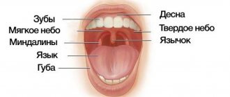
The oral cavity connects the body with the environment and can therefore be subject to a variety of negative influences. Such influences include the influence of too hot or too rough food, the influence of pathogenic microorganisms. In such cases, the first protective barrier is the mucous membrane, which lines the entire oral cavity and has, in addition to protective ones, many other functions. The mucous membrane is well supplied with blood, and its cells divide quickly and frequently. Therefore, wounds on the mucous membrane heal faster than on other parts of the body. However, for a number of reasons, the mucous membrane can be subject to inflammatory processes called stomatitis. Stomatitis is considered separately from such inflammatory processes in the oral cavity as gingivitis (inflammation of the gums; they have their own characteristics) and periodontitis (inflammation of the periodontium). The causes of stomatitis are very diverse.
Infectious causes
Stomatitis can be caused by infection with viruses or bacteria. In these cases, stomatitis is usually considered as an independent disease.
This is especially true for infection with herpes simplex viruses, which leads to very painful inflammation of the mucous membrane. There are two types of herpetic stomatitis - acute herpetic stomatitis (with primary infection) and recurrent herpetic stomatitis (which can pass through the acute stage or manifest itself as a general infection of the body in the presence of provoking factors). Herpetic stomatitis can lead to an aphthous form, in which erosions, aphthae, and even painful ulcers appear on the mucous membrane.
A specific type of stomatitis is chronic recurrent aphthous stomatitis, which begins almost immediately with the formation of aphthae, and the nature of which is not yet clear. But viral and bacterial infections and vitamin imbalances are also suggested as initiating factors.
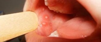
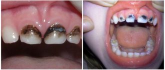
The specific infection that causes stomatitis includes a fungal infection (fungi of the genus Candida) and the disease itself is called candidiasis (thrush). Thrush is characterized by the formation of whitish dots on the mucous membrane, and as the fungal infection multiplies, a whitish coating. Candidiasis develops, as a rule, as a result of violations of hygiene rules, physiological disorders in the body, and the use of certain medications. It is often observed in children and its danger cannot be underestimated. This infection, which initially appears on the oral mucosa, can be spread to all organs and systems of the body, and in this case already poses a significant threat to health.
Purely bacterial causes of stomatitis are less common. As a rule, bacterial infections affecting the oral mucosa accompany viral types of stomatitis (for example, streptostaphylococcal lesions).
3. Symptoms and diagnosis
Iron deficiency anemia is characterized by dullness of taste, pallor of the mucous membrane, painful reaction of the mucous membrane to sour or spicy foods, various false sensations (tingling, tingling, etc.), swelling and, less often, bleeding of the tongue, as well as inflammation along the red border of the lips (cheilitis) and multiple caries.
With hypoplastic anemia, hemorrhagic foci, swelling and cyanosis of the interdental papillae, and a tendency to expose the periodontal pockets are observed; in more severe cases, erosions or ulcers and areas of local necrosis appear on the oral mucosa.
B12-folate deficiency Addison-Biermer anemia is characterized by a yellowish-pale tint of the mucous membrane, glossitis (inflammation of the tongue), and in some cases, pinpoint hemorrhages on the mucous membrane.
As for leukemia, their existing classifications (based on etiological, cytogenetic and other criteria) include several dozen clinical forms, the analysis of which is beyond the scope of this material. Even the traditional division in medicine according to the type of course - into acute and chronic leukemia - in this case takes on a slightly different meaning: in fact, these are two different diseases, and they never transform into each other.
If we do not touch upon the main symptoms and limit ourselves only to changes in the oral mucosa (as in the case of anemia, see above), then acute leukemia is manifested by severe bleeding of the mucous membrane on the gums, cheeks (along the line of dental occlusion), on the upper palate, and tongue. Loose hyperplastic growth of the gums in the form of ridges that bleed at the slightest touch or spontaneously is often observed. In addition, pale necrotic areas surrounded by a bluish halo with an off-white or gray foul-smelling coating are often observed. Swallowing is difficult, eating causes pain in areas of necrotic ulceration; trophic disorders are often accompanied by activation of chronic infections (herpes, candidiasis).
Chronic leukemia, which can be latent for a long time, is also characterized by bleeding of the oral mucosa, but it is less pronounced than in acute leukemia, and hemorrhages occur mainly due to trauma or microtraumatization. A diagnostically informative sign is the combination of bleeding, cyanosis, and pallor of the mucous membranes in the absence of signs of inflammation.
In myeloid leukemia, erosive and ulcerative lesions of the oral mucosa often predominate; in chronic lymphocytic leukemia, infiltrating nodes with a loose consistency and bluish tint predominate.
If, during an examination by a dentist, orthodontist, otorhinolaryngologist, therapist or other specialist, there is a suspicion that specific changes in the oral mucosa may be caused by diseases of the hematopoietic system, a consultation with a hematologist is strictly necessary, followed by an in-depth laboratory blood test , the results of which are the most informative and decisive in making a diagnosis. As necessary, other laboratory and instrumental studies are prescribed (imaging diagnostic methods, bone marrow puncture followed by histological analysis, etc.).
About our clinic Chistye Prudy metro station Medintercom page!
Other causes of stomatitis
In addition to infections, there is a whole range of other causes that can cause inflammation of the oral mucosa: • allergic reactions of a local and general nature (for example, to certain foods, medications, filling materials, oral care products) • the presence of soft and hard dental plaque, multiple caries • incorrectly installed dentures or orthodontic devices (braces) • irritation and trauma to the mucous membrane (mechanical injuries, burns) • deficiency conditions in the body (vitamins A, B and C, iron, folic acid); • intoxication with nicotine, alcohol, metal compounds.
Stomatitis can be manifestations and side effects of common infectious diseases (measles, diphtheria) or internal and general diseases and pathologies (blood and skin diseases, hormonal fluctuations, metabolic disorders).
Inflammation of the oral mucosa occurs especially often when the body's immune system is weakened and due to insufficient oral hygiene. The danger of stomatitis may also be associated with increased and constant dryness of the mouth, in which the protective functions of the mucous membrane are sharply weakened. The salivary glands produce on average up to 1.5 liters of fluid per day, and its deficiency leads to an imbalance in the acidity of the environment, an imbalance of inorganic components and, ultimately, has a depressing effect on the immune defense of the mucous membrane.
Traumatic lesions of the oral mucosa
- Chronic mechanical injury
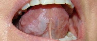
Clinical manifestations of a traumatic ulcer depend on the strength of the damaging factor, the general reactivity of the body, the state of the microbiocenosis of the oral cavity and other factors. As a rule, a traumatic ulcer is single. The mucous membrane around the ulcer is hyperemic and swollen. A traumatic ulcer is always painful, has uneven edges, and its bottom is covered with a fibrinous coating that can be easily removed. With the long-term existence of an ulcer, its edges and base become denser (due to the predominance of the phenomenon of proliferation). The edges of the ulcer are hyperemic, painful on palpation, the bottom is often bumpy, covered with necrotic plaque. The depth of the ulcer varies down to the muscle layer. Regional lymph nodes are enlarged, mobile, and painful on palpation.
Traumatic ulcers can be complicated by fusospirochetosis or candidiasis, and with a long course (2-3 months) they can become malignant.
- A traumatic ulcer must be differentiated from:
- ulceration of a cancerous tumor,
- tuberculous ulcer,
- syphilitic chancre,
- chronic ulcerative necrotic gingivostomatitis Vincent,
- trophic ulcer.
Removal of the traumatic factor also serves differential diagnostic purposes. Rapid healing of the ulcer within a few days indicates its traumatic origin.
The bone tissue of the alveolar process is resorbed in places, the alveolar edge becomes soft, mobile (“dangling”). Angular cheilitis often develops simultaneously. In the occurrence of such conditions, in addition to chronic trauma, a certain role is played by the influence of fungi of the genus Candida, which, as a rule, are found in large quantities on prostheses, and less on the mucous membrane of the prosthetic bed. You should also remember about the possibility of an allergic reaction of the oral mucosa to the base material of the prosthesis (usually acrylic plastic).
- Chemical damage
In case of acute injury, sharp pain usually occurs immediately and is localized at the site of contact with the chemical. The clinical picture depends on the nature and amount of the damaging substance and the time of action.
Burns with acids lead to the appearance of coagulative necrosis on the mucous membrane - a dense film of brown (from sulfuric acid), yellow (from nitric acid) or white-gray (from other acids) color. The films are tightly fused to the underlying tissues and are located against a background of pronounced inflammation of the mucous membrane with swelling and hyperemia.
A burn with bases (alkalies) causes liquefaction necrosis of the mucous membrane, while a dense film is not formed, necrotic tissue has a jelly-like consistency. The damage is deeper than with an acid burn. Necrosis from alkali burns can involve all layers of soft tissue, especially on the gums and hard palate. Burns, especially extensive ones, cause severe suffering to the patient. A few days after the necrotic tissue is sloughed off, slowly healing erosive or ulcerative surfaces are exposed. In mild cases, alkali burns can only cause a catarrhal inflammatory process.
Symptoms of “galvanism” should be differentiated from stomalgia, glossalgia, and allergic stomatitis.
Anamnesis (absence of symptoms of “galvanism” before prosthetics), as well as measurement of the magnitude of microcurrents in the oral cavity, are important in the differential diagnosis.
- Radiation sickness
Radiation sickness (morbus radialis) develops as a result of exposure to ionizing radiation on the entire body or its large parts: chest, abdomen, pelvic area.
Radiation damage can be caused by any type of ionizing radiation: X-rays, beams, neutron fluxes, etc. In irradiated tissues, the morphological structure of the walls of blood vessels changes, the barrier function of connective tissue decreases, and regeneration is suppressed. There are acute and chronic forms of radiation sickness.
Acute radiation sickness (morbus gadialis acutus) develops after a single irradiation of the body with massive doses (1 - 10 Gy, or 100 - 1000 rad). In the first period - the period of primary reactions, which begins soon after irradiation (1-2 hours) and lasts up to 2 days, dryness (or salivation) appears in the oral cavity, taste and sensitivity of the mucous membrane decrease. The mucous membrane of the mouth and lips swells, hyperemia appears, and pinpoint hemorrhages may occur. In the second, latent period, which lasts from several hours to 2 weeks, all these phenomena disappear. The period of pronounced clinical phenomena (third period) is the height of the disease. Against the background of a sharp deterioration in the general condition, changes in the oral cavity reach a maximum. A burning sensation appears, the mucous membrane becomes anemic and dry. A picture of radiation stomatitis appears.
Changes in blood vessels and blood composition during this period cause the phenomena of hemorrhagic diathesis: increased bleeding, multiple petechial hemorrhages. Due to a sharp decrease in tissue resistance, a rapidly developing autoinfection, especially putrefactive one, occurs.
Thus, the clinical picture of radiation stomatitis consists mainly of hemorrhagic syndrome and necrotic ulcerative process. The latter is most pronounced in places of injury with overhanging fillings, sharp edges of teeth, dentures, tartar, and where there were accumulations of microflora before the disease, i.e. First, the edge of the gums and tonsils are affected, and then the lateral surfaces of the tongue and the palate. In areas adjacent to the mucous membrane of metal prostheses and fillings, the damage may be more pronounced due to secondary radiation. The mucous membrane of the mouth, lips and face swell. The gingival papillae loosen, bleed, and then the gum edge becomes necrotic. The bone tissue of the alveolar process is resorbed, the teeth become loose and fall out. Multiple necrosis of the mucous membrane does not have sharp boundaries, the inflammatory reaction of surrounding tissues is mild. The bottom of the ulcers is covered with a dirty gray necrotic coating with a putrefactive odor. In severe cases, necrosis can spread from the mucous membrane to the underlying soft tissue and bone, radiation necrosis of the bone occurs with sequestration, and jaw fractures are possible. This is facilitated by the spread of infection from foci of chronic periodontitis and periodontitis.
The tongue swells, becomes covered with a thick coating, cracks, hemorrhages and necrosis appear, most often in the area of the root of the tongue; taste and sensitivity disappear. Severe necrotizing tonsillitis develops. If the patient does not die, then the fourth period begins - recovery: a slow reverse development of the symptoms of the disease occurs. In the oral cavity, everything also returns to relative normal. During this period, relapses of the disease are possible.
Chronic radiation sickness (morbus radialis chronicum) develops as a result of prolonged exposure to low doses of radiation on the entire body or a significant part of it. The oral cavity is sensitive to the effects of ionizing radiation, so at the onset of the disease, changes in the oral cavity can be especially pronounced. Dryness gradually increases due to damage to the salivary glands. Persistent catarrhal gingivitis occurs, which can subsequently develop into ulcerative gingivitis. Initially, erosions and ulcers may appear along the transitional folds on the vestibular side, followed by damage to the gums and red border of the lips.
Glossalgia may develop, and then glossitis with swelling of the tongue, cracks, and coating on the tongue. The long course of chronic radiation sickness usually leads to a picture similar to periodontitis - the so-called radiation periodontitis.
The reaction of the mucous membrane to radiation develops gradually - from hyperemia and swelling to the appearance of erosion. This reaction has its own characteristics in different areas of the mucous membrane. The first clinical signs on the mucous membrane, which does not have a keratinized layer in the epithelium (cheeks, floor of the mouth, soft palate), are manifested by mild hyperemia and swelling, which increase as the absorbed dose increases. Then the mucous membrane becomes cloudy, loses its shine, thickens, wrinkles appear, and when scraped, the surface layer is not removed. This occurs due to increased keratinization of the epithelium. Some affected areas resemble leukoplakia or lichen planus. If the radiation dose increases, the keratinized epithelium in some areas is rejected, desquamated, erosions appear, covered with a sticky necrotic plaque - focal membranous radiomucositis develops. Then the epithelium is rejected over large areas, the erosions merge and focal radiomucositis transforms into confluent membranous radiomucositis. The mucous membrane of the soft palate is highly radiosensitive, there is no keratinization stage when it is irradiated, and the reaction develops faster than in other parts of the oral cavity. In areas of the mucous membrane that are normally subject to keratinization, the radiation reaction proceeds more favorably and leads only to focal desquamation of the epithelium or single erosions.
The course of pathological processes in the oral mucosa is complicated by damage to the salivary glands (with remote irradiation methods). At the beginning (the first 3-5 days), increased salivation may be observed, which is quickly replaced by dry mouth up to complete xerostomia, which is practically impossible to stimulate.
The consequence of the death of the taste buds of the tongue is a violation of taste. At first, sensations in the tongue may manifest themselves as glossalgia, then a perversion of taste is noted, and later - loss of it.
Radiation changes in the oral cavity are largely reversible. After cessation of irradiation or during a break in treatment, the mucous membrane quickly returns to relative normal. This period lasts 2-3 weeks. With a large absorbed dose (more than 50-60 Gy, or 5000-6000 rad), irreversible changes in the salivary glands and mucous membrane (edema, hyperemia, telangiectasia, atrophy, radiation ulcers) may occur.
Radiation therapy should be preceded by sanitation of the oral cavity, since the reaction of the mucous membrane to radiation exposure is more severe in an unsanitized oral cavity.
Due to trophic disorders, the reactivity of the mucous membrane to mechanical trauma and infection is sharply reduced. The edematous mucous membrane with defective epithelial cover is easily injured by the sharp edges of teeth and dentures, which can lead to the appearance of sharply painful, long-term non-healing ulcers.
- Leukoplakia
The initial manifestations of leukoplakia are usually invisible, since there are no subjective sensations. Leukoplakia begins with the appearance of areas of clouding of the epithelium (grayish in color, with elements of keratinization on the surface). The lesion occurs against the background of an unchanged mucous membrane. Typical localization of the focus of leukoplakia is the mucous membrane of the cheeks along the line of closure of the teeth in the anterior section, the corners of the mouth and the red border of the lower lip without skin lesions. The dorsum and lateral surfaces of the tongue are somewhat less commonly affected. Smokers are characterized by lesions of the palate, described as “Tappeiner smokers' leukoplakia.”
There are the following forms of leukoplakia: flat or simple, verrucous and erosive. These forms can transform one into another. It is possible to combine different forms of leukoplakia in different areas of the oral mucosa in the same patient. The disease begins with the appearance of a flat shape.
Flat, or simple, leukoplakia (leukoplakia plana) is the most common. This form usually does not cause subjective sensations and is discovered by chance. Sometimes patients have complaints of a feeling of tightness, burning, and an unusual appearance of the mucous membrane. In the presence of extensive lesions on the tongue, taste sensitivity may be reduced. The main morphological element of the lesion in flat leukoplakia is a hyperkeratotic spot, which is an area of turbidity of the epithelium with clear contours. During the examination, foci of hyperkeratosis of various shapes and sizes are revealed, not rising above the level of the mucous membrane, but with clear boundaries of the lesion. Elements of flat leukoplakia resemble a burn of the mucous membrane with lapis, thin tissue paper pasted on, or a white coating that does not come off even with intense scraping. The keratinization can vary in intensity, so the color of the affected areas varies from pale grayish to intensely white.
The surface of the area of flat leukoplakia is usually slightly rough and dry. There is no compaction at the base of the lesion, just as there is no visible inflammatory reaction along its periphery.
There is a direct connection between the shape, color, size of keratinized areas and their location. Thus, with hyperkeratosis of the mucous membrane in the area of the corners of the mouth, symmetry of the lesion is observed in 85% of cases. The shape of the lesion has the shape of a triangle, the base of which faces the corner of the mouth, and the apex faces the retromolar space.
When the focus of leukoplakia is localized on the mucous membrane of the cheeks along the line of closure of the teeth, the elements of the lesion represent an elongated line, the continuity of which may be disrupted in certain areas. Leukoplakia on the red border of the lips looks like sticky tissue paper of irregular shape and grayish-white color (Fig. 11.5). Limited areas of lesions form on the mucous membrane of the tongue, hard palate, and floor of the mouth, sometimes resembling wide stripes or solid spots. More extensive lesions are also noted.
The relief of the mucous membrane and its turgor are also reflected in the appearance of the lesion. So, if leukoplakia develops against the background of folding of the tongue, the protruding areas become more keratinized and the surface of the tongue resembles a cobblestone street. A similar picture can be seen with a decrease in turgor and folding on the cheeks. If there is no pronounced folding on the tongue, flat leukoplakia looks like grayish-white, slightly sunken spots, the papillae of the tongue on them are smoothed.
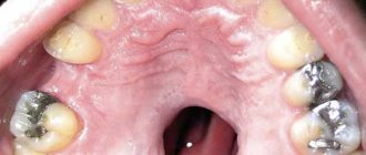
Tappeiner's leukoplakia (leucoplakia nicotinica Tappeiner, nicotinic leukokeratosis of the palate) occurs in heavy smokers (especially pipe smokers). The mucous membrane of the hard palate and the adjacent part of the soft palate is mainly affected. Sometimes the gum margin is involved. The mucous membrane in the affected area is grayish-white in color, often folded. Against this background, mainly in the posterior half of the hard palate, red dots stand out - the gaping mouths of the excretory ducts of cystically dilated salivary glands, looking like small nodules. They are formed due to blockage of the excretory ducts by hyperkeratotic masses. Damage to the palate in smoker's leukoplakia can be combined with the location of elements on the mucous membrane of the cheeks, corners of the mouth, lower lip, etc. This form of leukoplakia is an easily reversible process: stopping smoking (an irritating factor) leads to the disappearance of the disease.
Verrucous leukoplakia (leucoplakia verrucosa) is the next stage in the development of flat leukoplakia. This is facilitated by local irritants: trauma with sharp edges of teeth and dentures, biting areas of leukoplakia, smoking, eating hot and spicy foods, microcurrents, etc.
The main feature that distinguishes this form of leukoplakia from flat one is more pronounced keratinization, in which a significant thickening of the stratum corneum is detected. The area of leukoplakia rises significantly above the level of the mucous membrane and differs sharply in color. Upon palpation, a superficial compaction may be detected. Patients usually complain of a feeling of roughness and tightness of the mucous membrane, burning and pain when eating, especially spicy food.
There are plaque and warty forms of verrucous leukoplakia. In the plaque form, limited milky white, sometimes straw-yellowish plaques with clear contours are identified, rising above the surrounding mucous membrane. The warty variety of verrucous leukoplakia is characterized by dense grayish-white bumpy or warty formations, rising 2-3 mm above the level of the mucous membrane. The warty form of leukoplakia has a greater potential for malignancy compared to the plaque form. On palpation, the lesions are dense, painless, and not fused with the underlying areas of the mucous membrane. The thickness of the raised areas of hyperkeratosis varies from pronounced to barely noticeable during normal examination. With a slight elevation of the elements, the affected area is almost not determined by palpation. A leukoplakic lesion of considerable thickness becomes dense to the touch, and it is not possible to fold it.
Erosive leukoplakia (leukoplakia erosiva) is actually a complication of simple or verrucous leukoplakia due to trauma. With this form of leukoplakia, patients complain of pain, which intensifies under the influence of all types of stimuli (eating, talking, etc.). Usually, against the background of foci of simple or verrucous leukoplakia, erosions, cracks, and, less often, ulcers occur. Erosions are difficult to epithelialize and often recur. Patients are especially concerned about erosion in the keratinized area of the red border of the lips. Under the influence of insolation and other irritating factors, they increase in size without showing a tendency to heal. The pain intensifies.
Pathohistologically, thickening of the epithelium is revealed due to the proliferation of the horny and granular layers. The stratum corneum reaches considerable thickness, especially with the verrucous form of leukoplakia. In it, foci of hyperkeratosis often alternate with foci of parakeratosis. The granular layer of epithelium in the affected area has varying degrees of severity. Verrucous and erosive forms of leukoplakia are characterized by pronounced hyperkeratosis and acanthosis. Acanthosis in leukoplakia can be significant if the keratinization has the character of parakeratosis; conversely, with severe hyperkeratosis, acanthosis is minimal or completely absent. In the connective tissue stroma of the affected areas of the mucous membrane, diffuse chronic inflammation is determined with pronounced infiltration of the surface layers by lymphocytes and plasma cells, the manifestation of sclerosis. The latter explains poor healing and frequent relapses with the appearance of cracks and erosions, which is most typical for the plaque form of leukoplakia.
Leukoplakia is a precancerous condition, since all its forms can become malignant with varying degrees of probability, transforming into spinocellular cancer.
Flat leukoplakia malignizes in 3-5% of patients, and in some the process of malignancy occurs quickly (1-1.5 years), in others the disease can exist for decades without transforming into cancer. Most often, malignancy occurs in verrucous and erosive forms of leukoplakia (in 20% of cases).
Clinical signs of malignancy are increased keratinization processes; rapid increase in the size and density of the lesion; the appearance of compaction at the base of the plaque, erosion; papillary growths on the surface of erosions, bleeding due to injury; the appearance of non-healing cracks.
- Soft leukoplakia Pashkova
Characterized by the presence of areas of peeling, slightly swollen, pasty mucous membrane with unclear boundaries, without inflammation or compaction. The mucous membrane has a grayish-white color. Most often, soft leukoplakia is localized on the mucous membrane of the cheeks along the line of closure of the teeth and lips, and less often on the tongue and gums. The lesion can be limited or diffuse, when almost the entire mucous membrane, and sometimes the red border of the lips, is involved in the process. In patients with neuropathy, predominantly young, with habitual biting or sucking of the cheeks, lips, tongue, the epithelium along the line of closure of the teeth is unevenly desquamated, has a fringed appearance due to the presence of multiple small flaps, which causes the unevenness of the affected surface (it looks like it is moth-eaten). This altered epithelium is partially removed by scraping. In severe cases, painful erosions may occur from biting the epithelium.
Histologically, pronounced parakeratosis and acanthosis are noted. At all levels of the spinous layer there are so-called “light” vacuolated cells, the cytoplasm of which is almost not stained, and the nuclei are deformed (pyknotized). In the connective tissue layer, expansion of small blood vessels, thickening of collagen, and thinning of elastic fibers are detected.
- White spongy nevus of Cannon
White spongy nevus may appear in early childhood or later, then progresses and reaches its maximum development during puberty. The disease may then regress or remain unchanged.
There are no subjective sensations. Patients may complain of an unusual appearance of the mucous membrane.
The typical location of a white spongy nevus is the buccal mucosa. The lesion is always symmetrical. The mucous membrane of the cheeks is white or grayish-white, somewhat thickened, soft, as if spongy, strongly folded. In some cases, the folding and wrinkling of the mucous membrane is so pronounced that the folds hang into the oral cavity. The surface layers of the epithelium are removed by scraping. At the same time, similar lesions may appear on the mucous membrane of the genital organs and rectum.
White spongy nevus should be differentiated from:
- flat leukoplakia;
- candidiasis;
- lichen planus;
- soft leukoplakia.
There is great clinical similarity between white spongy nevus and soft leukoplakia. Some authors consider white spongy nevus and soft leukoplakia to be different clinical forms of the same disease related to nevi, but there are quite pronounced differences between them. White spongy nevus, unlike soft leukoplakia, occurs mainly in childhood and is often hereditary. The development of mild leukoplakia, as a rule, is associated with various kinds of neurotic reactions or with hormonal changes in the body (puberty, postmenopause).
Patients with Cannon's nevus have congenital defects in the structure and maturation of the epithelium of the oral cavity, and in some cases, the genitals and rectum. With mild leukoplakia, the oral mucosa initially has a normal appearance, and its pathological changes are associated with the action of irritating factors against the background of neurological disorders or hormonal dysfunctions. In addition, damage to the oral mucosa with white spongy nevus is usually generalized.
Main symptoms of stomatitis
Symptoms of stomatitis depend on its type and form, are very diverse and specific. General symptoms include redness, swelling of the mucous membrane, burning and pain in the mouth (including when eating hot, sour or spicy foods), an increase (or, conversely, a decrease) in salivation, bad breath, as well as the manifestation, in depending on the severity of the disease, specific lesions - erosions, af, ulcers.
Therapy for diseases of the oral mucosa
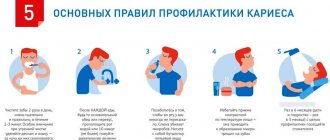
Prevention measures:
- Visiting the dentist twice a year to promptly identify and eliminate dental problems;
- Careful observance of local hygiene;
- To give up smoking;
- Avoiding too hot and spicy foods;
- Rinsing the mouth with disinfectant solutions and herbal decoctions;
Oral hygiene should be carried out regularly. Failure to comply with basic rules can provoke the occurrence of sore throat, problems with the gastrointestinal tract and other pathologies.
The health of the oral mucosa largely depends on the condition of a person’s teeth.
To successfully cure any disease of the oral mucosa, it is necessary to suppress the factors that influenced its development. If you suspect any problems in the oral cavity, you should contact a specialist. It is categorically unacceptable to get too carried away with self-medication and resort to non-standard methods of treatment. If you do not see a doctor for a long time, a minor problem can develop into a more serious one, lead to complications and worsen a person’s overall health. In addition, some oral diseases can develop into malignant ones. You should not put off visiting a specialist and expect the disease to go away on its own. Timely diagnosis and treatment will help avoid many problems in the future.
Treatment
The primary diagnosis of the disease based on clinical manifestations is carried out by a dentist, who also carries out local symptomatic treatment based on the primary diagnosis. But in complex and unobvious cases, laboratory diagnostics are carried out, and doctors of other specialties (allergists, dermatologists, immunologists) are brought in for treatment.
Local symptomatic treatment usually involves the use of rinses with solutions of painkillers and antiseptic drugs (including herbal origin), the use of drugs for tissue regeneration (after the acute phase subsides), if necessary, the elimination of irritating and provoking factors (removal of dental plaque, hard deposits, treatment dental caries).
Local drug treatment is carried out only after an accurate diagnosis has been established (the cause of stomatitis) and usually includes treatment with antiviral, antibacterial, antifungal drugs in the form of ointments and applications.
General treatment is carried out, as a rule, in a hospital and is aimed at eliminating the internal causes of stomatitis (treatment of internal pathologies), taking therapeutic drugs according to special regimens, and physiotherapy.
Prevention
In general, it is quite difficult to avoid this disease in the presence of predisposing causes; you can only take some general measures to possibly prevent the disease or complication in the form of stomatitis.
Since the herpetic virus, especially in acute primary forms, is extremely contagious, a certain isolation of the patient is required to prevent the spread of infection by airborne droplets and contact. The difficulty here is that there is a rather long incubation period, up to 26 days, and the first signs of stomatitis may escape attention.
Other preventive measures are of a general nature and consist of proper and complete oral hygiene, regular professional cleanings in a dental clinic (removal of soft and hard plaque), timely treatment of mucosal injuries, and measures to generally strengthen the immune system.
Posted in Useful information | Tags: teeth, diabetes, care