Syphilis is one of the most dangerous sexually transmitted diseases. But not everyone knows that it can manifest itself not only on the genitals, but also on the skin and in the mouth. Syphilis, formed in the oral cavity, is a very common disease that is caused by infection of the body with Treponema pallidum.
The mucous membranes of the mouth are very vulnerable, and this pathogen easily affects them. Because of this, you can become infected with oral syphilis not only through sexual contact, but also, for example, through unsterile medical equipment or utensils.
Insidious disease
This pathology is very insidious and is not completely cured in all cases. Lack of treatment leads to the development of very serious complications and can even cause the death of the patient. This disease can be either acquired or congenital. In the latter case, the infection is transmitted from mother to unborn child through the placenta. And with acquired syphilis, infection is transmitted through the skin or mucous membranes, as well as through household means.
In addition to infection of the mucous membranes of the oral cavity, there are lesions of the lips, larynx, throat, cheeks, palate, tongue, gums and tonsils. The course of this disease is characterized as chronic.
Known since ancient times
This pathology has been known since ancient times; mentions of it can be found in the records of such ancient healers as Avicenna and Hippocrates. But, despite the study of the disease, it is impossible to completely cure it, even in our age of high technology.
There are many reasons why a person becomes infected: kissing, oral sex, contact with sick people, household items or medical instruments. You can get the disease even at a dentist’s appointment if the latter uses instruments that are not properly disinfected.
Let's look at how syphilis manifests itself on the lips.
Oral syphilis
At all stages of this disease, rashes appear, localized on the mucous membranes of the oral cavity. The cause of infection can be unsterile medical devices, and if there are wounds on the skin or mucous membranes, this will make it easier for the infection to enter the patient’s body. Even invisible abrasions, microcracks, etc. are enough for infection. Treponema pallidum can also enter the bloodstream as a result of surgery or injection. The pathogen is transmitted from mother to child in the womb. Doctors, especially gynecologists and dentists, are at risk for increased risk of infection.
Almost always, secondary syphilis manifests itself in the mouth area - both inside and on the lips. The infection affects the tonsils, mucous membranes, gums, tongue, etc. The disease has several successive stages, and each of them has its own symptoms.
Syphilis
The examination methods are chosen by the doctor taking into account the patient’s general health, the presence of complications and the stage of the disease. Which doctors should I contact?
Women are consulted by a gynecologist, men by a urologist or dermatovenerologist. Consultations with other specialists are recommended in the following cases:
- pediatrician, ophthalmologist, neurologist, otorhinolaryngologist - for children with suspected congenital syphilis;
- ophthalmologist and neurologist - for all patients with syphilis;
- if a specific lesion of internal organs, the musculoskeletal system, etc. is suspected, consult specialists in accordance with complaints or pathological changes detected during an instrumental examination.
Treatment of syphilis
Treatment of syphilis is successful in the early stages. First, therapy is carried out in a hospital, then on an outpatient basis. Complete recovery is confirmed by laboratory tests. Treatment is given to all sexual partners with whom the patient has had intimate contact within the last 90 days, and in the case of the secondary form - 12 months. If partners test negative for syphilis, they are prescribed preventive therapy.
Treponema pallidum is sensitive to antibacterial drugs of the penicillin group. The first loading dose technique is practiced - at the beginning of treatment, the medicine is administered intramuscularly in large volumes at short time intervals. A high concentration of the active substance is constantly maintained in the body. The drug of choice for the treatment of syphilis is benzylpenicillin. Ceftriaxone gives positive dynamics in the later stages of syphilis and can be used in the treatment of pregnant women. The antibiotic suppresses the reproduction of treponema. The course of injections is at least six months. In the case of secondary syphilis, before prescribing long-acting penicillins, broad-spectrum antibiotics (tetracyclines, macrolides) are prescribed for 2 weeks.
The dosage and duration of medication are calculated individually for each patient. Syphilis in latent form is treated comprehensively with the use of immunomodulators and physiotherapy. If there is a chancre with purulent discharge, it is recommended to supplement the treatment complex with local lotions, powders and ointments with an antibiotic.
Patients are warned about the body's possible reaction to treatment. Medical organizations where therapy is carried out must have emergency care facilities.
Complications
Intrauterine infection of the fetus from the mother is considered the most dangerous - in 40% of cases, pregnancy ends in the death of the child before birth or a few days after. The risk of miscarriage, development of congenital growth disorders of the skeleton, teeth, deafness, blindness, and severe brain damage increases many times over.
Complications of a neurological nature in patients with advanced forms of syphilis may include meningitis, urinary incontinence, stroke, a significant increase in the pain threshold and lack of temperature sensitivity, impotence in men, and deafness.
Syphilis affects the functioning of heart valves and changes the cells of functional tissues.
Often, patients with advanced infection are diagnosed with inflammation of the aorta and other blood vessels, and aneurysms are detected.
The greatest danger is represented by gummas - ulcers that form on the skin or internal organs and are not visible during examination. The formations can heal on their own or progress into weeping syphilis ulcers. In the places where gummas are located, tissues soften and rotting processes begin. Even the bones of the skull are affected. Gummas are extremely difficult to treat, so therapy is mainly aimed at maintaining the patient’s quality of life.
Deformation of bones and joints leads to external deformities - first of all, the cartilage of the nose is destroyed.
Serious side effects of treatment are also known:
- The exacerbation reaction (Jarisch-Herxheimer) begins 2-4 hours after the first administration of the antibacterial drug, reaches its maximum severity after 5-7 hours, and the condition returns to normal within 12-24 hours. The main clinical symptoms are chills and a sharp increase in body temperature, general malaise. Occasionally, patients may develop psychosis, stroke, seizures, and liver failure.
- A reaction to intramuscular administration of long-acting penicillin preparations (Hine's syndrome) can occur after any injection of the drug. Characterized by dizziness, tinnitus, fear of death, pallor, paresthesia, blurred vision, increased blood pressure, short-term loss of consciousness, hallucinations or convulsions may occur immediately after injection. Lasts within 20 minutes. Symptoms can vary in severity, from mild to severe.
- Nikolaou syndrome is a symptom complex of complications after intra-arterial administration of penicillin or other drugs with a crystalline structure. It is characterized by sudden ischemia at the injection site, the development of painful bluish uneven spots (livedo) followed by the formation of blisters and necrosis of the skin, in some cases flaccid paralysis of the limb into which the drug was injected develops, in rare cases - transverse paralysis. To date, cases have been reported only in pediatric practice.
- Neurotoxicity - seizures (more often in children) when using high doses of penicillin, especially in the presence of renal failure.
- Electrolyte imbalance - in patients with heart failure, when large doses of benzylpenicillin sodium salt are administered, edema may increase.
- Allergic reactions - toxicoderma, urticaria, Quincke's edema, headache, fever, joint pain, eosinophilia, etc. The most dangerous complication is anaphylactic shock, which has a mortality rate of up to 10%.
Prevention of syphilis Prevention of
syphilis includes sanitary education, screening examination of certain groups of the population, as well as full specific treatment followed by clinical and serological observation.
You can reduce the likelihood of infection if you use barrier contraception; If you have unprotected sexual intercourse with a casual partner, you should immediately carry out emergency anti-venereal prophylaxis (treating the skin and mucous membranes of the genital organs with special disinfectant solutions (chlorhexine, miramistin)).
Sources:
- Clinical recommendations. Syphilis. All-Russian public organization “Russian Society of Dermatovenerologists and Cosmetologists” (approved by the Ministry of Health of Russia), 2022. – 102 p.
- Sokolovsky E.V., Krasnoselskikh T.V., Rakhmatulina M.R. Federal clinical guidelines for the management of patients with syphilis. Russian Society of Dermatovenereologists and Cosmetologists, 2015. - 45 p.
IMPORTANT!
The information in this section cannot be used for self-diagnosis and self-treatment. In case of pain or other exacerbation of the disease, diagnostic tests should be prescribed only by the attending physician. To make a diagnosis and properly prescribe treatment, you should contact your doctor.
Primary stage
The incubation period is followed by the primary stage, characterized by the formation of chancre. A chancre is a painless ulcer that lasts about one and a half to two months. Hard chancre begins its formation with the formation of an area of hyperemia on the mucous membrane or skin. This redness gradually turns into thickening; this occurs due to the formation of an inflammatory infiltrate. It increases in size, reaching several centimeters. And in its center a bright scarlet erosion forms. These are the manifestations of syphilis on the lips. At the stage of completion of its development, the chancre looks like a dense erosive island rising above the mucous membrane. If there has been injury, or a secondary infection has occurred, then a painless ulcer with clear boundaries of a round shape is formed in its place.
On the lips, chancre can have a round shape or form in the form of cracks in the corners of the lips.
Syphilis of the nose
Nasal syphilis occurs during different periods of the disease.
Nasal involvement in early congenital syphilis
With early congenital syphilis, the nose is affected in 75% of cases. The disease manifests itself either immediately after birth or during the first 4 to 5 weeks of the child’s life. A dry runny nose, accompanied by noisy exhalation and inhalation, is quickly replaced by severe rhinitis. The child begins to sniffle, snort and sneeze, and refuses to breastfeed. The mucous membrane at the entrance to the nose becomes hyperemic, thickens and becomes crusty. A viscous secretion is released from the nasal passages. Due to the development of the ulcerative process, blood appears in the discharge over time. From an epidemiological perspective, children during this period become extremely dangerous to others.
Sometimes newborns develop gummous infiltration in the nose. The disintegration of gummas is accompanied by damage to the bone structures, as a result of which a perforation of the nasal septum develops, and various types of retraction of the external nose are formed.
Syphilitic rhinitis of newborns must be distinguished from gonorrhea and nasal diphtheria. Persistent runny nose, thick nasal discharge, crusting, skin rash and enlarged spleen are factors that indicate a specific process.
Rice. 5. Diffuse infiltration of Hochsinger's skin and syphilitic rhinitis of the newborn.
Nasal lesions in late congenital syphilis
Nasal lesions in late congenital syphilis develop 5 or more years after birth. Foreign literature describes cases of the development of pathology at 23–28 years of human life.
The secretion of a viscous secretion, the formation of crusts, a feeling of dryness in the nose and throat, loss of smell and the appearance of pain in the nose, forehead and eye sockets are the main manifestations of syphilitic congenital runny nose in late congenital syphilis. As a result of tubercular infiltration, the mucous membrane grows, gradually obstructing the nasal passage.
The process often occurs like tertiary syphilis. Gummas are located on the nasal mucosa, in cartilage and bone, while the mucous membrane is involved in the pathological process for the second time. The nasal passages become overgrown, the wings of the nose, its back, tip, turbinates, bottom and nasal septum are destroyed. Recessed nose is a pathogonic symptom of late congenital syphilis. The nose may have the shape of a saddle (“saddle nose”), or be similar to the nose of a bulldog or have a lorgnette-like appearance.
Syphilis of the nose in the primary period of the disease
Nasal involvement in the primary stage of the disease is rare. Hard chancre most often appears on the wings of the nose, at the entrance to its cavity on the skin in the area of the septum. The chancre has the appearance of red erosion with a roller-like thickening along the periphery, a greasy coating is detected at the bottom, and a dense infiltrate is palpated at the base.
Nasal syphilis in the secondary period of the disease
Nasal syphilis in the secondary period of the disease develops 6-7 weeks after the appearance of chancroid and manifests itself in the form of roseola (erythema) or papules. Erythema (redness) spreads to the mucous membrane, which swells and begins to produce a secretion of a mucous or bloody-serous nature. A little later, papules appear, which are located on the skin at the entrance to the nasal cavity. Due to constant irritation, papules erode and the healing process is delayed.
Syphilis of the nose in the tertiary period of the disease
The nose is most often affected in the tertiary period of the disease. The lesion affects the mucous membrane, periosteum, bone and cartilage, where gummas with decay or diffuse infiltrates develop. Destroyed areas of bone tissue are sequestered and come out with pus. The decay of gumma is accompanied by a putrid odor. The gumma itself acquires a bluish-red color and becomes covered with purulent-bloody crusts. Removing crusts often leads to bleeding.
The bony septum and floor of the nose are the most commonly affected areas. When they are destroyed, communication occurs with the oral cavity and nasal passages. The nasal passages narrow. The patient experiences severe pain in the nose, eye sockets and forehead area.
Gummas can form in cartilage and bone, while the mucous membrane is involved in the pathological process for the second time. The nasal passages become overgrown, the wings of the nose, its dorsum, tip, turbinates, bottom and nasal septum are destroyed, the nose sinks and takes on the shape of a saddle (“saddle nose”). The act of breathing, swallowing and phonation is disrupted.
Patients with a gummous process in the nose during the period of tertiary syphilis are practically not dangerous to others from an epidemiological perspective.
Gumma in the nose should be distinguished from tuberculosis and a malignant tumor. Large deformations of the external nose after treatment are corrected with rhinoplasty.
Rice. 6. Forming nasal gumma.
Rice. 7. Gummous lesion of the wings of the nose.
Rice. 8. Nasal syphilis in the tertiary period of the disease - perforation (destruction) of the nasal septum.
Rice. 9. Recession of the nose is a pathogonic symptom of late congenital and tertiary syphilis.
Rice. 10. Syphilis of the nose. Destruction of the structures of the external nose by the gummous process.
Rice. 11. Syphilis of the nose. Tertiary period of the disease. Destruction of all structures of the external nose, its septum, bottom, anterior part of the alveolar processes, teeth and upper lip.
Rice. 12. Complete destruction of all structures of the nose.
Rice. 13. With the disintegration of gumma and destruction of bone between the nasal septum, nasal cavity and oral cavity, perforations occur, which makes it difficult for the patient to eat, phonation and the act of swallowing.
Secondary stage
In the secondary stage of syphilis, the patient develops roseola. This occurs two months after infection and only if the patient has not received treatment. During this period, papules and roseola appear in the patient's mouth. From a medical point of view, this stage is the most dangerous. Roseolas look like bright red spots, clearly visible against the background of the mucous membrane. If treatment is not carried out, this stage will last about a month.
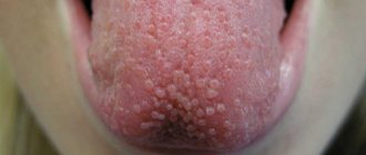
Papules are the most common symptom of the secondary stage of this disease. It can also occur due to relapse of the disease. These rashes are present over the entire surface of the oral mucosa.
Papules are round in shape, they are dense and of different sizes, they are covered with plaque, under which, when scraped off, red erosion is revealed. When papules appear, the papillae on the tongue disappear, and the skin becomes uncharacteristically smooth. Also, these formations can be concentrated locally, forming large foci. At the same time, in addition to erosions, jams may appear in the corners of the lips, which are accompanied by very painful sensations.
How to treat syphilis on the lips? We'll talk about this below.
Manifestations of syphilis during the secondary period of the disease
The rash on the face and scalp with secondary syphilis looks like roseola (spots), papules, vesicles and pustules. During the initial (fresh) rash, it is abundant, brightly colored, and often symmetrical. With secondary recurrent syphilis, the rash is not profuse. At the end of the secondary period, secondary syphilomas may be single.
Features of the rash on the face:
- high prevalence
- sudden appearance
- polymorphism (different types of rashes),
- clear boundaries
- lack of reaction of surrounding tissues,
- lack of subjective sensations,
- high infectiousness,
- benign course.
Rice. 3. Syphilis on the face - papular syphilide of the face.
Tertiary stage
At the tertiary stage, the patient develops so-called gumma and tuberculate rashes. Gummas spread throughout the oral cavity, but are more often localized on the lip, tongue or hard palate. They begin without pain, imperceptibly and gradually forming a deep knot in the mucous membrane. It increases in size and acquires a brown tint. The central part of this formation is destroyed at the stage of ulcer formation.
When gummas affect the lips and tongue, no pain symptoms are observed, and there are no acute inflammatory processes. And when the ulcer heals, a retracted scar appears in its place. This whole process takes place over several months.
Very often, tubercular syphilis appears on the lips. At the same time, these tubercles are dense, painless and quickly collapse, forming small but deep ulcers. This stage of the disease can last from several months to several years if the patient does not receive treatment. Once the ulcers heal, the scars remain forever.
Manifestations of syphilis in the tertiary period of the disease
Gummas and tubercular syphilide are the most common manifestations of tertiary syphilis. They appear on the skin of the face and scalp, mucous membranes of the oral cavity, pharynx and larynx, mucous membrane, cartilage and bone structures of the nose. The disease is characterized by a severe course.
In the case of untreated syphilis, gumma appears immediately after the secondary period. With insufficiently effective treatment, tertiary syphilis on the face appears after a hidden (latent) period, the duration of which is years and decades. Gummas compress and, when disintegrating, destroy organs in the places where they are located, disrupting their function and disfiguring a person’s appearance. The disease often leads the patient to disability and even death.
The rashes are often isolated and practically not contagious. Without treatment, they heal for a long time (within 4 - 6 months). In place of syphilides, retracted, star-shaped, disfiguring scars remain. Under the influence of specific treatment, gumma and tubercular syphilide heal quickly.
Rice. 4. The photo shows the complete destruction of nasal cartilage due to syphilis.
Manifestation of syphilis on the lips
The lips are one of the most typical localizations of syphiloma. As already mentioned, it all starts with a red, swollen spot, in the middle of which erosion gradually forms, and then an ulcer. The diameter of the latter can be from 0.5 to 2 cm. Its edges are smooth, bright red and rising above the level of the mucosa. The bottom of the ulcer is smooth, dense and red. Also, chancre can look like a long-lasting lesion that does not heal. Sometimes syphilis on the lips manifests itself as a thickening in the form of a mushroom cap, the size of which can reach two centimeters.
Syphilis on the face
The skin of the face is affected in all periods of syphilis.
Syphilis on the face during the primary period of the disease
Primary syphilomas (hard chancres) on the face look like ulcers and erosions. They are bright red in color, painless on palpation, with a smooth bottom, raised and compacted edge. In persons with reduced immunity, there is profuse purulent discharge and small hemorrhages at the fundus.
Rice. 21. Syphilis on the face - hard chancre in the chin area.
Rice. 22. Manifestations of syphilis on the face. Hard chancre on the upper eyelid.
Syphilis on the face during the secondary period of the disease
Papular syphilide of the face
In the secondary period of syphilis, papular syphilide appears on the face. Papules on the skin of the face are accumulations of cells in the upper dermis. They have a round, hemispherical or pointed shape, are located isolated and sharply demarcated from the surrounding tissues. Newly formed papules have a soft pink color, they are shiny, and over time they acquire a copper color or bluish-red color. Even without treatment, papules resolve after 1 - 2 months. In their place, brown pigmentation remains. In immunocompromised patients, papules often erode (erosive papules) or develop into ulcers (ulcerative papules).
Rice. 23. Manifestations of syphilis of the secondary period - papular syphilide of the face and scalp.
Rice. 24. In the photo, the lesion of the lower eyelid with secondary syphilis is papular syphilide.
Seborrheic papular syphilide
Seborrheic papules in secondary syphilis are located in areas of increased sebum secretion - most often on the forehead. The surface of the papules is strewn with fatty scales.
Rice. 25. Seborrheic papular syphilide on the skin of the forehead.
Rice. 26. Syphilis on the face - seborrheic papular syphilide on the skin of the forehead, face and head.
Acneiform (acne-like) syphilide
Acne syphilide is located at the mouths of the hair follicles. These are small round-shaped pustules with a conical apex, located on a dense base. When dried, a crust forms on their surface. After it falls off, a depressed scar remains. Acneiform syphilide is most often located on the neck, scalp and upper half of the body. Large numbers of rashes are recorded with fresh secondary syphilis; scanty rashes appear during subsequent relapses.
Rice. 27. Rash on the face due to syphilis - acne syphilide.
Pustular syphilide
Papulopustular syphilides more often appear in patients with weakened immunity and severe concomitant diseases. Pustular (pustular) syphilides can be acne-like, impetiginous, smallpox-like, manifest as syphilitic ecthyma and rupee. In patients with low immunity, pustular syphilide resembles severe pyoderma.
Rice. 28. Syphilis on the face - papulopustular syphilide.
Syphilide herpetiformis
Herpetiform (vesicular) syphilide is rare. It is usually recorded in patients with severe disease and reduced immunity.
Rice. 29. Vesicular syphilide of the face.
Impetiginous syphilide
Impetiginous syphilide of the secondary period manifests itself in the form of a dark red papule, the size of a pea or more. Next, the papule suppurates and becomes covered with a crust, from under which pus continues to ooze, and when it dries, a new crust appears. The layering can be large. After its rejection, the bottom of the syphilide is exposed. It is dark red in color, juicy, often with vegetative growths reminiscent of raspberries. When the ulcers merge, large areas of damage are formed (corrosive syphilide). Impetiginous syphilide of the scalp, nasolabial fold and beard growth area is similar to deep trichophytosis. On open areas of the face, the rash resembles impetiginous (superficial) pyoderma.
Rice. 30. In the photo there is syphilis on the face - impetiginous syphilide.
Syphilis on the face in the tertiary period of the disease
Syphilis on the face in the tertiary period of the disease manifests itself in the form of gumma or tubercular syphilide.
Gummas on the face
Gumma is a deeply located node, often single, located in the subcutaneous tissue and deep layers of the dermis. In the vascular endothelium, the accumulation of cells often leads to their overlap. Due to insufficient blood supply, tissue disintegration occurs in the center of the gumma. The resulting ulcer has steep edges and yellow-green purulent discharge. After its rejection, the bottom of the ulcer with grayish granulations is exposed. Healing of the ulcer occurs with a retracted disfiguring scar. When several gummas (mutilating gummas) are formed, healing with the formation of scar tissue leads to disfigurement of the patient’s appearance and deformities.
Rice. 31. Late syphilis. Disintegration of gum on the face. The formed ulcer has steep edges and a dense bottom with granulations. The discharge of gumma has a foul odor. In the peripheral sections there is a small number of treponemes, which quickly die when they decay.
Tuberous syphilide of the face
With tubercular syphilide, dense spherical tubercles appear on the face. Some of them are fading in their development, others are just emerging. On the face, tubercular syphilide is most often localized on the skin of the forehead and nose. The tubercles are hemispherical, copper-red in color, dense, prone to grouping, but never merge. When they decay, round ulcers form on their surface, with smooth edges, a smooth clean bottom and a dense base at the base. When ulcers heal, scars are formed, surrounded by a pigment border.
Rice. 32. Tubercular syphilide of the face.
Serping (creeping) syphilide
Creeping syphilide develops when the elements of the rash merge with its subsequent spread along the periphery. In the center of syphilide, cicatricial atrophy develops, and fresh rashes appear along the periphery. The edge of the syphilide is clear, scalloped. The disease lasts a long time - months and even years.
Rice. 33. Syphilis on the face - creeping syphilide in the forehead and neck.
Rice. 34. Manifestations of syphilis on the face. Gumma of the lower eyelid.
Rice. 35. Tertiary syphilis. Gummous infiltration of the face.
Rice. 36. Consequences of syphilis on the face.
What happens in later stages?
In the secondary stage of this disease, papules form in the mouth, affecting the pharynx, hard and soft palate, and larynx. The patient also experiences bouts of prolonged and dry cough. Damage to the larynx causes hoarseness and hoarseness.
At the tertiary stage, gummas on the lips look like purple bumps, which, after healing, turn into deep scars, often in the form of stars.
The problem is how to distinguish a cold sore from syphilis. Many people do not know how the latter manifests itself, confuse it with other diseases characteristic of the oral cavity, for example, herpes, and try to treat it themselves. Because this treatment is ineffective against Treponema pallidum, syphilis continues to develop.
What happens if syphilis is not treated?
If this disease is not treated, the consequences will be severe. The infection affects internal organs, most often the upper respiratory tract. The patient feels worse and worse. Necrosis begins at the site of syphiloma. Bones, internal organs and tissues are increasingly destroyed by Treponema pallidum. The vascular system is destroyed, facial and cervical tissues begin to rot, and the brain is also affected.
If all or some of the symptoms listed above appear, you should immediately consult a doctor. A specialist will be able to make an accurate diagnosis and prescribe the most effective treatment. The sooner it starts, the less complications there will be in the end. What tests should I take for syphilis on the lips?
Diagnostics
As a rule, the diagnosis is confirmed if the pathogen is detected on syphiloma or in the lymph nodes closest to it. Serological reactions will give a positive result for the presence of treponema, but only a few weeks after the onset of chancre, whereas during the incubation period and at the beginning of the primary, the results of this analysis will be negative. Diagnosis is also carried out using: the Wasserman reaction, the immune fluorescence reaction, the test for the presence of Treponema pallidum and the reaction to the immobilization of these microorganisms.
Primary syphilis on the lips is also manifested by weakness, headache, aching bones, leukocytosis and fever. To distinguish a chancre from a traumatic ulcer, it is examined for compaction (it is not present in case of injury). It will differ from herpetic erosion by the absence of pain, swelling, reactive flow and pimply formations. Also, sometimes chancre resembles pyoderma, but the latter is characterized by purulent discharge, pain and duration of occurrence. In addition, chancre can be similar to a disintegrating malignant tumor, but its infiltration is usually much deeper than syphilis on the inside of the lip of the primary stage.
Diagnosing secondary syphilis is usually much more difficult. One of the main signs is the absence of pain at the affected sites, resistance to the effects of drugs and the stability of the clinical picture of the disease. The main criterion for making a diagnosis will be the identified pathogen found in scrapings from plaque on the papules, as well as the corresponding serological reaction. Usually at this stage the doctor is faced with the task of differentiating syphilis from leukoplakia, lupus, and lichen ruber. This disease differs from candidiasis in the absence of eroded mucosa. It is also necessary to exclude exudative erythema and allergic stomatitis - in these diseases there are disturbances in the general condition of the patient, and erosions will be without infiltrates, superficial. Papular manifestations of syphilis may also resemble the course of leukoplakia and HIV.
The tertiary stage is even more difficult to diagnose. The main problem is that it is very difficult to detect the presence of Treponema pallidum in the contents of gummas. Therefore, doctors are guided by the readings of RIF and RIBT - if a microorganism is present, they will show the corresponding result. Gummas resulting from syphilis must be differentiated from diseases such as tuberculous or traumatic ulcers, tumor ulceration, and aphthous stomatitis.
Treatment of syphilis on the lips
Treatment of this disease, when chancre appears on the lips or in the oral cavity, is aimed at destroying the infection in the body. Therapy is also aimed at combating complications and symptoms. A set of measures aimed at treating the disease must be started as early as possible.
The main drugs in the fight against Treponema pallidum are bactericidal drugs of various groups. It is also important to increase the patient’s body’s resistance to infection. Treatment is carried out in stages, with long intervals between courses. The general condition of the patient is of great importance for its success. To increase resistance to infection, the patient is prescribed medications that help increase the body's immune strength. After each course, clinical and serological tests are carried out.
Manifestations of syphilis in the primary period of the disease
At the end of the incubation period, primary syphiloma (chancroid, hard ulcer) appears at the site of introduction of pale treponema. Most often single (in 60 - 90% of cases).
Hard chancre in the form of erosion has a smooth bottom, flat edges and clear boundaries. The infiltrate at its base is weakly expressed, which is why primary syphiloma is hardly noticeable upon examination.
Hard ulcerative chancre in syphilis has a hard infiltrate at the base, for which it is called a “hard” ulcer or hard chancre. With a deep ulcer, the infiltrate at the base is powerful, has a cartilaginous structure, the bottom of such ulcers is dirty yellow, often with small hemorrhages, and the discharge from the ulcers is copious. When localized on open areas of the skin, hard chancre quickly drys out, becomes crusty and becomes similar to pyodermic (pustular) elements. When localized in the corners of the mouth, the chancre resembles a crack.
Rice. 2. The photo shows a hard chancre on the lips.
Complications
Why are ulcers on the lips caused by syphilis dangerous? If left untreated, the disease affects not only the tissues, but also the patient’s organs, which naturally affects the general condition of the body for the worse. After some period, the symptoms may subside, which creates the illusion of a cure, but in the future the disease progresses sharply. Necrosis, deep damage to bones, organs, tissues, bleeding resulting from the destruction of blood vessels, and brain damage may be observed.
Now you know what syphilis looks like on the lips.