In this article you will find answers to questions such as:
- why does caries appear between teeth?
- how is interdental caries treated?
- photos of the main stages of treatment.
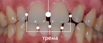
Caries between teeth is a carious lesion that is localized on the lateral surfaces of the teeth (in the interdental spaces). It must be said that this zone is the most favorite for the occurrence of dental caries, which is explained by two reasons. Firstly, after eating, food debris constantly accumulates in the interdental spaces, and secondly, very few people use dental floss every time after eating to clean the interdental spaces.
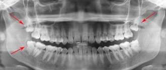
As a result, carious lesions of tooth enamel occur in the interdental space, which is accompanied by minor pain when eating sour/sweet foods, as well as thermal irritants (cold or hot). As the carious defect deepens, these symptoms then disappear. It should be noted that interdental caries can be difficult to diagnose in time, because this area is difficult to visually observe, and patients usually do not see a dentist in the early stages of caries.
Caries between teeth: photo
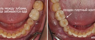
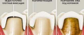
In dentistry, the treatment of interdental caries is not a simple task, because... here, in addition to restoring the side wall of the tooth, it is necessary to restore the contact point between the teeth. This last task is quite difficult for most dentists. Filling a tooth without creating good contact between the teeth in the interdental space leads to the fact that food will actively get stuck in it, and this, in turn, will provoke the development of secondary caries near the filling, as well as periodontal pockets.
Causes of caries between teeth
The causes of interdental caries are determined by the individual characteristics of the tissue structure and certain conditions - previous diseases, diet, location, crowding, hygiene:
- "metabolic explosion"
. Excessive sugar consumption provokes active production and accumulation of acid on the tongue, surfaces and pits. Demineralization occurs, the saturation of saliva with salts decreases, which contributes to the destruction of tooth enamel and the occurrence of caries; - tissue resistance
. The degree of resistance to the action of factors causing pathology depends on heredity, health status, and fluoride content in food/water. Manifests itself in the age-related dynamics of the onset of the lesion, the aggressiveness/prevalence of the process; - ignoring the rules of oral hygiene and dental care.
Clinical researches
Clinical studies have proven that regular use of professional toothpaste ASEPTA REMINERALIZATION improved the condition of the enamel by 64% and reduced tooth sensitivity by 66% after just 4 weeks.
Sources:
- Report on determining/confirming the preventive properties of toothpaste “ASEPTA PLUS” GENTLE WHITENING” Author: doctor-researcher A.A. Leontyev, head Department of Preventive Dentistry, Doctor of Medical Sciences, Professor S.B. Ulitovsky First St. Petersburg State Medical University named after. acad. I.P. Pavlova, Department of Preventive Dentistry
- Clinical and laboratory assessment of the influence of domestic therapeutic and prophylactic toothpaste based on plant extracts on the condition of the oral cavity in patients with simple marginal gingivitis. Doctor of Medical Sciences, Professor Elovikova T.M.1, Candidate of Chemical Sciences, Associate Professor Ermishina E.Yu. 2, Doctor of Technical Sciences Associate Professor Belokonova N.A. 2 Department of Therapeutic Dentistry USMU1, Department of General Chemistry USMU2
- Report on the determination/confirmation of the preventive properties of personal oral hygiene products “ASEPTA PLUS” Remineralization doctor-researcher A.A. Leontyev, head Department of Preventive Dentistry, Doctor of Medical Sciences, Professor S.B. Ulitovsky First St. Petersburg State Medical University named after. acad. I.P. Pavlova, Department of Preventive Dentistry
- Clinical studies of antisensitive toothpaste “Asepta Sensitive” (A.A. Leontyev, O.V. Kalinina, S.B. Ulitovsky) A.A. LEONTIEV, dentist O.V. KALININA, dentist S.B. ULITOVSKY, Doctor of Medical Sciences, Prof. Department of Therapeutic Dentistry, St. Petersburg State Medical University named after. acad. I.P. Pavlova
- The role of anti-inflammatory rinse in the treatment of periodontal diseases (L.Yu. Orekhova, A.A. Leontyev, S.B. Ulitovsky) L.Yu. OREKHOVA, Doctor of Medical Sciences, Prof., Head of Department; A.A. LEONTIEV, dentist; S.B. ULITOVSKY, Doctor of Medical Sciences, Prof. Department of Therapeutic Dentistry of St. Petersburg State Medical University named after. acad. I. P. Pavlova
- Report on determining/confirming the preventive properties of toothpaste “ASEPTA PLUS” COFFEE and TOBACCO Author: doctor-researcher A.A. Leontyev, head Department of Preventive Dentistry, Doctor of Medical Sciences, Professor S.B. Ulitovsky. First St. Petersburg State Medical University named after. acad. I.P. Pavlova, Department of Preventive Dentistry
- Report on determining/confirming the preventive properties of commercially produced personal oral hygiene products: Asepta toothpaste used in combination with Asepta mouthwash and Asepta gum balm Head. Department of PFS Doctor of Medical Sciences Professor S.B. Ulitovsky St. Petersburg State Medical University named after Academician I.P. Pavlova. Faculty of Dentistry. Department of Preventive Dentistry.
Symptoms of interdental caries
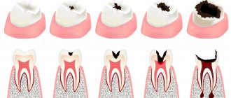
The features of the development and treatment of interdental caries are not very different from other types of carious lesions. There are 4 stages in total:
- Initial interdental caries - a small spot forms on a unit, invisible to the naked eye. To identify the initial stage, a hardware method is used.
- Superficial – the disease destroys the enamel. Darkening forms on it, it turns yellow and becomes rough, and sensitivity increases. But bacteria do not penetrate into deeper layers.
- Medium - at this stage the dentin layer is affected, the tooth reacts to irritants: cold, hot, sweet. The patient experiences severe pain.
- Deep - an advanced form of carious damage, pathogenic microorganisms penetrate into the root part. There is a risk of damage to healthy tissues and the development of periodontitis. At a deep stage, filling or restoration of the tooth is necessary.
Types of disease
Depending on the location of the disease, the dentist determines treatment methods, as well as the necessary medications.
The main types of interdental caries are:
- Disease of the chewing teeth. Typically, some food particles clog the space between these units. It is difficult for the patient to notice this disease and is often diagnosed when the infection reaches the root of the tooth.
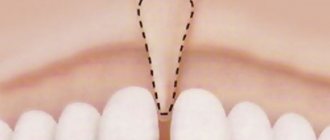
- It is much easier to determine caries if bacteria have formed between the front teeth. It is impossible to hide the damaged appearance of the units; immediate treatment and elimination of the infection should be resorted to.
- Carious bacteria between baby teeth are the most dangerous because they can damage the root of the tooth, which in the future will affect new, permanent teeth.
As mentioned earlier, the bacteria of this disease progress very quickly. If proper measures are not taken in time, the adjacent tooth will also become ill.
In any case, if you detect the slightest signs of interdental caries, immediately seek help from a dentist, who will determine a special course of treatment.
Stages of development of caries between teeth:
- Spot. As you know, enamel loses its natural shine due to insufficient mineralization. Tooth decay begins the moment it loses the required amount of minerals. In the initial stage, carious bacteria take the form of a small matte spot. Modern technologies, together with careful oral hygiene, can eliminate infection in the bud.
- Superficial caries. Unpleasant, painful sensations after contact with sour, sweet, hot, cold. Most likely, carious bacteria are already developing in this oral cavity. The possibility of dentin damage should not be excluded.
- Deep caries. It manifests itself as a powerful, aching pain. It happens when softened dentin fills a carious cavity. It is fraught with the formation of pulpitis.
Diagnostics
- visual examination of the oral cavity
. It is performed using special instruments to assess the condition of the oral cavity. A dental mirror allows the specialist to carefully examine the distal surfaces: to detect the localization of spots, stripes/dots of black, brown color, and a carious lesion in the gap. The probe helps differentiate the depth of the cavity; - percussion method
. Intended for diagnosis and identification of pathological segments by tapping. The diseased unit reacts with a muffled sound, the patient experiences pain and severe discomfort; - radiography
. The x-ray image shows the distance of the lesion from the pulp, the level of prevalence of the process; - transluminescence
. Luminescence (transmission through a filter with an ultraviolet beam) and fiber optic exposure (transmission with a light beam) are practiced. Damaged areas appear darker than healthy areas; - coloring
. Application of staining markers (potassium iodide/methylene blue solution). The technique allows you to diagnose pathology and distinguish it from fluorosis/hypoplasia.
In difficult cases, fissurotomy (opening the grooves on the surface of the molars) and electroodontometry are used to determine the degree of pulp damage.
Diagnosis of dental discoloration in CELT
Before making a diagnosis, our dentists conduct comprehensive diagnostic tests, which include:
- Taking anamnesis and performing a dental examination;
- X-ray (allows you to identify violations of the internal structure);
- Rheodentography (determines the condition of the pulp and the depth of the lesion);
- Computed tomography (providing images of teeth in three-dimensional format);
- Microscopy (multiple magnification makes it possible to assess the condition of the enamel);
- Electroodontodiagnostics (determines the condition of the tissues located around the tooth root).
Pigmentation must be differentiated from necrosis and erosion of tooth enamel, as well as from Capdepont disease.
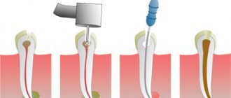
Treatment of caries between teeth
Treatment of caries between teeth is long and complex, since the doctor has to work with two units - to regenerate their anatomical structure and eliminate the carious lesion. When sanitizing the front teeth, it is also important to achieve an aesthetic result:
- remineralizing therapy
. Remineralization is carried out at the spot stage and involves restoration with the help of fluorine/calcium-containing preparations; - Icon technology
. The modern infiltration method is effective for minor dentin deformations (no more than 1/3): a sealing polymer composition is applied to the damaged area, restoring the density of the enamel; - filling
. The classic method involves installing a filling; in case of serious damage, it may be necessary to install an inlay/pin; - prosthetics
. Restoration with crowns/inlays is practiced for volumetric damage, in the absence of alternative options.
Before and after
Contraindications:
- First trimester of pregnancy;
- Bleeding gums;
- It is not recommended to carry out treatment during the period of illness with herpes and acute respiratory infections. It is better to postpone a trip to the dentist until you have fully recovered.
Painfulness of the procedure
Most people are afraid of the procedure precisely because of toothache, but modern technology and the use of anesthesia eliminate this ailment.
The session is completely painless. To reduce the likelihood of discomfort, all manipulations are carried out after the administration of an anesthetic. If there is no nerve, the procedure is performed without anesthesia. The patient may feel slight pressure on the gum.
An exception is intolerance to certain medications used for anesthesia. The choice is made in favor of a similar drug with a different active substance.
After the injection, no painful sensations are observed.
It hurts if there is a strong inflammatory process at the root apex and in the canals. Unpleasant sensations occur only when the drug gets into the periodontal tissues. They go away instantly after increasing the dosage of the painkiller.
Some pain is possible if there is a deep stage of carious lesions, depulpation or canal treatment is performed, or sensitivity is increased.
Features of the treatment of caries between the front teeth
When treating front teeth, it is important to preserve not only their functionality, but also their aesthetics. Therefore, the dentist is required to have extensive experience and skills in artistic restoration. Treatment of anterior teeth has several nuances:
- Carious lesions develop quickly due to the thin layer of dentin and enamel.
- During sanitation, it is necessary to achieve high aesthetic indicators, which is quite difficult with interdental caries.
The dentist carefully removes the affected tissue from the walls, and then restores the damaged teeth and interdental space. All medical procedures are performed under local anesthesia. There is an opinion that caries between the anterior units of the lower jaw is much easier to treat than on the upper jaw, but this is only a myth. There are no particular differences in the treatment protocol for the lower/upper dentition. To preserve beauty, a durable filling with high aesthetic indicators is installed on the front and side units. The most popular option is artistic restoration using compomer and reflective filling materials that preserve the aesthetics and natural appearance of the tooth.
What kind of caries is this and why does it occur?
The front teeth include the incisors (denies incisivi) and canines (dentes canini), the purpose of which is to bite off individual pieces of food. The main difference from the lateral teeth (premolars (dentes praemolares) and molars (dentes molares)) is the absence of a tuberous chewing surface for grinding. Therefore, caries forms on the crown in the form of a chisel (incisors) or slopes (fangs), in the interdental space, at the junction of the visible part of the tooth and the gum (cervical caries of the front teeth). The tooth is gradually destroyed, “pockets” form at the “neck” - caries spreads deeper, destroying the root.
Modern dentistry is based on the theory of W. D. Miller (1883), who, while conducting experiments, noticed that if an extracted tooth was placed in a fermenting mixture of bread and saliva, something similar to caries appeared on it. At the same time, the density and structure of dental tissue provide protection of teeth from destruction.
Caries is, first of all, problems in the neurohumoral regulation of the body. Normally, the hypothalamus, with the help of the pituitary gland, controls the parotid salivary glands, which secrete parotin. With the help of this substance (in medicine “hormone”), calcium metabolism in the blood-bone tissue-organs system is controlled and stimulates the movement of dental lymph through microscopic tubules inside the teeth. This cleanses and strengthens the dental tissue. Therefore, the main cause of caries is a delay in the production of dental lymph and subsequent demineralization of the tooth. Then the weak tooth in the aggressive environment of the oral cavity is destroyed from the outside.
Caries on front teeth
The dental lymph contains monoblasts, with the help of which the enamel and dentin are nourished. In order for the tooth to be cleaned from the inside, the microscopic structures of monoblasts work like a pump. The nutritious fluid released from the blood is pumped through the dentinal tubules and, under capillary pressure, is pushed towards the enamel, strengthening it. In case of disorders, when capillary pressure decreases, the amount of mineral substances decreases, plaque is literally attracted to the tooth walls (the enamel is like a fine-mesh sieve and allows oral fluid to pass into the tooth).
According to classical dental textbooks, caries is classified as an infectious bacterial disease. Recent research confirms that caries:
- odontoporosis (decreased density of dental tissue);
- odontoclasia (destruction of all layers of the tooth).
Factors influencing the intensity of caries development on the front teeth:
- Excessive consumption of confectionery products without cleaning the mouth for 4 hours (maximum time for the formation of soft plaque). In addition, excess sugar inhibits the normal functioning of the pituitary gland.
- Inflammatory process or removal of the salivary glands (saliva contains a complex of antimicrobial factors, neutralizes the acids of dental plaque and is involved in enamel calcification).
- Environmentally unpleasant factors and work in the metallurgical, mining, coal, chemical industries.
- Reduced general resistance (resistance) of the body.
- Improper development of the jaw, crowded teeth, making it difficult to clean the mouth.
Features of the treatment of caries between chewing teeth
Although molars are located in the lateral part of the jaw and are practically invisible when smiling, interdental caries is especially dangerous for them, as it is often diagnosed already at the stage of deep damage, when the dentin layer of one or more chewing units is affected. Very often the disease affects the pulp of two teeth. This situation is the most unfavorable, since endodontic treatment of canals is a long and expensive process and is performed only by an experienced dentist. If you do not start fighting the disease in a timely manner, the patient may encounter complications in the form of periodontitis, pulpitis (nerve inflammation), etc.
When filling molars, special attention is paid to the strength and durability of the filling material. To restore them, high-quality glass ionomer fillings are used.
Case from practice
I remember a patient who came to see me with advanced caries between her teeth. The destruction was significant, the girl was nervous, afraid not only of the dissection, but also of the injection. It is difficult to work with frightened patients; fears must be dispelled. Before administering the anesthetic, I treated my gums with Lidocaine, the girl realized that it wouldn’t hurt, and she calmed down a little. Fortunately, the pulp was not exposed, and the treatment was successful. But due to deep lesions on the lateral surfaces, both dental units had to be restored with ceramic crowns.
Prevention
Preventing the development of interdental caries is quite simple. To do this, it is enough to follow the recommendations of dentists:
- It is recommended to clean your teeth after each meal, moving the brush up and down to remove as much food debris as possible in hard-to-reach places. For care, use pastes that improve the protective properties of enamel.
- Rinse your mouth several times a day. It is not necessary to use special solutions; boiled water will be enough for cleaning. Additionally, use an irrigator.
- It is better to avoid foods such as chips, chocolate, and crackers, as their particles settle tightly in the interdental space and become a haven for bacteria. As a last resort, carry dental floss with you.
- Visit your dentist's office every six months for a checkup and professional cleaning.
To find out the cost of treating caries between teeth, sign up for a consultation at Dr. Razumenko’s dental clinic in Moscow by phone.
Is it possible to independently determine the disease?
Lateral dental caries can be determined by personally completing the following steps:
- With the help of a mirror and good lighting, you can see the color differences between the main and infected parts of the tooth. The dark spot is visible through less transparent enamel.
- Painful sensations after contact with thermal and chemical irritants.
- Unfavorable odor from the mouth. Carious bacteria, together with food debris, “trigger” the process of decay, which is accompanied by a bad aroma.
Prices for treatment of caries between teeth
| Code no. | Name of procedures | Unit of measurement | Cost, rub. |
| 101 | Repeated treatment and diagnostic appointment of the patient | 650,00 | |
| 300 | Application anesthesia | 150,00 | |
| 301 | Infiltration anesthesia, conduction | 550,00 | |
| 303 | Treatment of medium caries (ultrasound, cavity formation) | 650,00 | |
| 307 | Placement of a light-composite filling (Sonicfil, Estelite) | 3 950,00 | |
| 310 | Recovery from significant damage is complex | 1 unit | 5 500,00 |
| 313 | Artistic restoration (front, side teeth) simple | 1 unit | 11 000,00 |
* The prices indicated on the website are not a public offer. The exact cost can only be determined by visiting a doctor.
Prices for treatment in Moscow full price list
Share on social media networks:
Article Expert:
Ustinova Elena Fedorovna
Doctor of the highest category. Root canal treatment (complex, repeated retreatment), microprosthetics with ceramic restorations.
Work experience 15 years
How it manifests itself
Among the main signs of caries between teeth:
- increased sensitivity of units;
- intolerance to cold and hot food/drinks;
- frequently occurring sore throat;
- presence of bad breath.
It is important to remember that in the first stages the disease usually does not manifest itself in any way. Discomfort occurs in a person only when the structure of the enamel has already been damaged.
We recommend that you read
Dental treatment during pregnancy
Treatment of anterior teeth
Dental filling
Canal filling