Medicines for the treatment of diseases of the mucous membrane of the mouth, tongue and lips
Diagnosis, treatment and prevention of diseases of the mucous membrane (OM) of the mouth, tongue and lips remain an important problem in clinical medicine. Changes in the CO of the mouth, tongue and lips in many systemic diseases occur long before the appearance of general clinical symptoms. Therefore, the correct interpretation of such changes and the choice of appropriate medications are of great importance not only for dentists, but also for other specialists.
When presenting the material, we used the classification of diseases of the oral cavity by E.V. Borovsky and A.L. Mashkilleyson [7], which provides for the identification of 9 groups of this pathology, taking into account etiological and pathogenetic factors, which, due to the impossibility of citing them, will be omitted in this publication. We limited ourselves only to issues of medical tactics, drug treatment of major diseases of the oral mucosa, tongue and lips.
The authors have made every effort to ensure the accuracy of the information presented, including drug dosages. Aware of our responsibility associated with the preparation of the publication, and taking into account the constant changes occurring in medical science, we recommend that the dosage of medications be updated according to the appropriate instructions.
Basic principles of treatment of diseases of the mucous membrane of the mouth, lips and tongue:
- Rational treatment requires contact between the dentist and other dental and non-dental specialists.
- Treatment must be carried out in compliance with the principles of bioethics, these diseases must be considered from the standpoint of the state of the whole organism, therefore in most cases one cannot limit oneself to local influences only.
- An axiom for the dentist should be the elimination of all unfavorable irritating factors in the oral cavity in the patient, which can support and provoke the development of the pathological process. It is unacceptable to use so-called cauterizing agents and long-term use of the same mouthwashes.
- Treatment should begin only after at least a preliminary diagnosis has been established and meet the following requirements: be comprehensive; provide a pathogenetic approach; do not disturb the anatomical and physiological characteristics of the oral mucosa; eliminate the pain factor; promote rapid epithelization of lesions; provide for the active involvement of the patient in performing medical procedures at home.
Traumatic lesions
Medical tactics for traumatic lesions of the mucous membrane due to the action of mechanical factors, high and low temperatures, radiation, unfavorable meteorological factors, and chemicals boils down, first of all, to eliminating the effect of the traumatic factor, which not only facilitates differential diagnosis, but is also a therapeutic intervention.
Decubital ulcer
Heals, as a rule, 10-12 days after eliminating the mechanical cause. The surface of the ulcer is treated with oxygen-containing antiseptics (hydrogen peroxide, potassium permanganate, sodium hypochlorite), and after cleaning its surface from necrotic plaque, keratoplasty agents are used. It should be remembered that with prolonged existence (more than 2-3 months), especially in older people, a decubital ulcer can become malignant.
Chemical injury CO
It occurs as a result of its immediate immediate contact with concentrated solutions of acids, alkalis, some medications and agents sometimes used by dentists (silver nitrate, arsenic paste, resorcinol-formalin mixture). In case of acute chemical injury, the CO drug that caused the burn is immediately neutralized (in the absence of a neutralizer, the mouth is washed with water). Subsequently, the affected areas are treated with anesthetic substances, weak antiseptic solutions, proteolytic enzymes, and then keratoplasty agents are applied.
Physical trauma CO
It can also be acute and chronic. Painkillers, antiseptics, and drugs that promote tissue regeneration are used. In some cases, it is necessary to resort to excision of the affected area.
Leukoplakia
A fairly common disease of the oral mucosa and red border of the lips, pathomorphologically characterized by chronic inflammation with pronounced hyperplasia and keratinization of the epithelium.
The scope of treatment measures is determined both by the form of the disease and the size of the lesion and the speed of development of the process. Among the medications for flat form of leukoplakia, applications are made to the lesion and vitamin A is prescribed orally (3.4% oil solution of retinol acetate or 5.5% retinol palmitate, 10 drops 2-3 times a day for 1. 5-2 months). Courses are repeated 2-3 times a year. For verrucous and erosive forms, the same is done within one month, and in the absence of positive dynamics, the lesion is excised, followed by mandatory histological examination, which determines further medical tactics.
The manifestations and severity of changes in the mucous membranes of the mouth, tongue and lips during infectious diseases depend both on the type of pathogen and on the individual characteristics of the patient’s body, which determines the dentist’s tactics.
Adenoviral diseases
In case of influenza and adenoviral diseases, the lesions of the mucosa are not specific; after a few days from the onset of the disease, hyperemia with “granular” rashes on the mucous membrane is replaced by petechial rashes, and by the 7-9th day the pathological changes disappear.
Chronic recurrent herpes
It appears at any age in persons previously infected with the herpes simplex virus.
Medical tactics for viral infections include the use of drugs from several groups that affect various parts of the etiology and pathogenesis of diseases: antivirals, immunocorrectors and immunomodulators, vaccines, interferons and their inducers. The most pronounced therapeutic effect is observed with the combined use of 2-3 drugs of general and local action with different mechanisms of antiviral effect.
Ointment and cream are started to be used when the first signs of infection activation appear and continue until the erosions are epithelialized. Early treatment can prevent the development of vesicles. A large group of antiviral drugs includes nucleoside analogues, similar in structure to intermediate products of DNA and RNA biosynthesis (iododeoxyuridine, chlordeoxyuridine, fluorodeoxyuridine), effective against generalized forms of herpes and herpes zoster. An effective antiviral agent is acyclovir (Zovirax, Virolex). In addition to acyclovir, the group of abnormal nucleosides includes: valacyclovir (Valtrex) in tablets of 500 mg; take 2 times a day for 5-7 days; famciclovir (famvir) tablets 250 mg; take 3 times a day for 7 days.
It should be noted that the therapeutic effect of antiviral drugs is most effective when prescribed in the first hours and days of development of the lesion elements and in the prodromal period of the viral disease.
Local antiviral drugs include: adimal, bonafton, florenal, riodoxol in the form of ointments and liniments (gossypol). Ointment and cream are started to be used when the first signs of infection activation appear and continue until the erosions are epithelialized. Moreover, early treatment can prevent the development of vesicles.
The leading role in the development of antiherpetic immunity belongs to interferon. The antiviral drug "Infagel" (it includes recombinant interferon alpha-2) has a wide spectrum of antiviral activity, bacteriostatic and anti-inflammatory effects, as well as antitumor and immunomodulatory activity. It is used in the form of applications to the lesions 2 times a day with an interval of 12 hours and dried for 10-15 minutes to form a protective film [10].
Interferon inducers (poludan, larifan) and antiviral vaccines (in the form of a cycle of intradermal injections to prevent relapses and during remission of the disease) have found widespread use in dental practice. The therapeutic effectiveness of the vaccine increases significantly when combined with poludanum or other interferon inducers.
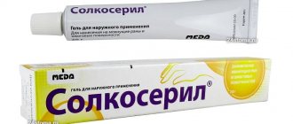
In addition to these drugs, salicylates, analgesics, antihistamines and antiseptic solutions, multivitamins, EF radiation and helium-neon laser radiation are used in the treatment of herpes infection. To stimulate the processes of regeneration of the mucous membrane of the mouth and the red border of the lips, applications of oil solutions of vitamins A, E, ointment and jelly of solcoseryl and Actovegin, aerosols “Livian”, “Spedian”, “Gipozol” are used. After eliminating the elements of the lesion, sanitation of foci of chronic infection is mandatory.
In case of herpes zoster, along with the measures mentioned above, the doctor’s task includes eliminating the pain syndrome and preventing postherpetic neuralgia. For this purpose, analgesics, ganglerone, and B vitamins are prescribed.
Vincent's ulcerative-necrotizing gingivostomatitis (YANGS)
This is an inflammatory disease of the mouth, characterized by necrotic decay of the affected areas. Medical tactics for YANGS in some cases (especially in severe cases) include hospitalization of the patient, local and general measures, and medical examination.
Local treatment is important and begins with the patient’s first visit to the doctor, and then is carried out daily and in stages:
- Pain relief (applications, oral baths).
- Treating the mouth with antiseptic solutions (more effective - those that release atomic oxygen and chlorine-containing preparations: hydrogen peroxide, potassium permanganate, hypochlorite).
- Mechanical (and after application of proteolytic enzyme solutions to the lesion) removal of necrotic tissue, removal of mineralized dental deposits and elimination of other local traumatic factors.
- After cleansing the lesions from necrotic plaque, it is possible and advisable to use (in the form of applications) keratoplasty agents and preparations that promote tissue regeneration (oxycort aerosol, methyluracil ointment, oil solutions of vitamins A and E).
- It is better to postpone the removal of damaged teeth and roots, treatment of chronic forms of pulpitis and periodontitis until the ulcers are completely epithelialized.
- Subsequently, final sanitation of the oral cavity (planned treatment of caries, its complications, periodontal and mucosal diseases).
General treatment (especially for severe disease) involves the use of antimicrobial drugs. Orally prescribed: metronidazole (Flagyl, Klion) 0.25 g 2 times a day for 7-10 days, in some cases it is necessary to use broad-spectrum antibiotics. General strengthening and stimulating therapy is also carried out: ascorutin, vitamin C in large doses (up to 3.0 g per day), and antihistamines are prescribed. The nutrition of patients with YANGS should be complete and balanced.
Fungal infections of CO
Fungal infections of the mouth (oral mycoses) are most often caused by fungi of the genus Candida albicans.
General treatment includes:
- Ingestion of antifungal drugs for 10 days: diflucan (capsules) 50-100 mg 1 time per day; the use of polyene antifungal drugs (nystatin, levorin, amphotericin B) is traditionally included in general treatment, although it is known [11] that polyenes are practically not absorbed in the gastrointestinal tract, thus exerting a local effect on oral mucosa, and therefore It is recommended to dissolve the tablets in the mouth after meals (nystatin 1 tablet 500,000 units every 6-8 hours, levorin 1 tablet 500,000 units every 8-12 hours); Amphotericin B preparations are used mainly for severe systemic mycoses due to their high toxicity [11].
- Vitamins C, PP, group B taken inside.
- Use of antihistamines.
- Treatment together with other specialists of concomitant and background diseases.
Local treatment includes:
- Antifungal drugs for application and lubrication of lesions: 0.5% decamine ointment, 1% ointment and clotrimazole solution (canesten); 1% pimafucin ointment; for yeast infections and cheilitis, it is advisable to use 5% levorin ointment.
- Aqueous solutions of aniline dyes: 1-2% solution of gentian violet, 2% solution of methylene blue.
- Agents that alkalize the oral cavity: rinse with a 2% sodium bicarbonate solution; applications and lubrication of lesions with a 20% solution of borax in glycerin.
- A decoction of wild rosemary for mouth rinsing and oral baths.
Lesions of the oral mucosa are observed in all periods of syphilis, and dentists with similar manifestations have become much more common. Treatment of patients with syphilis is carried out in specialized venereological institutions. Dental interventions are usually associated with the sanitation of the oral cavity and are carried out in strict compliance with the disinfection and sterilization regime in health care facilities.
Treatment of fixed and widespread allergic and toxic-allergic diseases includes the “exclusion” of the allergen, for example, a drug, the use of painkillers, antiseptics, desensitizing and keratoplasty agents, if necessary. Sanitation of odontogenic foci of chronic infection is mandatory.
Erythema multiforme exudative
A fairly common dermatosis affecting the oral mucosa, observed mainly in young and middle-aged people. Stevens-Johnson syndrome is a severe variant of exudative erythema multiforme (also called acute mucocutaneous-ocular syndrome).
Medical tactics depend on the severity of the disease. In some cases, patients need to be hospitalized.
General treatment is usually prescribed by other specialists (dermatologist, immunologist) and includes:
- Detoxification therapy: drinking plenty of fluids or infusion therapy if it is impossible to take liquids orally.
- Taking glucocorticoids: prednisolone in an initial dose of 20-60 mg per day orally for 5-7 days (the dose depends on the severity of the process and the form of the disease, with Stevens-Johnson syndrome higher dosages are required), then the dose of prednisolone is reduced by 5 mg every 2 —3 days before complete withdrawal of the drug.
- Immunomodulatory therapy: for example, Lavomax (Tilorone) is prescribed 125 mg (1 tablet) orally in the first two days, then 125 mg after 48 hours. The dose of the drug per course is 2.5 g [15].
- Prescription of antihistamines, B vitamins, ascorutin.
- Antibiotics are prescribed when there is a threat of purulent-inflammatory complications and the doctor is confident that there is no connection between the development of the disease and the use of these antibacterial agents.
Local treatment is aimed at relieving inflammation and accelerating epithelization of the affected areas of the mucous membrane of the mouth and lips. It includes:
- The use of painkillers (1-2% solutions of trimecaine, lidocaine, anesthetics in aerosols) before each manipulation in the oral cavity (baths, rinses, applications) and before meals.
- Antiseptic treatment of the mouth (solutions of hydrogen peroxide, potassium permanganate, chloramine).
- To improve the rejection of necrotic tissues, apply solutions of proteolytic enzymes (trypsin, chymotrypsin) to the lesions.
- After cleaning the eroded surfaces from purulent-fibrinous plaque, use keratoplasty preparations (sea buckthorn and rosehip oil, oil solutions of vitamins A and E, the drug “Gipozol”).
- EF irradiation of the oral mucosa.
After the elimination of acute phenomena, the foci of infection are sanitized. In the inter-relapse period - specific desensitizing therapy.
Chronic recurrent aphthous stomatitis
One of the most common diseases with an autoimmune component of pathogenesis, it is characterized by periods of remission and exacerbation with the formation of aphthae (a superficial painful defect of the mucous membrane of a round or oval shape, covered with fibrinous plaque and surrounded by a bright inflammatory rim).
General treatment is prescribed after consultation with an immunologist:
- Immunocorrective and immunomodulatory therapy with thymogen, levamisole.
- Metabolic drugs for 10 days (calcium pantothenate orally 0.1 g 4 times a day; potassium orotate 0.5 g 3 times a day an hour before meals, lipamide 0.025 g 3 times a day after meals).
- Sedatives and adaptogens.
- Exclusion from the diet of hot, spicy foods, prohibition of smoking and drinking alcohol.
- In the inter-relapse period - specific and nonspecific desensitizing therapy.
- Vitamins: C, group B.
Local treatment:
- If there are aphthae, apply painkillers to the aphthae.
- Injections under the base of the aphthae with a mixture of 0.1 ml of 0.1% atropine sulfate and 1 ml of 0.25-0.5% solution of novocaine or trimecaine.
- Applications to aphthae of biopolymer soluble films (for example, oblekol films).
- Applications to aphthae of oil solutions of vitamins A, E, Oblekol, “Aevita”.
Pemphigus
Among the pathological processes of the oral mucosa with the formation of blisters (vesical dermatoses), the central place is occupied by pemphigus - an autoimmune process in which intraepithelial blisters appear on the skin and mucous membranes.
Treatment is coordinated with a dermatologist and begins in the hospital. Glucocorticoid therapy (first in shock and then in minimal, maintenance doses) interrupts the progression of pemphigus and leads to the disappearance of clinical symptoms of the disease. It should be remembered that local use of glucocorticoid drugs is ineffective. Sometimes cytostatics (mainly methotrexate) are also used to treat pemphigus.
Local treatment is aimed at preventing secondary infection of erosions and ulcers. Non-irritating CO solutions of antiseptics and keratoplastics are used.
Lichen planus
Mucocutaneous reaction - lichen planus occurs in 1-2% of the population. When treating lichen planus, an integrated approach is required, the basis of which is psychotherapy, especially at the initial stage, supplemented by the prescription of tranquilizers, sedatives, adaptogens, desensitizing agents, vitamins (A, PP). An effect was detected when prescribing the anxiolytic “Tenoten” (from 1 to 4 tablets per day for 3-6 weeks) and the antioxidant “Kudesan” [9].
Smoking and taking allergenic products is prohibited. It is necessary to exclude dissimilar metals in orthopedic structures and replace metal fillings. With exudative-hyperemic and erosive-ulcerative forms, it is sometimes necessary to resort to steroids (prednisolone) and drugs such as delagil.
Local events include:
- Local anesthetics.
- Applications of 1% emulsion of dibunol, vitamins A, E, Solcoseryl ointment.
- Irradiation of lesions with a helium-neon laser in combination with oral administration of tigazone capsules (10 mg 3 times a day for a course of up to 6-8 weeks).
- Electrophoresis of vitamin PP from dimexide medium.
In his medical practice, the dentist must proceed from the fact that lichen planus can become malignant.
Changes in the mucosa of the mouth and the red border of the lips often occur in pathologies of various organs and systems of the body and metabolic disorders.
It should be noted that in most cases, the manifestation of systemic diseases in the oral cavity is not specific, however, some changes in the oral cavity clearly indicate one or another type of organ or systemic disorder and are of great clinical significance.
Vitamin A deficiency
Causes disturbances in the epithelial structures of the skin and oral mucosa. Retinol is prescribed (in the form of a concentrate or pill) up to 50,000-100,000 IU per day.
Vitamin B1 deficiency
Accompanied by hyperplasia of the fungiform papillae of the tongue, paresthesia and allergic reactions of the oral mucosa. For treatment, preparations of thiamine chloride and thiamine bromide are used in courses of up to 30 days. It is unacceptable to mix a solution of vitamin B1 with other B vitamins in one syringe or to administer them sequentially through one needle.
Vitamin B2 deficiency
It manifests itself as a peculiar change in the skin, red border of the lips, ulceration in the corners of the mouth (angular stomatitis), weeping, maceration of the epithelium. For medicinal purposes, riboflavin is prescribed orally at a dose of 0.01 g 3 times a day for 4-6 weeks.
Vitamin B6 deficiency
With a deficiency of vitamin B6, symptoms of disorders of the nervous system (polyneuritis) and gastrointestinal tract, angular stomatitis, cheilitis, and glossitis are observed. The daily therapeutic dose of pyridoxine for adults, administered primarily parenterally, is 0.05-0.1 g.
Vitamin B12 deficiency
Accompanied by neurological disorders, changes in hematopoiesis (B12-deficiency anemia). Hunter-Meller glossitis is characteristic. For therapeutic purposes, 1000 mcg of vitamin B12 is administered intramuscularly daily until hematological disorders are completely eliminated, during the same time (4-6 weeks) manifestations in the oral cavity are stopped. Subsequently, lifelong maintenance therapy is prescribed (one injection of vitamin B12 per month).
Vitamin PP deficiency
Severe PP (nicotinic acid) deficiency is described as “pellagra,” which is characterized by a triad of symptoms: dementia, dermatitis, diarrhea. Treatment consists of taking nicotinic acid orally after meals (0.1 g 2-3 times a day for a course of 2-3 weeks). At the same time, thiamine, riboflavin and pyridoxine are usually prescribed, since in most cases polyhypovitaminosis occurs.
Vitamin C deficiency
It manifests itself as hemorrhagic syndrome (petechial hemorrhages in various parts of the oral mucosa, swelling and bleeding of the gums) and complications caused by the addition of a secondary infection (ulcerative necrotizing gingivitis). For therapeutic purposes, ascorbic acid is prescribed up to 1.5 g per day (after meals) with simultaneous intake of rutin (50-100 mg 2-3 times a day).
Most diseases of the blood and hematopoietic organs have typical manifestations in the oral cavity: changes in the color of the mucous membrane, the phenomenon of hemorrhagic diathesis, paresthesia, ulcerative-necrotic processes. The mucous membrane of the mouth in acute leukemia is almost constantly affected. In more than half of the patients, changes in the mucosa are ulcerative-necrotic and hemorrhagic in nature.
The dentist’s task when treating such patients (usually in a hematology hospital) remains pain relief, proper professional antiseptic treatment of the oral cavity, careful removal of necrotic plaque (mechanically and with the help of proteolytic enzymes), and stimulation of epithelization of lesions.
Salivary dysfunction
Disorders of this kind are of great clinical significance, especially associated with a decrease in the secretion of the salivary glands, up to hyposialia or even xerostomia.
Medical tactics:
- Distinction between “subjective” (i.e., developing against the background of somatic pathology without the death of the glandular epithelium) and “objective”, in which, due to the death of salivary gland tissue, there is a decrease in the secretory activity of the salivary glands.
- Identification and elimination of organ disorders; correction of disorders of “general” and “local” immunity.
- Elimination of foci of chronic odontogenic infection.
- Rational prosthetics.
- Medications include vitamins A, E, C, group B, and antidepressants.
- To stimulate salivation - orally (2-4 drops 15 minutes before meals) 1% solution of pilocarpine; galvanization of the salivary glands.
The doctor’s tactics for neurogenic diseases of the tongue, lips and mucus (glossalgia, glossodynia, stomalgia, burning mouth syndrome) include:
- Sanitation of the mouth to eliminate irritating factors (galvanism phenomena, defects in fillings and prosthetics) and eliminate chronic odontogenic infection; treatment of fungal infections of the mouth and tongue.
- Treatment of somatic diseases.
- Normalization of the function of the central and autonomic nervous systems; for somatized depression - antidepressants (for example, amitriptyline according to the scheme starting with one-fourth tablet at night, increasing weekly by one-fourth tablet, leading to a whole tablet per day by the end of the month).
- B vitamins.
- Vasoactive drugs and vegetotropic drugs.
- Applications of local anesthetics.
- Galvanization of the upper cervical sympathetic nodes, in some cases - ultratonotherapy, ozone therapy, etc.
If conservative treatment of erosive and ulcerative lesions (for example, in chronic trauma, leukoplakia, lichen planus) within 10-14 days is ineffective and there is no tendency to their healing, surgical or cryosurgical excision of the lesion with mandatory histological examination should be used.
It must be remembered that all precancerous conditions must be treated surgically. There is no need for wait-and-see tactics and long-term drug therapy. This is permissible, as mentioned above, only in cases of damage to the mucosa of the mouth, tongue, and lips, where reverse development of the pathological process is possible under the influence of only therapeutic treatment.
This article is intended both for practicing dentists and for students, interns, residents, and young teachers of medical universities. However, it will be a shame if the reader only absorbs some of the information and concepts presented in it. To expand your own knowledge and for the benefit of our patients, it is advisable to read other publications on this issue [1-8, 12-14, 16-18].
LITERATURE
- 1. Banchenko G.V. Combined diseases of the oral mucosa and internal organs. - M.: Medicine, 1979. - 190 p.
- Banchenko G.V., Maksimovsky Yu.M., Grinin V.M. Language is the “mirror” of the body: Clinical guide for doctors. - M., 2000. - 408 p.
- Borovsky E. V., Danilevsky N. F. Atlas of diseases of the oral mucosa. — 2nd ed., revised. and additional - M.: Medicine, 1991. - 320 p.
- Danilevsky N. F., Leontyev V. K., Nesin A. F., Rakhniy Zh. I. Diseases of the oral mucosa. - M., 2001. - 271 p.
- Diagnosis, treatment and prevention of dental diseases / V. I. Yakovleva, E. K. Trofimova, T. P. Davidovich, G. P. Prosveryak. — 2nd ed., revised and supplemented. - Mn.: Higher. school, 1994. - 494 p.
- Dmitrieva L. A., Glybina N. A., Glybina T. A. et al. Experimental substantiation of the use of a new antioxidant drug in the treatment of erosive and ulcerative lesions // Periodontology. — 2012, No. 3 (64). - pp. 52-58.
- Diseases of the mucous membrane of the mouth and lips / Ed. prof. E. V. Borovsky, prof. A. L. Mashkilleyson. - M.: Medicine, 1984. - 400 p.
- Diseases of the oral mucosa: Textbook / Ed. L. M. Lukinykh. - N. Novgorod: Publishing House of NGMI, 1983. - 212 p.
- Karakov K. G., Vlasova T. N., Oganyan A. V., Pisarev G. Yu. Optimization of complex therapy of lichen planus of the oral mucosa // Dentist-practitioner. - 2012, No. 1. - P. 35-37.
- Karakov K. G., Vlasova T. N., Oganyan A. V., Polyakova O. V. The role of complex therapy in the treatment of herpetic infection of the dental system // Practitioner dentist. - 2012, No. 2. - P. 48-49.
- Practical guide to anti-infective chemotherapy / Ed. L. S. Strachunsky, Yu. B. Belousov, S. N. Kozlov. - M.: Borges, 2002. - 384 p.
- Rybakov A. I., Banchenko G. V. Diseases of the oral mucosa. - M.: Medicine, 1978. - 232 p.
- Therapeutic dentistry: textbook: 3 hours / ed. G. M. Barera. - M.: GEOTAR-Media, 2055. - Part 3. - 288 p.
- Tretyakovich A. G., Borisenko L. G., Pishchinsky I. A. Differential diagnosis and principles of treatment of diseases of the oral mucosa: educational method. allowance. — 2nd ed., revised. and additional - Mn.: BSMU, 2005. - 66 p.
- Udzhukhu V. Yu., Minatulaeva M. A., Kubylinsky A. A. Pathogenetic rationale and clinical effectiveness of the use of Lavomax in patients with exudative erythema multiforme // Farmateka. - 2008, No. 9. - P. 60-62.
- Tsepov L. M., Nikolaev A. I. Medical tactics for erosive and ulcerative lesions of the mucous membrane of the mouth, tongue and lips (Training manual). - Smolensk: SGMA, 2005. - 16 p.
A complete list of references is in the editorial office.
Definition of disease. Causes of the disease
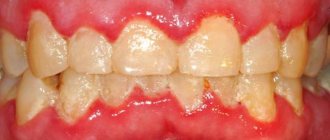
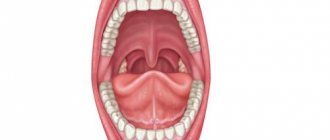
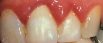
Stomatitis is an inflammation of the oral mucosa.
This disease is characterized by all the signs of an inflammatory process: redness, pain, swelling, dysfunction, increased local and general body temperature. Stomatitis can occur on the mucous membrane of any part of the oral cavity: on the gum, on the tongue, on the mucous membrane of the palate, in the vestibule of the oral cavity, in the retromolar fossa, on the mucous membrane of the cheeks and lips. The disease can manifest itself in a variety of forms: it can be redness of the mucous membrane in a certain area or the appearance of ulcers in the mouth. Stomatitis manifests itself with decreased immunity and chronic diseases, and stomatitis can also develop as a protective reaction of the body to the action of various irritants (for example, mechanical trauma to the mucous membrane from the sharp edges of teeth).
Inflammation of the oral mucosa occurs in most cases in children [12]. But recently, stomatitis has also appeared in the adult population. This is mainly due to the weakening of a person’s immunity, for example, with acute respiratory viral infections, chronic diseases, and HIV infection.
Where can I buy
The online store “Russian Roots” offers ready-made products and ingredients for their preparation, used in folk medicine for the treatment of stomatitis. Courier delivery of goods is available in Moscow and the Moscow region. Orders to other regions are sent by mail. You can buy goods in Moscow at any of our herbal pharmacies. Call us or order products in the online store!
Attention! All materials published on our website are protected by copyright. When re-publishing, attribution and a link to the original source are required.
Symptoms of stomatitis
The manifestation of stomatitis can be different.
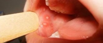
This depends on the form and stage of development of the disease. The first symptom is redness of the oral mucosa in a certain area. The spot does not rise above the level of the mucous membrane, has a round or oval shape and may be pinkish or whitish in color. Next, the area around the affected area becomes swollen, hyperemic, and pain appears. Aphtha appears at the site of the spot. Aphtha (from the Greek aphtha - ulcer) is a superficial defect of the mucous membrane, having a round shape and a grayish fibrinous coating. If you remove necrotic plaque from the surface of the aphthae, the ulcer will begin to bleed [5]. Most often, aphthae are located along the transitional fold, on the lateral surface of the tongue, on the mucous membrane of the cheeks and lips.
In severe forms, the amount of fibrinous plaque on the surface of the ulcer increases, and an infiltrate appears at the base of the aphthae (an accumulation of cellular elements of blood and lymph in the mucous layer). With such a clinical picture in the oral cavity, the patient experiences pain, burning, itching, bad breath, increased salivation, and eating becomes difficult. Body temperature rises to 38 ℃, weakness appears.
A feature of the disease is frequent relapses. Repeated rashes may appear after a few days or every few months. It depends on the ability of the immune system to fight infection.
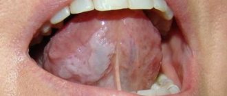
In the most severe cases, stomatitis manifests itself in the form of ulcers on the mucous membrane. An ulcer is a deep defect of the mucous membrane, in which the entire thickness of the mucous membrane is affected. The ulcer is surrounded by an inflammatory infiltrate, the edges of the ulcer are uneven, scalloped (jagged) [5]. When ulcers appear in the oral cavity, the general condition also worsens. The submandibular lymph nodes on the affected side may become inflamed. On palpation they will be enlarged and painful.
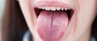
With candidal stomatitis, a white coating appears on the tongue and gums; when trying to remove the plaque, the affected areas begin to bleed. There is also an unpleasant taste in the mouth and a burning sensation. If candidal stomatitis develops against the background of diabetes mellitus or immunodeficiency conditions, it always has a chronic course.
With viral lesions of the oral mucosa, pain occurs, which intensifies during eating and when talking. The mucous membrane turns red and swells, then small blisters appear on it. The bubbles quickly open and aphthae form in their place. In moderate and severe forms, body temperature rises to 37-40 ° C, general malaise, weakness, and headache are noted.
Is stomatitis contagious?
The disease can be recognized and treated in a short time.
At the appointment, the doctor must correctly determine the causes and type of stomatitis, because drawing up a treatment plan depends on this. Depending on the classification, appropriate drugs are selected. Is stomatitis contagious?
Inflammatory ulcers that arise as a result of:
- allergic reaction;
- mechanical damage to the mucosa;
- during intoxication.
The disease is contagious if it develops during an infection in the body.
This class includes: fungal, herpetic, infectious stomatitis. Stomatitis in children can be caused by the herpes virus, chickenpox, adenovirus, influenza or parainfluenza. Transfer methods:
- general toys;
- household items;
- dishes and cutlery;
- hygiene items;
- towels
It can also appear with a weakened immune system or while taking antibiotics.
Whether stomatitis is contagious can only be determined by a doctor upon examination of the oral cavity. You cannot self-medicate and wait until the inflammation goes away on its own.
How to get rid of stomatitis?
Stomatitis is perhaps the most common and unpleasant disease of the oral cavity. When an infection enters the body, ulcers form in the mouth. They cause discomfort and can subsequently lead to serious complications. How to treat stomatitis ?
Treatment of any infectious diseases first of all involves going to the doctor. Antibacterial therapy should be carried out under medical supervision. Bacterial disease most often goes away in 1-2 weeks. If the stage is not advanced, it can be treated independently. How to treat stomatitis at home :
- Rinse your mouth with soda solution. Add 1 spoon of soda to a glass of warm water. You can also use boric acid (1 teaspoon per 150 ml of water) or hydrogen peroxide (1 teaspoon per half a glass of water).
- Chamomile decoction. Heals wounds quickly.
- Sea buckthorn oil has an antibacterial effect. You can use antiseptics: chlorhexidine or miramistin.
- Anesthetic dental gels will help relieve severe pain. They should contain lidocaine.
Soda solution can also be used to treat stomatitis in infants .
Wipe the mouth and tongue with moistened gauze. If signs of stomatitis (bacterial) do not disappear, you should consult a doctor.
In case of aphthous form of the disease, you should urgently go to the clinic. How to determine:
- multiple large and deep ulcers;
- grayish coating in the center;
- red rim around the wound.
This type of stomatitis should not be treated at home. It is forbidden to cauterize ulcers with alcohol, iodine, or brilliant green. An advanced disease can lead to suppuration and tissue death.
How to get rid of stomatitis in children?
Basic methods of local therapy:
- Treating the oral cavity with baking soda. For preparation you will need 1 tablespoon per glass of boiled water. Apply 3-6 times a day.
- Rinse with Blue solution. Or 2% boric acid.
- Use of antifungal agents. This includes medicated creams and ointments. For example, Clotrimazole, Pimafucin, Nystatin, Candide.
Symptoms of stomatitis in children and treatment can be determined by a doctor. Treatment is carried out in accordance with the doctor's prescriptions. If the dosage is not followed correctly or the course is interrupted, side effects may occur.
You should not self-medicate, as this can harm the baby.
Only a pediatric dentist will prescribe the correct therapy. He can also advise treating stomatitis in children at home .
Types of stomatitis
Depending on the depth of tissue damage and the cause of inflammation, experts distinguish the following types of stomatitis:
- catarrhal;
- aphthous;
- candida;
- ulcerative;
- herpetic;
- traumatic;
- allergic.
The catarrhal form of stomatitis is the mildest and rarely causes complications. But treatment is still required, mainly local. The most severe course is ulcerative necrotizing stomatitis, which often causes bacterial complications.
The candida type occurs more often in young children.
Each type of disease has its own characteristics, causes of development and symptoms. It is difficult to determine the shape yourself.
Sometimes even specialists do not have enough clinical picture to make an accurate diagnosis. Additional laboratory tests are often required to determine why stomatitis occurs in the mouth and whether it is associated with other diseases.
Prevention
To ensure that candidal stomatitis never bothers you, you should follow a few simple but very useful rules:
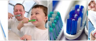
- Follow basic hygiene rules. Namely: wash your hands well with soap, especially when returning from the street, brush your teeth, use dental floss at least once a day, and also rinse your mouth with special means.
- You should definitely buy a new toothbrush. For those who have dentures, it is recommended to place them in a disinfectant solution overnight.
- After consuming liquid antibiotics, you should immediately rinse your mouth with water.
- When using spray corticosteroid drugs, you should wear a special nozzle.