The main methods of treating missing lower teeth
In the case where a tooth is completely missing (even the root is missing), restoration of the lower jaw teeth is carried out using one of the methods below:
- Installation of a fixed bridge prosthesis. The method is used when one or more teeth are destroyed or lost; the remaining healthy teeth serve as supports for the bridge structure. Fixed dentures cannot redistribute the chewing load on the jaw; therefore, when chewing, the load is not distributed evenly, but only on the supporting teeth to which the denture is attached. The fixation of fixed dentures is permanent, and it is impossible to remove such dentures yourself without the help of a dentist. The advantages of the method are aesthetic characteristics, restoration of chewing function, fast treatment time and relative low cost compared to dental implantation. Disadvantages - the need to depulp possibly healthy teeth, uneven distribution of the load, ongoing atrophy of the bone tissue of the alveolar crest in places where teeth are missing.
- Use of removable dentures. Most often they are used for long-term defects in the dentition (the absence of 3 or more teeth in a row). However, sometimes they are used in the absence of only 1-2 teeth (butterfly dentures) - if the patient does not want to undergo implantation or grind adjacent teeth for crowns. It is recommended to remove removable dentures once a day for hygienic procedures. Such dentures are less comfortable than fixed ones, because... occupy a significantly larger volume. Due to anatomical features, removable dentures for the lower teeth are smaller in size than the upper ones. Advantages: low cost, minimally invasive treatment, acceptable aesthetics. Disadvantages - does not restore chewing function, does not prevent bone tissue atrophy, the need for daily removal and periodic relining.
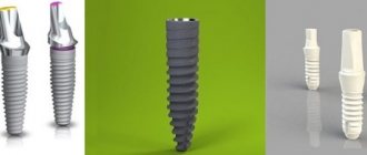
- Dental implantation. Implants are installed both when one or several teeth are lost in a row. Their purpose is to replace the biological root of a tooth; in prosthetics, implants are used as supports for artificial crowns: metal-ceramic or ceramic with a zirconium dioxide frame. Three main stages of implantation: installation of the implant into the bone tissue, formation of aesthetic gums, fixation of abutments on the implants and installation of crowns on the abutments. Implants can also be used in cases of complete absence of teeth in the lower jaw. The main advantage of implantation is the almost complete restoration of chewing function, and, as a result, the cessation of bone atrophy; such prosthetics also provide excellent aesthetics.
If the root of the tooth is not removed and there is a possibility of preserving it for use under a fixed prosthesis, the stump of the tooth is first restored, onto which a crown - metal-ceramic or completely ceramic - is fixed.
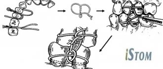
Bone or wire suture
In 1825, surgeon Rogers from Dublin performed the world's first operation using silver wire. With its help, he connected the fragments of the lower jaw. Later, in 1863, Russian surgeon Yu.K. Szymanowski successfully used a bone suture. From this point on, the bone suture was successfully used for osteosynthesis for many years. The main material was initially stainless steel, later it was replaced by titanium, nichrome, tantalum, etc.
There are various modifications of the bone suture (loop-shaped, cruciform, figure-of-eight, trapezoidal, double, etc.). The choice depends on the nature and location of the fracture.
The application of a bone suture occurs according to certain rules. It is important to make holes for the material in those areas where damage to the mandibular canal and tooth roots is excluded, and no closer than 1.0 cm from the fracture line. Ideally, the suture should cross the fracture line in the middle of the distance between the edge of the mandible and the base of the alveolar process.
Implantation of lower teeth is the best solution
Dental implants are artificial tooth roots, most often made of titanium (can also be ceramic or tantalum), a biocompatible metal, or its alloys. The word is made up of the Latin "im" meaning "inside" and "planta" meaning "sapling". Thus, implantology deals with replacing lost tooth roots with artificial ones through the installation of dental implants.
Implants for the lower jaw in the absence of teeth are inserted into the bone tissue, and then the dentist fixes a crown, fixed bridge or removable denture on them. Dental implants not only maintain the strength of your chewing muscles, but also strengthen your jaw bones. They help maintain facial proportions. Dental restoration with implants helps prevent the signs of aging. After implantation, patients look younger.
According to foreign statistics accumulated since the 1960s, the service life of a correctly installed implant ranges from 20 years to life. The service life depends significantly on the patient’s discipline in oral hygiene.
First aid for fractures of the lower jaw
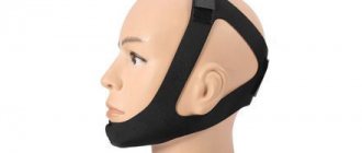
It can be carried out either at the scene of the incident or in an ambulance. It can be provided by both medical and non-medical workers in the form of mutual assistance.
The victim’s damaged jaw is temporarily (for several hours) fixed (the upper jaw is pressed to the lower) using bandages or other devices in order to be able to deliver him to a medical facility.
For fixation, the following can be used: a circular bandage parietal-mental bandage, a soft chin sling (Pomerantseva - Urbanskaya), an Entin splint (standard transport rigid bandage), as well as various types of intermaxillary ligature binding. If the patient has a traumatic brain injury, ligature binding is used with extreme caution - fixed jaws do not allow opening the mouth, and can lead to aspiration of vomit or blood.
Features of implantation of lower chewing teeth
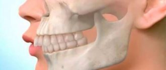
The chewing teeth of the lower jaw bear the highest load. Implantation of the lower teeth should be carried out after preliminary planning and modeling of future teeth, since in some cases, the artificial root will have to withstand the load, which was previously distributed over 3-4 natural tooth roots. And the chewing force is calculated to be at least 30 - 40 kg per tooth.
Another argument in favor of planning is that the diameter of the implant neck is smaller than the conventional neck diameter of the biological posterior tooth, which means that the shoulder between the crown and the implant will be larger, and any deviation of the implant axis from the direction of the application axis will lead to greater negative consequences.
Therefore, classical implantation with preliminary planning, in two stages with delayed loading, is an ideal option for restoring lower chewing teeth. That is, first an implant is installed, and then, a few months later, a permanent crown is fixed onto it.
Sometimes, before dental implantation, bone grafting is required - a surgical operation to build up bone tissue at the site where the implant is supposed to be installed. The fact is that for implantation to be effective, certain parameters of bone tissue are required - height, width and density, and if tooth extraction was traumatic or a lot of time passed between tooth loss and implantation, then most likely the bone has atrophied.
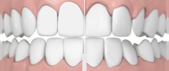
Which teeth bite is not considered correct?
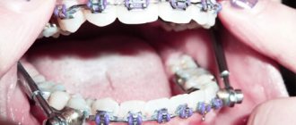
The presence of deviations from the norm is at least the basis for a dental diagnosis, based on the results of which a decision is made on the need for treatment. The best option is orthodontic correction in childhood, since the dentofacial apparatus, which is in the stage of formation, is more easily amenable to the targeted influence of a special machine and takes on the desired structure without surgical intervention. The most commonly diagnosed abnormal forms have certain signs that allow us to understand whether a person’s bite is correct or not, what is going wrong, and how this deviation can affect the further development of the jaw:
- Distal - diagnosed with excessive development of the upper jaw, noticeably protruding forward and distorting facial features.
- Mesial is a reverse pathological condition in which the mandibular row protrudes, almost completely covering the incisors and canines.
- Cross - characterized by the presence of free areas between the teeth, as well as a “scissor” intersection of elements in a random order.
- Deep - significant, more than 30%, overlap of the lower section.
- Open - contact during closure is observed only between individual chewing units, while in the incisal region there are pronounced gaps, creating the impression of a constantly slightly open mouth.
Any pathological manifestation requires timely medical intervention. Not all patients know what a correct bite in a person is and what it is needed for - but this indicator affects the quality of respiratory and speech function, ensures the conditions for normal food processing and the functioning of the gastrointestinal tract. By contacting Dentika dentistry, you can undergo a comprehensive diagnosis, receive recommendations from qualified specialists and, if necessary, resort to orthodontic correction services.
Features of implantation of lower front teeth
In modern humans, the chewing load on the front teeth, due to a specific diet, is small. People rarely use their incisors to bite tough foods; in recent centuries, they have been using a knife and fork more often. Rather, healthy and beautiful teeth in the smile area emphasize its beauty and certain life values of their owner.
Therefore, the decisive factor in choosing a method of prosthetics for the lower front teeth will be the most complete imitation of the appearance of a natural tooth.
The most aesthetic result is achieved with the help of metal-free ceramic prostheses on a zirconium dioxide frame, which are most often used in the manufacture of anterior dental implants. The abutment must also be made of zirconia.
An excellent result can be achieved by using metal-ceramic crowns, but in certain lighting, this may be noticeable to others... Although this applies more to the upper front teeth.
Recommendations
Treatment of mandibular fractures involves long-term immobilization of the jaws, which becomes a significant psychological problem for the patient. The success of treatment directly depends on how closely both the doctor and the patient act together and take the process seriously.
For the entire period of healing of the fracture, the patient is prescribed a diet (maxillary table No. 1 and No. 2); food is allowed to be taken only in a creamy consistency (well boiled and passed through a blender). In most cases, opening the mouth is impossible, because... The patient has an intermaxillary rubber traction device installed. Food is supplied through tubes, tubes and sippy cups.
From the moment of double-jaw splinting or osteosynthesis, the splints are left in place for an average of 21–30 days. If the doctor has confidence in the favorable course of the recovery process, then it is possible to reduce the period of wearing the intermaxillary rubber traction.
Even after removing the splints, the patient cannot fully open his mouth for 1-2 weeks. To restore the function of the chewing and facial muscles, he is prescribed myogymnastics.
Symptoms of jaw cancer
When a cancerous tumor originates in the upper jaw, there are practically no symptoms or they are identical to the manifestations of other types of pathologies, for example, sinusitis. That is why a malignant tumor can rarely be diagnosed in the initial stages.
With jaw cancer, patients experience:
- headache;
- numbness of the cheek;
- aching pain in the jaw;
- pungent odor from the mouth;
- purulent discharge from the nose.
Later, the following symptoms begin to appear:
- swelling of the neck;
- pain or numbness in the teeth closest to the tumor;
- loose teeth (a symptom of osteoporosis);
- alveolar processes may increase;
- a tumor from the bone of the upper jaw can grow into the orbit, causing displacement of the eyeball;
- neuralgic pain;
- headaches radiating to the forehead and temples;
- nosebleeds for no apparent reason;
- when the tumor reaches the trigeminal nerve, ear pain appears;
- mobility of the lower jaw is limited.
Sarcoma of the lower jaw is accompanied by the following specific symptoms:
- lower lip goes numb;
- contact teeth hurt;
- teeth become loose and fall out for no reason;
- bleeding ulcers appear on the mucous membrane;
- pain on palpation.
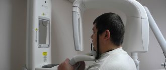
Diagnostics
Most often, the disease can be diagnosed only at the stage of metastasis due to the erased symptoms at the beginning of the disease.
X-ray diagnostics
Direct and lateral radiography of the jaw helps to detect a tumor in the bone. A neoplasm of odontogenic cellular material is also clearly visible on an x-ray. If there is a tumor, the image will show that the teeth are not in contact with the bone, and the alveolar edge does not have a clear enough contour.
General clinical examination
A patient with suspected jaw cancer must undergo a general blood test, urine test, and biochemical tests.
CT scan, biopsy, other diagnostic methods
To get an idea of the location and distribution of the tumor, a computed tomography scan of the jaws is done. A puncture biopsy of the submandibular lymph nodes and positron emission tomography are needed to detect metastases.
The most accurate diagnosis can be made by histological examination of the tumor material. Depending on the nature of the cancer cells, the analysis can be taken through the socket of a lost tooth or through trepanation of the jaw.
Additionally, you may need to consult an ophthalmologist, otolaryngologist, dentist, or surgeon.
Aesthetic consequences of loss of bone volume due to missing teeth
The facial changes that naturally occur with age can be exacerbated and accelerated by tooth loss. Pronounced aesthetic consequences result from loss of alveolar bone. Patients do not even suspect that all these changes in soft tissues are associated with tooth loss:
- Reduction in facial height occurs due to disturbances in the vertical dimension of the alveolar bone.
- A change in the labiomental angle and deepening of the vertical lines in this area give the face a rougher appearance.
- A malocclusion develops. As a result, the chin turns forward.
- The corners of the lips droop, the patient's face has an unhappy expression.
- Due to poor support of the lip by dentures and loss of muscle tone, the border of the red border of the lips becomes thinner.
- Age-related deepening of the nasolabial philtrum and other vertical lines on the upper lip is more pronounced with loss of bone volume.
- In edentulous patients, the decrease in the tone of the facial muscles that support the upper lip occurs faster, and the lengthening of the lip occurs at an earlier age. As a result, the smile ages.
- Bone atrophy has a negative effect on the attachment of the mental and buccal muscles to the body of the mandible. The fabric sags, creating a double chin. This effect is caused by decreased muscle tone when teeth are lost.
Reasons for the development of micrognathia
Anomalies of jaw development can be congenital or acquired.
Congenital ones include:
- Severe acute illnesses of a woman during pregnancy,
- Anomalies of intrauterine development of the fetus,
- Congenital pathologies of the development of the dental system,
- Genetic predisposition.
An acquired anomaly can result from:
- Maxillofacial trauma,
- Artificial feeding and improper sucking in infancy,
- Diseases of the endocrine system,
- Past infectious diseases and inflammatory processes,
- Early loss of baby teeth,
- Problems with nasal breathing.