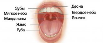
Epulis or central giant cell granuloma is a neoplasm in the oral cavity that appears on the gum as a result of exposure to various traumatic factors. This benign tumor grows from periodontal tissues and is most often located near the incisors, canines and small molars in the upper jaw. In rare cases, epulis appears on the lower jaw from the cheek side.
The main risk group is women under 30 years of age, however, some forms of epulis occur more often in children - at the stage of occlusion change. In newborns, epulis is usually not diagnosed, since it arises from periodontal tissue, so jaws with unerupted teeth are rarely susceptible to this disease.
What is epulis?
Epulis is a formation on the gum in the form of a tumor. It has synonymous names: supragingival, epulid, giant cell granuloma. Most often, epulis occurs on the alveolar jaw process, in the area of small molars; There are cases of occurrence on the body of the jaw (usually the lower one). Occurs both in adults (several times more often in women) and in children
Clinical picture
Since no clinical signs are observed in the initial stages, the disease is often detected at later stages. At this time you can see the following:
- A strange, lumpy mass gradually grows along the outer edge of the gum.
- There is a displacement of teeth. In severe cases, the dog's jaw resembles a saw.
- With advanced disease, even deformities of the facial part of the skull can develop.
- Excessive drooling. Drooling constantly and non-stop flows from the dog's mouth.
- Bad breath. In dogs, of course, it is not very pleasant anyway, but with the pathology we are describing, it becomes simply unbearable.
- Dysphagia. The dog cannot chew or swallow food.
- Weight loss.
- The gums begin to bleed. In addition, deep ulcerative lesions may appear on the gums.
Causes: why does a tumor form?
Epulis occurs under the influence of the following factors:
- Injury to the gums through: an overhanging filling;
- edges of a decayed tooth;
- tartar;
- burn;
- bruise;
- poor quality prosthesis.
What causes epulis
The cause of the development of epulis is injury to the mucous membrane by the sharp edge of a decayed tooth, an incorrectly installed filling or orthodontic equipment. Long-term irritation of epithelial tissue provokes the growth of new cells, which gradually form a growth. The risk of developing epulis increases in the presence of additional factors, including:
- hormonal changes (diabetes mellitus, menopause);
- pregnancy and postpartum period;
- immunity weakened by disease;
- period of eruption of baby teeth in children [3].
Symptoms and signs: what does epulis look like?
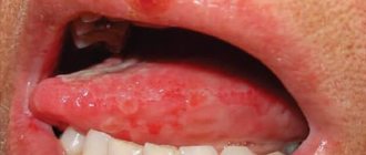
In general, epulids in diameter can reach from several mm to 3 cm or more. The color of the supragingival can be reddish-brown, brown, bluish , and also match the color of the gums . Often, ulcers may appear on the surface of a giant cell granuloma, which is most often caused by permanent trauma (sharp edge of a tooth, denture).
The shape of epulid is mushroom-shaped. The formation is located on a stalk, sometimes on a broad base. When pressed, it may move slightly. Epulis can be both soft and hard to the touch. Sometimes the teeth in the area of formation are mobile and slightly displaced.
There are two clinical forms of epulis: benign and malignant. Each of them has its own symptoms. Signs of a benign epulis tumor are:
- Slow growth;
- Diameter up to 2 cm;
- Usually asymptomatic course of the disease.
Malignant supragingival tissue is characterized by:
- Swelling of the gums;
- Rapid growth;
- Soreness;
- Destruction of dental root canals in tissues close to inflammation.
Signs
Epulis is distinguished by the following characteristics:
– the body is soft or compacted;
– location – on the jaw on one or both sides of the gums;
– gives mobility to closely spaced teeth;
– growing in size, changes the symmetry of the face;
– Bleeds when accidentally bitten.
Malignant tumors differ from benign ones in their symptoms.
It grows dynamically, becomes large, causes pain, and destroys nearby teeth. Malignant epulis is the cause of swelling of the oral tissues and bleeding gums.
The circumference of a benign formation is up to 3 centimeters. There is no pain or swelling. Since the tumor develops at a low speed, it remains undetected for a long period. Patients complain only of aesthetic discomfort.
Diagnostics
The diagnosis is made based on examination by a specialist and histological examination. A differential diagnostic method is used to exclude the presence of hypertrophic gingivitis in the patient. It has similar symptoms to epulis. An x-ray is also used for diagnosis, where, with epulide, rarefaction and destruction of bone tissue due to inflammation are observed.
Treatment of epulis in dogs
Treatment of epulis in dogs is predominantly surgical. You should not call a doctor at home. Treatment is carried out directly in the clinic and requires special knowledge and experience of a veterinarian. It is necessary to completely remove the pathological tissue while including healthy tissue to avoid relapse. In this regard, the doctor often has to remove part of the altered periosteum and affected teeth along with the epulis. When surgery is performed correctly, the prognosis is usually favorable. In some cases, radiation therapy or chemotherapy may be needed.
Classification
There are several types of epulis. Each of them has its own number in ICD-10 (International Classification of Diseases, 10th revision).
Fibrous (K06.82 - benign neoplasms):
Location area: vestibular side of the gum;
- Round or oval shape;
- Slow growth;
- Surface smooth/lumpy surface;
- Doesn't bleed;
- Wide base;
- Thick consistency;
- Light pink (to match the gum color).
Angiomatous:
- Location area: neck of the tooth;
Giant cell (K06.81 - other specified changes in the gums):
- Location area: alveolar part;
Causes
In most cases, the cause of epulide lies in injury to the oral mucosa. Sources of injury are a tooth fragment, an incorrectly installed filling, a removable denture, braces or bite defects. Other reasons include the following:
– jaw injuries from a fall, impact, or playing contact sports;
– hormonal disorders caused by diabetes, pregnancy, menopause, adolescence;
– the presence of mineralized plaque on the teeth and gums;
– burns of thermal and chemical nature;
– weakened immunity;
– period of mixed dentition.
The rapid growth of epulis is promoted by regular irritation of the mucous membrane.
Kinds
Dentists distinguish three types of epulide, differing in symptoms, structure and color.
- Angiomatous. The formation has a reddish-blue hue because it is penetrated by capillaries. Appears mainly in children, localized in the cervical area.
The pathology is characterized by a wide base, density, pronounced hypertrophy of the mucosa, and deformation of the alveolar process. Damage to the body causes bleeding.
This type of epulis is characterized by rapid growth and the likelihood of relapse. When it occurs in a small child, it prevents teeth from erupting, causing them to shift and form bite defects.
- Fibrous. The most common type of pathology is a small round/oval lump on the gum or hard palate. The painless, blue-purple formation has a wide base and a smooth surface.
Epulid of this type is characterized by slow growth and lack of bleeding. In some cases, ulcerations appear on the mucous membrane.
- Giant cell. Epulis of this type is characterized by rapid growth, a wide stalk and low density. Impressions of antigonizing teeth remain on the brown tumor.
This is the most dangerous type of epulide, causing displacement and destruction of teeth, prone to becoming malignant.
Treatment
Pathology is eliminated in several ways:
- Operationally. Under local anesthesia or general anesthesia, the tumor is excised, sometimes with the periosteum. In some cases, jaw resection is performed.
- Using a laser. The method is bloodless, while simultaneously disinfecting the surgical area. The prognosis is favorable due to a reduced risk of complications and a shorter postoperative period.
- Cryotherapy. Despite insufficient research on this method, the result is 97% favorable.
Prevention
To avoid negative consequences and the risk of cancer, you must follow the preventive recommendations:
- Regularly visit the dental clinic to undergo a medical examination, professional cleaning, and sanitation of the oral cavity.
- Do not wear anatomically inappropriate appliances to correct malocclusion or for aesthetic purposes.
- Maintain oral hygiene. Brush your teeth twice a day, and after each meal, rinse out any remaining food.
• Periodically examine the oral cavity for the appearance of tumors. If detected, immediately seek dental care.
Treatment methods, how the tumor is removed
Surgical removal
Treatment with epulid is carried out by removing the tumor. An incision is made at a short distance from the epulis, after which it is removed along with the periosteum. If surgery is performed on a giant cell granuloma, an area of bone tissue is also removed. The procedure is carried out using a bur or cutter. If the formation is more than 1 cm, then removal is often carried out under anesthesia with pharyngeal tamponade. The operated area is covered with iodoform gauze. Teeth are removed only when there is great mobility and root exposure. The dentist is the doctor you should contact if you have symptoms of epulis.
After stopping the bleeding, cauterization with alcohol, piocide, thermocautery, or an electrical regulator may be suggested. Treatment of angiomatous epulis can be carried out through sclerotherapy (urethane-quinidine mixture).
Laser treatment
One treatment option is laser removal of the granuloma. The procedure is performed under infiltration anesthesia. Excision of the body of the epulis is performed using a calibrated laser; removal of the body is done with tweezers.
Folk remedies
It is impossible to cure the supragingival tissue with folk remedies (without surgery). Folk remedies will only help in tissue healing. The composition of rinses for quick recovery should be agreed upon with the dentist. Effective recipes:
- Chamomile / sage / eucalyptus / calendula. Two tablespoons of dry plant + a glass of boiling water. Wait until the solution cools slightly. Rinse several times daily.
- A teaspoon of soda + a glass of warm water (prevents suppuration);
- A teaspoon of salt + a glass of hot water. Wait until it cools down (anti-swelling).
How does it occur in children?
In babies, epulis may appear during teething. Most often this is an angiomatous type of disease, but others can occur less frequently. It is advisable to contact a specialist as soon as possible to prevent the danger of tumor growth. Epulis must be removed through surgery. Sometimes the angiomatous appearance is treated with sclerotherapy
Epulis during pregnancy
Giant cell epulide is most common among pregnant women. Other types of disease are also common in expectant mothers. The body of epulis in pregnant women is subject to accelerated growth due to hormonal changes in the body (occurs for the same reason). Epulis should not be confused with a fistula (a white pimple-like formation with an opening from which white pus flows). In this case, folk remedies will only help speed up healing after excision. There is evidence that after pregnancy, epulis decreases significantly.
How to care for an operated animal at home?
Let us note right away that treatment with folk remedies is impossible . But still, at home, you can follow some rules that will help speed up your pet’s recovery:
- You should take your dog to the veterinarian at least once a quarter. If he belongs to representatives of brachycephalic breeds, a complete veterinary examination of the animal's oral cavity should be carried out each time during admission.
- It is very important to follow the instructions given to you by your veterinarian. During the postoperative period, a soft diet and regular rinsing of the oral cavity with antiseptic compounds are required. If for some reason it is impossible to purchase specialized products, you can use weak solutions of soda and table salt.
- The prognosis varies depending on the type, location, size, and resectability of the tumor.
- Unfortunately, to this day no specific prevention has been developed for this disease. We might advise against buying puppies whose ancestors suffered from the disease we describe.
FAQ
Epulis and bleeding: normal or not?
Yes, this is the norm . Angiomatous epulis most often bleeds heavily, while giant cell epulis bleeds moderately. If the supragingival body is injured, bleeding may occur.
Can the tumor go away on its own?
No, reverse development is almost impossible. It is necessary to consult a doctor to remove epulid. Most often, experts recommend excision of the tumor.
Can epulis reappear after cauterization?
Yes, relapses are not excluded . If a formation occurs, you should contact your dentist again.
How to distinguish epulis from hypertrophied gingivitis?
With hypertrophied gingivitis, the gums swell and enlarge, acquiring a scarlet color. There is also bleeding of the gums, heavy plaque on the teeth, an unpleasant odor, and discomfort when chewing solid food. With epulis, such changes in the gums do not occur.
Is it possible to place implants if you once had epulis?
Yes, you can.
How is epulis different from gum cancer?
Symptoms and consequences. Signs of gum cancer are:
- Large swelling;
- Whitish lesions;
- Painful sensations when touched;
- Pigmentation of the seal;
- Ulcers and cracks around the formation;
- Heavy bleeding.
Fatalities are extremely rare, but they do happen. Sometimes the signs of gum cancer are blurred, so if swelling occurs, you should consult a doctor. In the early stages, the disease is treated much faster and easier, but an advanced form can lead to many complications.
Will my gums heal after surgery?
Yes, it is overgrown. It is necessary to follow the wearing of the bandage and all the doctor’s recommendations for good healing.
Epulis on the gum under the denture
The reasons for the appearance of lumps under the prosthesis can be:
- Expired prosthesis;
- Improper dental care (lack of hygiene);
- Gum injury;
- Taking strong medications.
Diagnostic measures
As a rule, the veterinarian only needs to analyze the visible signs. They are quite characteristic, and therefore there are no particular difficulties in making a diagnosis. Despite this, it is important for a specialist to exclude or confirm the presence of other diseases that can give a similar clinical picture.
Click to view in a new window. Attention, the photo contains images of sick animals!
Therefore, veterinarians resort to the following diagnostic studies:
- A complete microscopic blood test , its biochemistry is checked, and urine is also examined. However, with “classic” epulis, all indicators are usually normal, their indicators do not differ in any way from the standard values.
- Visual examination of the oral cavity.
- Radiographs of the oral cavity . Useful in advanced cases of the disease, as they can be used to assess the extent of damage to bone and ligamentous tissues.
- X-rays of the chest. They are carried out when the malignant nature of the tumor is suspected or confirmed, since it is important for the veterinarian to make sure there are no metastases.
- Computed tomography (in some cases). This is done extremely rarely. This method is very useful from a diagnostic point of view, but it is very expensive, and specialized equipment is not available in every “human” clinic, not to mention veterinary hospitals.
- Finally, it is mandatory to study the tumor mass , a sample of which is obtained through a biopsy.