Sometimes situations arise when it is necessary to refill a dental canal. The reasons for such cases may be different. For example, poor-quality root canal filling, incorrect identification of the source of infection during initial treatment. In addition, the structure of the patient’s tooth or a violation of the tightness of the installed filling also affects the result of tooth treatment. We will tell you about the reasons for the need to refill a tooth and the rules for its implementation to obtain a reliable result.
Features of the procedure for re-treatment of a dental canal
Refilling the canal of a previously treated tooth is a special case in the practice of dentists. Before doing this, it is necessary to remove the old filling, clearing the entire dental canal of the filling material. During such work, the flexible rod may break. This will somewhat complicate the dental treatment process.
Typically, this problem occurs with a very dense filling that is closely adhered to the walls of the canal of the tooth being treated.
Difficulty also arises if it is necessary to refill curved dental canals. When cleaning such channels, you can damage their walls or even break the instrument. Such situations require increased attention from the specialist to ensure high-quality completion of the canal filling. Specialists are required to inform the patient about possible difficulties during treatment before the procedure.
How is a foreign body removed from the maxillary sinus?
To prevent the development of complications in the presence of foreign objects in the maxillary sinus, minimally invasive surgical intervention is performed. Its main purpose is to remove filling material. The operation can be performed using optical instruments such as an endoscope or laparoscope. When carrying out such interventions, significant trauma to bone tissue and incisions is not required. These minimally invasive operations are pain-relieved with local anesthesia and take no more than half an hour. Traditionally, endoscopic techniques are used to remove foreign bodies from the maxillary sinuses. To perform the operation, a small incision is made above the upper lip through which an endoscope is inserted. This optical device helps the surgeon detect a foreign body. The dental material is then removed. When performing laparoscopic surgery, tissue puncture is performed in the place where the bone tissue is thinnest. A special instrument is inserted into the laparoscope, which captures and removes the foreign particle. This technique is used in cases where endoscopic intervention cannot be performed for some reason. Sometimes operations are supplemented by such manipulation as curettage. This method is necessary in cases where, due to the filling material, a granuloma has formed on the tissues of the cavity. After surgical removal of a foreign particle, the patient is prescribed anti-inflammatory drugs to prevent inflammatory and purulent processes. To achieve the desired result, antibiotics, immunostimulants and other drugs may be prescribed. In addition, the affected sinus is washed with antibacterial, antiseptic and anti-inflammatory solutions. To increase the protective functions of the body, the patient is prescribed vitamin and mineral complexes. The best prevention of such complications is regular visits to the dentist. In addition, it is necessary to exclude facial injuries, try to avoid stress, eat right and lead an active lifestyle.
Why undergo a dental canal refilling procedure?
There are several reasons why you need to re-treat a dental canal:
- persistence or resumption of pain after filling;
- X-ray shows the presence of inflammation;
- incomplete closure of dental canals.
In addition, complications may result from errors or inaccuracies that arose during treatment. For example, when preparing the canal for treatment, an infection was introduced, access to the base of the canal is impossible, or there are pathological holes in the walls of the tooth or its bottom. In addition, no one is immune from the mistakes of the doctor performing root canal treatment.
When treating a dental canal before filling, the following problems most often occur:
- Dentim gets into the sawmill,
- the middle part of the canal is greatly expanded,
- tool failure,
- the integrity of the root walls is compromised.
When filling a canal, the main problems arise if it is not completely filled, the filling material extends beyond the hole, or there is a longitudinal fracture at the root of the tooth. Medical errors are also possible at this stage - the doctor may incorrectly estimate the length of the canal or not completely clean it. This may contribute to the development of inflammation.
Extension of filling material beyond the apex: endodontic failure versus clinical success
Many dentists believe that procedural errors in daily practice, such as extrusion beyond the apical foramen and contact of filling material (most often gutta-percha and zinc oxide eugenol sealer) with periapical tissues after treatment, are a direct cause of endodontic failure.
Some authors show the negative impact of extending the filling material beyond the apex on the predictability of endodontic treatment and emphasize the following:
- High incidence of postoperative pain with exacerbation of inflammation
- Toxicity of materials
- Constant irritation of the periapical tissues with manifestations ranging from mild inflammation to a general reaction of the body.
In contrast, other authors have argued that apex extrusion in itself does not directly contribute to endodontic failure, although it should be avoided. Ingle says that endodontic treatment can be successful despite extrusion of material beyond the root apex. Schilder notes that he has not encountered complications specifically associated with excessive filling of the canal. Weine states that periapical tissues, fortunately, tolerate contact with gutta-percha well and therefore endodontic failure due to its removal beyond the apex is rare. In most cases, there is no pathological radiological evidence, and sometimes there is even a decrease in the amount of excreted material due to phagocytosis.
Image 1 – Mandibular first molar with amalgam filling and insufficient root canal obturation.
After retreatment, the radiograph shows three-dimensional filling of the root canals with a slight extension beyond the apex in the mesial canals obturated with Thermafil #25 and in the distal canal obturated by vertical condensation of heated gutta-percha.
After 5 years, the excess material had disappeared and the periapical tissue remained healthy.
These contradictory results can be explained by differences in research methodology and different composition of the derived materials. In general, the most common causes of discrepancy are related to differences in apical anatomy, limiting the effectiveness of endodontic treatment in various clinical situations.
Image 2 - The main factors leading to the removal of material beyond the root apex:
External inflammatory apical root resorption associated with apical damage to a previously treated tooth.
Incompletely formed root.
Excessive instrumentation with deviation from the natural course of the canal and apical perforation.
Inadequate handling of plasticized or unplasticized gutta-percha and sealants during obturation.
It is extremely important to identify and distinguish between the removal of filling material in three-dimensionally cleaned, formed and obturated root canals and in canals with obstructive overflow in combination with incomplete internal obturation.
Image 3 – Typical view of an exposed gutta-percha point: note the scratches on the point.
Image 4 – The apical foramen is completely filled.
Image 5 – Proper expansion for obturation, but insufficient filling of the canal. Note the transport of the apex during canal formation. (Courtesy of Dr. M. Vitullo).
Image 6 - Postoperative radiograph of the mandibular first molar showing a large apical lesion of endodontic origin. The mesial canals were obturated with Thermafil. A follow-up examination after 4 years shows osteoreparation. Excess filling material did not interfere with bone restoration.
Image 7 – Thermafil carrier extended beyond the apex in the distal root. Thermafil is a good method of obturation if the protocol is followed. The media was removed and the canals were refilled. After 6 months, the lesion was smaller than on the preoperative radiograph, and the patient noted the disappearance of pain when chewing.
Image 8 - These images show that even MTA can be released into the periapical tissue. In this case, the rule is the same as in the previous examples: if the canal is well cleaned, excess MTA cannot interfere with tissue restoration. 6-year follow-up kindly provided by Dr. R. Tonini.
Conclusion
Obturation is the last stage of endodontic treatment. Bringing either sealer or gutta-percha beyond the apical foramen will not necessarily be a mistake when performing obturation. However, this may result in an error in canal formation when parallel canal walls were created instead of conical ones, apical resorption or expansion of the apex.
Therefore, it is necessary to determine the direct causes of excessive root canal filling. “Is the excess material remaining after a complete three-dimensional obturation or is the material brought beyond the apex in combination with incomplete internal obturation of the canal?” Excess material beyond the apex is clearly not the goal of endodontic treatment, but rather the result of safely generated hydraulic forces in achieving three-dimensional obturation of the root canal system.
Numerous cases of root apex removal that have been successful, as evidenced by clinical and radiological signs of long-term restoration of periapical tissues, should reassure patients, doctors and insure against any legal charges.
Source: forum.stomatologija.su, styleitaliano.org
How does the process of refilling dental canals take place?
The current level of development of medicine in the field of dentistry is quite high. But dentists make mistakes, which is why filling dental canals again is quite common. At this point in time, doctors use several methods for performing this procedure.
Mechanical methods involve the use of a special instrument - an endodontic motor and an apex locator. In this case, antiseptic agents are added to the filling material. To prevent the development of inflammation, the doctor must very carefully and completely fill the dental canal with filling material.
Medicinal methods involve the use of products with organic solvents. They are able to soften and destroy the filling in a few minutes.
The cement filling can be removed from the canal using ultrasound and an endo-nozzle. This canal cleaning is usually carried out in one procedure.
How can a complication be identified?
External signs of dental materials getting into the maxillary cavities and patient interview data may not be enough to make a correct diagnosis, and additional studies are carried out to confirm complications of the dental procedure:
- x-ray of the maxillary sinus;
- MRI;
- CT;
- puncture.
The diagnostic plan may include several procedures. Their number is determined by each doctor individually.
Modern technologies in the field of tooth refilling
Dental treatment under a microscope is one of the newest services that has appeared in dentistry. High-tech equipment allows specialists to fill the dental canal with better quality, more accuracy and efficiency. Thanks to the use of such a device, the likelihood of causing harm to healthy tissue is close to zero. In addition, the treatment is carried out very delicately. Microscope glasses allow you to view the tooth at multiple zooms. This will allow you to see all the affected areas that may simply not be visible during normal examination.
A microscope allows dentists to perform root canal treatment faster and safer for the patient.
What symptoms may indicate a foreign body in the maxillary sinuses?
After dental treatment, a person may suspect the development of this complication if the following signs appear:
- the appearance of pain when performing light taps on the facial bone under the eyes near the nose;
- severe, aching headache;
- prolonged and persistent runny nose;
- aching pain in the upper jaw, aggravated by chewing.
One of these symptoms or a combination of several may indicate that dental medications have entered the maxillary sinus cavity. This condition is a reason for mandatory consultation with a doctor and the visit should not be postponed. If the patient does not see a doctor on time or has a weakened immune system, the disease becomes more complicated. Because of this, a person develops thick, purulent and profuse nasal discharge with a specific odor. In addition, the progression of the inflammatory process causes an increase in temperature and a deterioration in general health. In some cases, immediately after dental mixtures enter the sinuses, inflammation does not develop. However, this condition is also unsafe for his health in the future, since various negative factors (for example, hypothermia, stress, injury or hypovitaminosis) can provoke the development of inflammation and a deterioration in general well-being.
Removing the seal under the anchor pin
Obturation of the tooth root canal is done in parallel using anchor pins. If the filling in the lower part of the root is placed correctly and hermetically, then it is left to install the pin structure. This procedure is carried out over two trips to the dental office.
On the first day, the doctor treats the dental canal mechanically, softens the filling with medications and cleans the passage with an endo-instrument. The doctor then soaks a cotton swab in the medicine and inserts it into the canal. The patient takes the medicine for a couple of days and then makes another visit to the doctor, during which the cotton swab is removed and the canal is refilled.
Why is this complication dangerous?
The maxillary sinuses located above our upper jaw (their area extends from the inner corner of the eye to the outer) perform important functions. They can warm the air and thereby protect the respiratory tract from hypothermia and foreign particles. The sinuses are located under the bone tissue and if its thickness is too small, when cleaning the dental canals, filling masses can fall into these cavities. Once the filling is completed, it is no longer possible to remove the foreign material on your own, since this procedure can only be carried out by a specialized specialist who has the necessary tools. The doctor can perform this procedure without difficulty and it will not take much time. It is numbed with local anesthesia and after removing the foreign body from the maxillary sinus, the patient will need to stay in the clinic for several hours. In some cases, discharge occurs the day after this minimally invasive procedure.
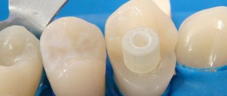
Preparation for filling
Before filling the canals with filling material, the doctor must thoroughly clean them and prepare the cavity in a special way. This process takes place in several stages:
- Removal of affected tissue
Most often, the need for root canal treatment arises as a consequence of another pathological process, in particular caries. To destroy the source of infection and open access to the mouths of the canals, the dentist removes dead and diseased tissue using a bur. - removal
Pulp is the sensitive tissue of the tooth, a plexus of blood vessels and nerves located in the coronal part and inside the roots. The method of filling root canals involves removing the pulp using a special tool. Most often, this procedure, like the previous one, is performed under local anesthesia to eliminate pain and discomfort when removing the pulp. - Measuring canals
In order to perform a quality filling, the doctor needs to obtain as accurate information as possible about the properties and current condition of the canals. For this purpose, before treatment, as a rule, a targeted radiograph is performed. An important procedure is also measuring the length of the channels. The length is individual and depends not only on the size of the root, but also on the degree of curvature of the canal. - Mechanical processing
Before filling, the channels are cleaned and expanded using special tools. This is necessary to completely remove the affected tissue, as well as to more densely and evenly fill the canal cavity with filling material. Mechanical processing is performed using special thin tools - files. With the help of a file, the canal is passed completely, from the mouth to the apex.
Only after completing all the preparatory steps can the doctor proceed to filling.
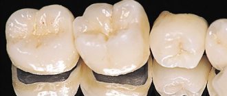
Methods for filling root canals
There are several basic techniques used in the treatment of pulpitis and periodontitis:
- One-paste method
The channel is filled with a plastic, subsequently hardening material. The method is outdated and gives a large number of complications. - Single pin method
First, the root canal is filled with a special paste, and then a gutta-percha pin is inserted into it. The percentage of complications is lower, but this method is also gradually being phased out. - Filling with “thermophile”
A more modern method, also based on the use of an initially plastic material - heated gutta-percha. The fluid state allows the material to densely fill all cavities inside the canal, which significantly reduces the risk of complications. The disadvantages of the method include high requirements for the doctor’s qualifications and relatively high cost. - Gutta-percha condensation method
This method ensures even more dense filling of the canal lumen with filling material. Currently, the method is one of the most common. The procedure is performed in several stages.
Errors when filling
Endodontic treatment is a rather complex procedure. For successful implementation, you should remember three principles: proper processing, high-quality asepsis, reliable sealing.
At each stage, the doctor is capable of making a considerable number of mistakes:
Tooth perforation - in the absence of proper instrumentation and expansion, there is a risk of damaging the wall or bottom of the root canal. This happens, in most cases, due to a poor understanding of the anatomical structure of the roots and cavity of the tooth. Poor machining is the result of incorrect tool size selection. If the diameter of the file or reamer does not match the diameter of the canal, it can damage the apical foramen or leave an accumulation of dentinal filings on the walls.
Perforation of a tooth with a tool
Breakage of an endodontic instrument - this complication occurs when the technique and sequence of using files and reamers is violated, or when a blunt, damaged instrument is used. Breakage can occur during both manual and machine processing. To prevent this type of complication, the doctor should check all instruments before the appointment and make sure they are functional.
A piece of instrument in the channel
Incorrect expansion of the canal and its filling. To achieve high-quality obturation, the canal must be expanded to the optimal diameter. To avoid the formation of voids and further infection, the canal must be given a shape that is convenient for filling with material. Do not forget that canals filled with one paste will not have sufficient sealing, and over time, pores will form in the material.